


Benefits of AI Interoperability in Dental Radiology
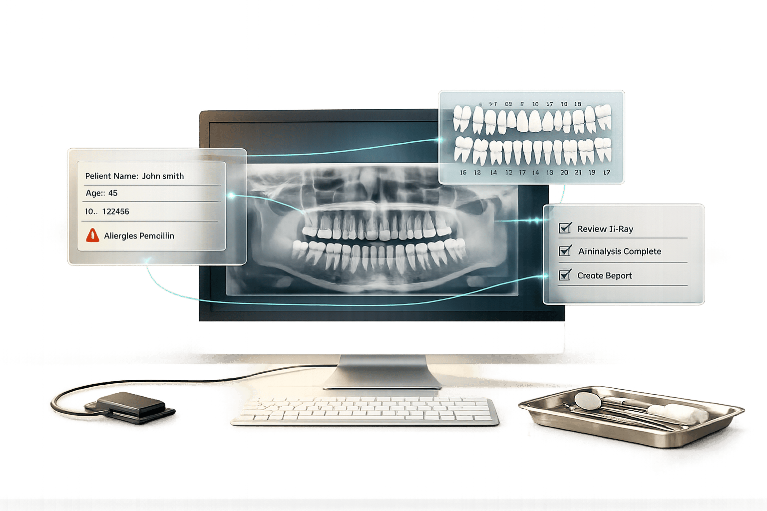
AI interoperability in dental radiology addresses key challenges like data silos, manual workflows, and diagnostic inconsistencies. By enabling AI systems to seamlessly connect with existing platforms, dental practices can improve diagnostic accuracy, streamline processes, and enhance patient outcomes. Here’s what you need to know:
- Data Integration: AI interoperability ensures patient data flows smoothly between systems, reducing manual re-entry and errors.
- Improved Diagnostics: AI systems trained on diverse datasets outperform traditional methods in detecting conditions like periodontitis, caries, and osteoporosis.
- Faster Workflows: Automated processes cut review times from 20–25 minutes to 5–10 minutes, freeing up clinicians for critical tasks.
- Patient Safety: AI reduces diagnostic errors, with tools achieving up to 92% sensitivity in detecting oral diseases.
- Standardisation: Universal formats like DICOM and HL7 ensure compatibility across systems, preventing vendor lock-in and enabling broader data use.
AI interoperability sets the foundation for better care and teledentistry opportunities, particularly in underserved areas. While implementation requires planning, compliance with standards, and clinician involvement, the potential benefits for both practices and patients are clear.
Dental Interoperability and Data Exchange Panel
sbb-itb-2be92ed
Main Benefits of AI Interoperability in Dental Radiology
When AI integrates seamlessly into dental platforms, it can significantly improve diagnostic precision, streamline workflows, and enhance patient outcomes. By addressing challenges like data silos and manual data entry, effective AI integration transforms dental radiology. Below are the key benefits that interoperability brings to the field.
More Accurate Diagnoses
AI-powered systems, when trained on diverse datasets, deliver a level of diagnostic precision that surpasses traditional methods. For example, in February 2026, researchers tested HC-Net+, a deep-learning model trained on 10,881 orthopantomograms from institutions like the Hong Kong University Faculty of Dentistry, Shanghai Ninth People’s Hospital, and Sapienza University of Rome. The AI achieved an impressive AUROC of 94.2% for detecting stage II–IV periodontitis, compared to 85.6% for periodontal specialists, and maintained accuracy above 92.4% across multiple international locations [7].
AI systems have also demonstrated exceptional accuracy in other areas: 93.67% for teeth identification, 91.5% for AI vs. traditional caries detection, and 89.29% for osteoporosis screening. Pooled caries detection results showed a sensitivity of 0.86, specificity of 0.91, and an overall area under the curve of 0.94 [5] [6]. These advancements even enable junior dentists to match the diagnostic abilities of seasoned professionals [7].
Traditional methods often lack consistency. For instance, manual bone loss identification has shown Cohen’s kappa values between 0.454 and 0.482, revealing poor agreement among practitioners [7]. Interoperable AI addresses this by standardising data formats, ensuring consistent diagnostic performance regardless of the imaging hardware or software used [8].
Faster Workflows
Beyond improving diagnostic accuracy, interoperability significantly speeds up clinical processes. Automated radiographic analysis reduces review times from 20–25 minutes to just 5–10 minutes. Similarly, CBCT tasks that traditionally take tens of minutes can now be completed in seconds with fully automated segmentation [4] [10].
"The use of DIAGNOCAT artificial intelligence can help dentists to achieve faster and more objective quality control in diagnostics and treatment." – MDPI Electronics [4]
AI systems also eliminate repetitive tasks by generating detailed dental and orthodontic reports that highlight issues like caries, bone loss, and periapical lesions automatically. This frees up dental assistants and radiology technicians to handle complex preprocessing tasks, such as cephalometric analysis and nerve pathway identification, while allowing specialists to focus on clinical decisions rather than manual data entry [4].
Better Patient Care and Safety
Interoperable AI not only improves efficiency but also enhances patient safety by reducing diagnostic errors. In traditional dental radiology, errors of omission have led to undertreatment in 72% of cases and legal complications in 82% of instances. By acting as cognitive aids, machine learning algorithms achieve a diagnostic odds ratio of 20.3, drastically cutting down on interpretive errors [9].
"ML algorithms serve as valuable assistive cognitive aids, reducing interpretive errors and enhancing clinician confidence in dental radiology." – Shwetha Hegde, PhD, University of Sydney School of Dentistry [9]
AI-assisted tools for oral cancer detection have shown a pooled sensitivity of 92% and specificity of 91.9%. Similarly, AI models for periodontal disease assessment achieve 87% accuracy, rivalling expert periodontists [10]. Robotic implant placement systems, guided by real-time AI feedback from CBCT scans, achieve a mean coronal deviation of just 0.7 ± 0.3 millimetres – far more precise than freehand methods [10].
Interoperability also ensures that critical patient data, such as medical history and prior radiographs, is accessible to clinicians. Without this integration, vital information can remain trapped in isolated systems, leading to incomplete decision-making. Breaking down these barriers not only improves care quality but also reduces the estimated $36 billion wasted globally on manual data re-entry and repeated searches [1].
How AI Interoperability Solves Radiology Problems
Dental radiology faces several hurdles, from incompatible software to time-intensive manual tasks. These challenges are tackled through three main interoperability approaches: standardised data formats, automated image processing, and collaborative human-AI workflows. Together, these strategies improve diagnostic accuracy while streamlining operations.
Standard Data Formats for Compatibility
Interoperability relies on universal standards like Digital Imaging and Communications in Medicine (DICOM) and Health Level 7 (HL7). These standards allow dental images and patient data to flow seamlessly between systems. While DICOM focuses on imaging data and metadata, HL7 FHIR (Fast Healthcare Interoperability Resources) uses XML and JSON for sharing clinical information, ensuring compatibility across various practice management platforms [2][11].
The Integrating the Healthcare Enterprise (IHE) initiative goes a step further by offering implementation guides for applying these standards to real-world scenarios. Dr Kent Hutson, AI Lead Technical Consultant at Radiology Partners, explains:
"IHE doesn’t create standards, it tells us how to apply existing standards – DICOM, HL7 and others – to specific use cases. In doing so, it provides a script that developers can use to design interoperable solutions."
To ensure software complies with these standards, vendors participate in Connectathons – events where tools are rigorously tested before being used in clinics [2]. Additionally, calibration phantoms (standardised reference materials) help align measurements across different imaging machines. This ensures that AI models trained on one scanner can deliver consistent results on another, eliminating compatibility issues that often arise in isolated systems [11].
Automated Image Processing
Interoperable AI takes over repetitive tasks, like CBCT segmentation, dramatically reducing the time spent on manual processes. Tools such as Diagnocat can automatically segment CBCT scans, identifying teeth, bones, nerves, and airways in seconds rather than minutes [4]. This efficiency addresses the bottlenecks that many dental practices face.
Dr Ali Tejani, a radiologist at UT Southwestern Medical Centre, notes the impact:
"Interoperability makes things simpler, scalable and translatable for both the vendor and the consumer. It’s the difference between manually reporting measurements or AI results versus auto population of information in our radiology reports."
By auto-populating reports, AI eliminates transcription errors and frees clinicians from tedious data entry. This allows them to focus on patient care and decision-making. Beyond saving time, automated systems also improve diagnostic accuracy by facilitating better human-AI collaboration.
Combined Human-AI Models
Interoperable systems enhance diagnostic reliability by integrating human expertise with AI precision. Acting as a second reader, AI helps catch pathologies that clinicians might miss. This is particularly important for addressing errors of omission, which contribute to undertreatment in 72% of cases and legal issues in 82% [9].
Transparency is key in these systems. Explainability features ensure clinicians understand how AI arrives at its conclusions, maintaining trust and allowing practitioners to make informed final decisions [9]. This feedback loop not only improves AI performance but also ensures the technology adapts to specific practice needs over time.
| Challenge | AI Interoperability Solution |
|---|---|
| Interpretive Errors | Machine learning aids improve sensitivity to 79% [9] |
| Workflow Inefficiency | Automated CBCT segmentation reduces task time to seconds [4] |
| Data Incompatibility | DICOM and HL7 standards enable seamless data sharing [2] |
| Measurement Uncertainty | Calibration phantoms standardise values across devices [11] |
| Privacy Risks | Automated de-identification safeguards patient data [11] |
Implementation and Future of AI Interoperability in Dental Radiology
AI-Integrated vs Standard Dental Radiology Systems Comparison
Requirements for Integration
For dental practices to effectively integrate interoperable AI systems, adopting global standards and updating procurement processes is essential. Protocols like DICOM and HL7 are crucial in ensuring different software platforms can exchange data smoothly. Strategic planning and early involvement of clinicians are also key to overcoming challenges such as data silos and manual workflows [2][12].
When acquiring new radiology systems, practices should include clear specifications in their Request for Proposal (RFP) documents. Dr Kent Hutson, Co-chair of the IHE Radiology Planning Committee, explains:
"If a developer follows the provided script and data format, which IHE refers to as ‘profiles’, then the developed solution will be interoperable with any system… that supports that same profile." [2]
Additionally, machine-readable codes like RadLex, SNOMED CT, and LOINC should be adopted to streamline automated report generation and minimise manual data entry [2][12]. For clinics using multiple AI tools, employing an AI orchestrator can simplify the management of data traffic [12].
Post-market monitoring is another critical step. AI systems can experience concept drift, where accuracy fluctuates due to changes in patient demographics or software updates. To address this, a control sample approach – using reserved test cases for routine evaluation – can help detect performance issues early [3]. The Royal Australian and New Zealand College of Radiologists (RANZCR) underscores the importance of rigorous evaluation to ensure AI systems meet ethical standards and support local patient populations [3].
Australian practices must also comply with the Office of the Australian Information Commissioner (OAIC) guidelines. The Australian Dental Association highlights the importance of privacy and data security:
"Any data should be obtained with appropriate permissions, privacy controls, checked for accuracy and relevance, only used for the stated purpose, and stored securely as per the OAIC guidelines." [13]
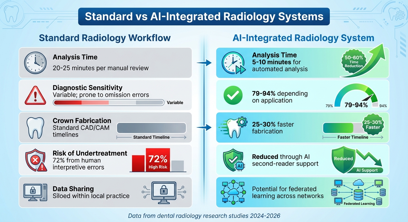
Comparison of Standard vs AI-Integrated Systems
The table below outlines how AI integration can significantly enhance standard radiology workflows:
| Metric | Standard Radiology Workflow | AI-Integrated Radiology System |
|---|---|---|
| Analysis Time | 20–25 minutes per manual review [10] | 5–10 minutes for automated analysis [10] |
| Diagnostic Sensitivity | Variable; prone to omission errors [9] | 79%–94% depending on application [10][9] |
| Crown Fabrication | Standard CAD/CAM timelines | 25–30% faster fabrication [10] |
| Risk of Undertreatment | 72% from human interpretive errors [9] | Reduced through AI second-reader support [10][9] |
| Data Sharing | Siloed within local practice | Potential for federated learning across networks [10] |
Future Opportunities in Teledentistry
AI interoperability is not just about improving current workflows – it also sets the stage for advancements in teledentistry and its legal and ethical considerations. Interoperable systems can act as a "safety net" in high-volume screenings or in areas with limited access to specialists, providing much-needed diagnostic support [3][9].
Federated learning offers a promising solution for Australian dental practices, allowing institutions to collaboratively develop AI models without sharing sensitive patient data. This approach enhances diagnostic accuracy while maintaining privacy [10]. AI integration in public health surveillance also enables real-time monitoring of oral disease trends, benefiting population-level health initiatives [10].
Looking ahead, Explainable AI (XAI) methods like SHAP (SHapley Additive Explanations) could play a pivotal role. By offering visual explanations for AI-generated diagnoses, XAI empowers clinicians to validate findings instead of relying on opaque "black box" outputs [9].
For successful teledentistry implementation, addressing infrastructure gaps is crucial. Practices need reliable hardware and robust internet connectivity, particularly in rural areas. The shift from occasional dental visits to continuous monitoring – enabled by AI-powered biosensors and smart devices – could revolutionise care delivery, especially for remote communities [10].
Conclusion
AI interoperability is transforming dental radiology by revolutionising how diagnostic data is captured, shared, and utilised across healthcare systems. By adopting standardised protocols, data silos can be dismantled, saving billions currently lost to manual data re-entry. This structured framework ensures AI algorithms have access to high-quality data with clear semantics, which improves accuracy and reduces risks[1].
Machine learning algorithms, when used as cognitive aids, demonstrate 79% sensitivity, significantly cutting down omission errors that lead to undertreatment in 72% of cases[9]. Additionally, when patients see AI-generated annotations on their radiographs, 92% indicate they are more likely to proceed with recommended treatments[14]. This shows how interoperable AI tools not only enhance diagnostic precision but also increase patient engagement – an impactful combination for better clinical outcomes.
For Australian dental practices, strategic planning is essential. Dr Kent Hutson, Co-chair of the IHE Radiology Planning Committee, emphasises the importance of ensuring vendors adhere to specific IHE profiles:
"require that any vendor applying to the RFP support a specified IHE profile"
[2]. This approach ensures smooth integration and avoids vendor lock-in, setting the stage for long-term innovation.
Beyond improving diagnostics and workflows, interoperable AI holds promise for advancing teledentistry. It can facilitate seamless data exchange and expand access to dental care in rural and underserved areas[15]. While the technology is ready, effective implementation requires careful planning, comprehensive clinician training, and ongoing monitoring to address performance drift. By focusing on standardised solutions that meet real clinical needs, dental practices can fully leverage AI’s potential while keeping patient care as the top priority.
FAQs
What does “AI interoperability” mean in dental radiology?
AI interoperability in dental radiology means different AI systems, imaging devices, and electronic health record platforms can smoothly share, interpret, and use data. This compatibility allows for better diagnostic precision and more efficient workflows by ensuring these technologies communicate effectively using standardised methods.
What standards should my dental imaging and practice software support (DICOM, HL7/FHIR, IHE)?
Your dental imaging and practice software needs to align with standards like DICOM, HL7/FHIR, and IHE profiles. These standards are crucial for ensuring smooth data exchange and integration across healthcare systems. Frameworks like the IHE Radiology and Dental Technical Frameworks outline how these standards work together to create interoperability. By supporting these protocols, your practice can streamline diagnostic workflows and foster better collaboration between dental and medical professionals.
How can clinics check that an AI tool stays accurate over time (performance drift)?
Clinics can keep AI tools reliable by putting consistent quality checks in place. This means regularly monitoring how well the tool performs and making updates to its algorithms when necessary to address any changes or "drift" in performance. Routine evaluations are key to ensuring the tool stays dependable and maintains its diagnostic precision over time.
Related Blog Posts
- AI-Powered Dental Imaging: Future Trends
- AI-Powered Radiology: What Dentists Need to Know
- AI Dental Radiology: Benefits for Patient Care
- AI vs. Traditional 3D Dental Imaging
Important Notice: Any surgical or invasive procedure carries risks. Before proceeding, you should seek a second opinion from an appropriately qualified health practitioner.
Individual results may vary. The information provided in this article is for educational purposes only and does not constitute medical advice.
Checkout Related Blogs


Get in touch with us
For more information, call us now to start feeling better. Or fill the form below to make appointment
The Latest News from Complete Smiles
How to Clean Clear Plastic Retainers
Checklist for Choosing Wearable Dental Devices
Checklist for Choosing Cloud AI Platforms in Dentistry
Complete Smiles Bella VistaAccepts All Major Health Funds, Including


