


CBCT Technology: Mechanisms Behind 3D Imaging

CBCT (Cone-Beam Computed Tomography) is a 3D imaging system used in dentistry to provide detailed views of teeth, jaws, and surrounding structures. Unlike 2D X-rays, it uses a cone-shaped X-ray beam and a rotating scanner to generate hundreds of 2D images, which are processed into a 3D model. This allows dentists to examine complex areas from multiple angles.
Key Features:
- Detailed Imaging: Provides sub-millimetre resolution for precise diagnostics.
- Radiation Efficiency: Uses 3–20% of the radiation dose compared to conventional CT scans.
- Fast Scans: Takes 5–40 seconds, reducing patient discomfort.
- Applications: Useful in orthodontics, dental implants, root canal therapy, and more.
Why It’s Useful:
- Eliminates overlapping structures seen in 2D X-rays.
- Offers accurate measurements for treatment planning.
- Safer and more efficient than conventional CT for dental purposes.
CBCT has become an important tool in dentistry, improving diagnostic accuracy and aiding in procedures like implant placement, orthodontic planning, and endodontic evaluations. Its ability to combine precision with reduced radiation exposure makes it a preferred choice for dental professionals.
How CBCT Technology Works
X-Ray Beam and Rotation
CBCT technology operates using a cone-shaped X-ray beam paired with a flat-panel detector, both mounted on opposite sides of a rotating C-arm or gantry. During a scan, the patient’s head is stabilised with chin rests, forehead supports, or bite blocks to reduce movement. The gantry then rotates 180° to 360° around the maxillofacial area. As it rotates, the cone-shaped beam emits X-rays, while the flat-panel detector captures 150–200 high-resolution 2D images from various angles. This detector, made with amorphous silicon and a scintillator, converts the transmitted X-rays into digital signals [2]. Each image reflects the combined attenuation of X-rays as they pass through teeth, bones, and soft tissues. Unlike traditional CT scans that use a fan-shaped beam to capture sequential slices, CBCT captures the entire area of interest in one sweep. This approach reduces scan time and mechanical complexity while gathering all the data needed for 3D reconstruction [3].
To optimise image quality and minimise radiation exposure, the cone beam is collimated to a specific field of view. Clinicians can adjust this field of view depending on the diagnostic needs. A smaller field of view provides higher spatial resolution and reduces scatter, making it ideal for focused assessments like examining a single tooth or planning a localised implant. This also lowers the patient’s radiation dose, adhering to Australian dental practice guidelines for radiation protection [2]. These captured images serve as the foundation for the 3D reconstruction process, which is described in the next section.
Image Reconstruction Algorithms
Once the images are captured, specialised algorithms transform the raw data into accurate 3D models. The most commonly used method in dental CBCT is the modified Feldkamp–Davis–Kress (FDK) algorithm [3]. This process begins with pre-processing each image to correct for blurring and intensity inconsistencies. The algorithm then back-projects these filtered images into a 3D grid of voxels, assigning each voxel an attenuation value based on how much radiation was absorbed along intersecting X-ray paths. By repeating this for all projection images, the system creates a complete 3D dataset. This dataset can be reformatted into axial, coronal, and sagittal planes, as well as custom cross-sections and 3D surface renderings.
Dental CBCT systems can achieve sub-millimetre voxel sizes, often ranging from 0.08 to 0.2 mm. This allows for the detailed visualisation of intricate structures like root canals and trabecular bone [2]. Modern software enables the reconstruction of the full 3D volume in under a minute, giving clinicians almost immediate access to multiplanar images. Advanced practices, such as Complete Smiles Bella Vista (https://completesmilesbv.com.au), leverage these capabilities for precise treatment planning in areas like orthodontics, implantology, and endodontics.
Short scan times, secure head stabilisation, and motion-correction algorithms work together to minimise artefacts caused by patient movement, ensuring high-quality imaging results.
Technical Features of CBCT
Radiation Dose and Beam Limitation
CBCT machines are designed to use much lower radiation doses compared to conventional medical CT scans – typically only around 3–20% of the dose needed for similar head and neck imaging [2]. This is made possible by the use of a cone-shaped X-ray beam and collimation, which narrows the field of view (FOV) to the specific area being scanned. With adjustable FOVs, clinicians can focus only on the area of interest, reducing unnecessary radiation exposure and limiting scatter.
The technology also operates with lower tube currents (1–15 mA at 90–120 kVp) and employs pulsed exposures during a single 360-degree rotation. This results in effective doses ranging from 50–200 µSv, which is significantly lower than medical CT scans (up to 2,000 µSv) and closer to intraoral or panoramic X-rays (1–8 µSv). These features allow CBCT to achieve a balance between reduced radiation exposure and high-quality imaging.
Resolution and Accuracy
CBCT doesn’t just minimise radiation – it also delivers impressive spatial accuracy. By using isotropic voxels (which are equal in all dimensions), CBCT systems achieve sub-millimetre spatial resolution. Dental CBCT devices typically offer voxel sizes between 0.075 and 0.4 mm. Smaller voxels are ideal for detailed imaging, such as endodontic assessments, while larger voxels are used for broader evaluations. This precision allows for highly accurate measurements, often with an accuracy range of 0.1–0.2 mm, making it possible to assess bone density, nerve canal locations, and tooth alignment with confidence.
During a single rotation, CBCT captures between 150 and 600 high-resolution 2D images using a flat-panel detector. These images are then processed into a true 3D dataset by advanced reconstruction algorithms. This capability enables clinicians to examine intricate anatomical details, such as periodontal ligaments, root fractures, and trabecular bone patterns – features that standard 2D radiographs might miss. Tools like maximum intensity projection (MIP) further enhance the visibility of dense regions, aiding in complex treatment planning.
Scan Time and Motion Artefacts
CBCT scans are quick, typically lasting between 10 and 40 seconds. These short scan times not only improve patient comfort but also help reduce motion artefacts caused by head movements, swallowing, or breathing. This is especially helpful when working with children or anxious patients. To further minimise artefacts, stabilisation tools like chin rests, forehead pads, and bite blocks are often used, along with motion-correction algorithms that refine image quality.
These advanced features make CBCT a critical tool in modern dental care. In Australian practices such as Complete Smiles Bella Vista (https://completesmilesbv.com.au), CBCT imaging plays a key role in planning procedures like implant placements, orthodontic treatments, and endodontic therapies, ensuring precision and efficiency in treatment outcomes.
CBCT in Dentistry: What Is A Cone Beam CT
sbb-itb-2be92ed
CBCT Compared to Other Imaging Methods
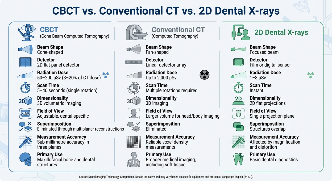
CBCT vs Conventional CT vs 2D X-rays Comparison Chart
CBCT vs. Conventional CT
CBCT and conventional CT both utilise X-rays to produce images, but their approaches differ significantly. CBCT employs a cone-shaped X-ray beam along with a 2D flat-panel detector, capturing the entire dental area in a single rotation lasting between 5 and 40 seconds. In contrast, conventional CT uses a fan-shaped beam and a linear detector, requiring multiple sequential rotations to create axial slices. This process takes longer and involves larger equipment.
One of the standout differences is the radiation dose. CBCT operates with reduced tube currents (1–15 mA at 90–120 kVp), delivering only 3–20% of the radiation dose typically required by conventional CT. This reduced exposure makes CBCT a practical choice for in-office dental use, offering the detailed imaging necessary for procedures like treatment planning, while prioritising patient safety.
| Feature | CBCT | Conventional CT |
|---|---|---|
| Beam Shape | Cone-shaped | Fan-shaped |
| Detector Type | 2D flat-panel detector | Linear detector array |
| Radiation Dose | Approx. 50–200 µSv (3–20% of CT dose) | Up to around 2,000 µSv |
| Scan Time | 5–40 seconds (single rotation) | Multiple rotations required |
| Field of View | Adjustable, dental-specific | Larger volume for head/body imaging |
| Primary Use | Maxillofacial bone and dental structures | Broader medical imaging, including soft tissue |
While conventional CT excels in soft-tissue imaging and provides reliable voxel density measurements, CBCT is tailored for high-resolution imaging of bone and dental structures. This makes it ideal for dental clinics such as Complete Smiles Bella Vista, where it plays a key role in implant planning, orthodontic assessments, and endodontic evaluations. Next, we’ll explore how CBCT stacks up against traditional 2D dental X-rays.
CBCT vs. 2D Dental X‑Rays
The shift from 2D to 3D imaging has revolutionised diagnostic accuracy. Traditional 2D radiographs provide flat images, which can result in overlapping structures. This overlap can obscure critical details such as root fractures or the spatial positioning of teeth and nerves.
CBCT, on the other hand, offers true 3D volumetric imaging. It eliminates superimposition by allowing clinicians to view images in multiple planes, including axial, coronal, sagittal, and oblique. This detailed perspective helps in assessing complex anatomical relationships, such as the bucco-lingual width of bone for implant placement, the path of the inferior alveolar nerve, and intricate root canal anatomy.
| Feature | CBCT | 2D Dental X‑Rays |
|---|---|---|
| Dimensionality | 3D volumetric imaging | 2D flat projections |
| Superimposition | Eliminated through multiplanar reconstructions | Structures overlap |
| Measurement Accuracy | Sub-millimetre accuracy in three planes | Affected by magnification and distortion |
| Field of View | Volumetric with selectable regions | Single projection plane |
| Radiation Exposure | Approx. 50–200 µSv | Typically 1–8 µSv |
Although CBCT involves higher radiation exposure – often more than ten times that of 2D imaging – it’s justified when the added 3D information significantly influences diagnosis or treatment planning. For example, CBCT is particularly valuable for dental implant planning, evaluating impacted teeth, or detecting root fractures that may not be evident on standard periapical radiographs.
Applications of CBCT in Dentistry
Orthodontics and Treatment Planning
CBCT provides orthodontists with detailed 3D models of dental structures, enabling them to assess impacted teeth, evaluate root positions, measure bone thickness, and analyse skeletal relationships in all three dimensions – something traditional 2D X-rays simply can’t achieve [2][3]. This advanced imaging is indispensable for identifying impacted canines, gauging the risk of root resorption, and planning tooth movements within the available alveolar bone. It’s also helpful in evaluating airway volume in patients suspected of having obstructive sleep apnoea. By offering a clear view of root positions and surrounding bone, clinicians can plan less invasive extractions, reducing risks like dehiscence and fenestration, and making treatment more efficient.
CBCT is particularly useful for cases involving impacted or ectopic teeth, craniofacial anomalies, asymmetries, Class II/III skeletal discrepancies, TMJ bony changes, and suspected root resorption or alveolar defects that are hard to interpret on 2D images [1][2][3]. In Australian dental practices, limited field-of-view scans are often used to focus on specific areas of interest. The DICOM data from these scans can be imported into orthodontic planning software for 3D cephalometrics and virtual treatment setups. By combining CBCT with intraoral scans, clinicians can create highly accurate digital models for designing custom appliances [2][3].
Dental Implants and Surgical Guides
CBCT is equally vital in implant dentistry, offering high-resolution 3D imaging to evaluate bone height, width, density, and angulation. It also maps critical anatomical features like the inferior alveolar nerve, mental foramen, maxillary sinus, and nasal floor [1][2][3]. This level of detail ensures accurate measurements of ridge dimensions, sinus assessments, and evaluations of root proximity. Additionally, it allows for virtual simulations of implant placement, helping to determine the appropriate angulation and length. This detailed planning minimises risks such as nerve damage, sinus perforation, or cortical plate compromise, while also guiding decisions on whether bone grafting or sinus lift procedures are necessary.
CBCT also plays a key role in creating surgical guides that translate virtual implant plans into precise physical placements [2][3]. By integrating CBCT scans with intraoral scans, clinicians can produce exact digital models. These models enable virtual implant placement that aligns with both prosthetic and anatomical requirements. A 3D-printed guide is then fabricated, either in-house or through a lab. Clinics like Complete Smiles Bella Vista use this workflow to reduce chair time, enhance predictability, and even allow for immediate provisionalisation in complex cases, ultimately improving patient outcomes.
Endodontics and Root Canal Therapy
In endodontics, CBCT improves diagnostic accuracy by identifying root canal anatomy, missed canals, vertical root fractures, and early periapical lesions that may not be visible on 2D radiographs due to overlapping structures [2][4]. It’s particularly valuable for examining teeth with complex root morphology, detecting separate MB2 canals in maxillary molars, measuring periapical radiolucencies, identifying root resorption (both internal and external), and evaluating perforations or posts before retreatment [2][4].
The European Society of Endodontology advises using small field-of-view CBCT when traditional radiographs fail to provide sufficient information – for instance, in cases of dentoalveolar trauma, complex root canal systems, suspected root fractures, root resorption, or pre-surgical planning for apical surgeries [2]. Beyond these uses, CBCT aids in surgical planning for impacted teeth, TMJ evaluations, diagnosing jaw pathologies like cysts and tumours, assessing sinus disease linked to dental infections, trauma evaluations, and planning craniofacial reconstructions [1][2][3]. In multidisciplinary clinics offering oral surgery, restorative, and cosmetic services, CBCT enables coordinated care, leading to more predictable outcomes in both function and aesthetics. This technology has become a cornerstone in advancing diagnostics and treatment across various dental fields.
Conclusion
CBCT technology has revolutionised dental imaging by providing high-resolution 3D visualisation through a single, efficient scan. By using a cone-shaped X-ray beam that rotates around the patient’s head, it rapidly captures 2D images and transforms them into detailed 3D reconstructions. This approach eliminates the challenges of superimposition seen in traditional 2D radiographs and serves as a more efficient option compared to conventional CT scans.
One of the standout benefits of CBCT is its ability to reveal anatomical details that cannot be captured with 2D imaging, all while keeping radiation exposure significantly lower – ranging from just 3% to 20% of the levels associated with traditional medical CT scans. This balance between accuracy and safety has made CBCT indispensable in areas like orthodontics, implant planning, endodontics, and oral surgery, where precise diagnostics are crucial for effective treatment. Additionally, its integration with digital treatment planning enhances its utility in clinical practice.
CBCT goes beyond diagnostics, enabling virtual treatment simulations, the creation of surgical guides, and real-time multiplanar viewing [5]. As advancements continue to refine its diagnostic capabilities and further minimise radiation exposure, CBCT is poised to play an even greater role in dentistry, transforming clinical decisions from experience-based judgments to data-driven precision.
With its ability to enhance accuracy and efficiency, CBCT empowers clinicians to make informed, evidence-based decisions, reducing uncertainty and improving treatment outcomes. It has firmly established itself as an essential tool in modern dental care.
FAQs
How does CBCT imaging minimise radiation exposure compared to traditional CT scans?
CBCT imaging uses cutting-edge technology to minimise radiation exposure while delivering high-quality diagnostic images. Unlike traditional CT scans, CBCT targets only the specific area under examination. This precision reduces the dose of ionising radiation by as much as 80–90%.
This approach makes CBCT a safer and highly effective choice for dental procedures, providing detailed 3D images with a focus on patient safety.
What are the benefits of using CBCT technology for planning dental implants?
CBCT technology delivers detailed 3D imaging, making it a powerful tool for planning dental implants. Dentists can use it to evaluate bone quality and volume, pinpoint important anatomical features, and map out implant placement with incredible accuracy.
With its ability to provide a clear view of the treatment area, CBCT plays a crucial role in minimising risks like nerve damage or sinus perforation. This leads to safer procedures and more reliable outcomes. By enhancing decision-making, CBCT contributes to better surgical results and higher patient satisfaction.
How does CBCT imaging identify dental issues that traditional X-rays might miss?
CBCT, or Cone Beam Computed Tomography, offers highly detailed 3D images of your teeth, jawbone, and surrounding structures – something traditional 2D X-rays simply can’t match. This cutting-edge imaging technology is invaluable for identifying tricky dental problems such as hidden fractures, impacted teeth, and even pinpointing bone density with remarkable accuracy. These are issues that standard X-rays often struggle to reveal.
Because CBCT provides a complete view of your oral anatomy, it plays a crucial role in planning treatments like dental implants, root canals, and orthodontic procedures. Its precision helps ensure more accurate diagnoses and tailored care for each patient.
Related Blog Posts
- CT Scans in Bone Graft Planning
- CBCT in Endodontics: Accuracy and Limitations
- Guide to Periapical X-rays for Root Canal Therapy
- AI and Cone-Beam CT: A Diagnostic Revolution
Important Notice: Any surgical or invasive procedure carries risks. Before proceeding, you should seek a second opinion from an appropriately qualified health practitioner.
Individual results may vary. The information provided in this article is for educational purposes only and does not constitute medical advice.
Checkout Related Blogs


Get in touch with us
For more information, call us now to start feeling better. Or fill the form below to make appointment
The Latest News from Complete Smiles
How to Clean Clear Plastic Retainers
Checklist for Choosing Wearable Dental Devices
Checklist for Choosing Cloud AI Platforms in Dentistry

Complete Smiles Bella VistaAccepts All Major Health Funds, Including


