





AI in Oral Pathology: Reducing Diagnostic Errors
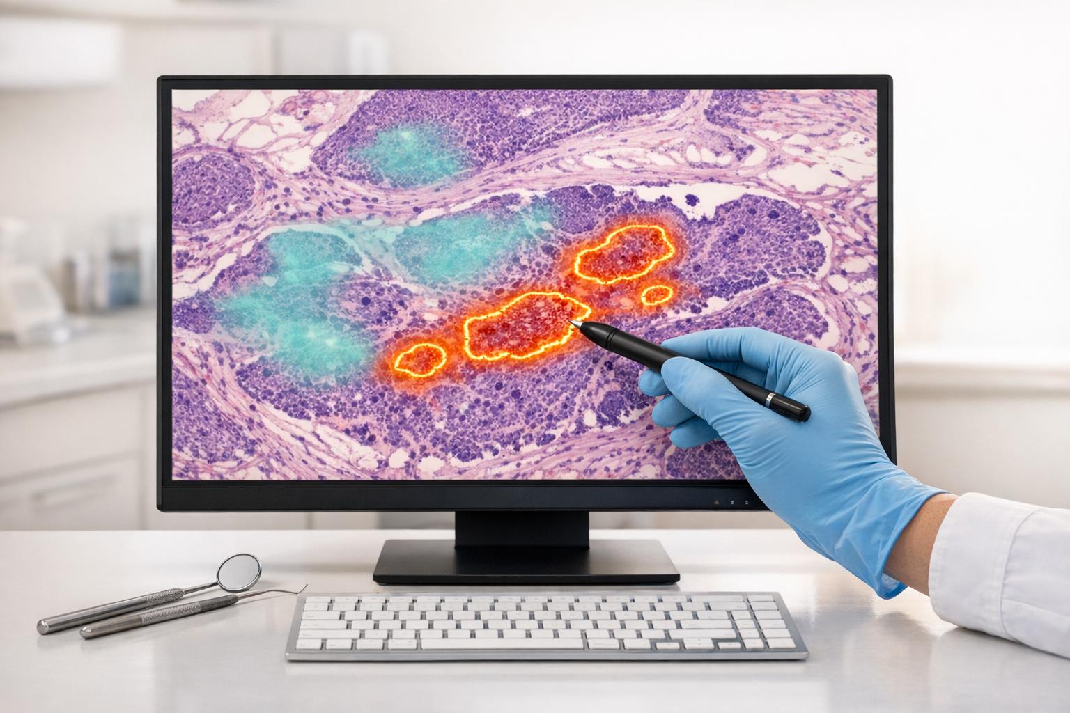
AI is transforming oral pathology by addressing diagnostic challenges like variability, fatigue, and late detection of diseases. By analysing clinical images with precision, AI tools can detect oral cancers, periodontal diseases, and rare conditions earlier and more accurately. For example, AI models like HC-Net+ and YOLOv11 outperform human specialists in identifying oral diseases, achieving accuracy rates as high as 99.3%. These tools also enhance efficiency, processing large datasets in minutes, and assist clinicians in making more informed decisions. However, challenges like data quality, regulatory hurdles, and the need for clinician oversight remain. AI serves as a powerful support tool for pathologists, improving outcomes while ensuring human expertise remains central to the process.
Unleashing AI in Oral Pathology and Oral Medicine: Challenges and Opportunities #oralpathology360
sbb-itb-2be92ed
How AI Reduces Diagnostic Errors
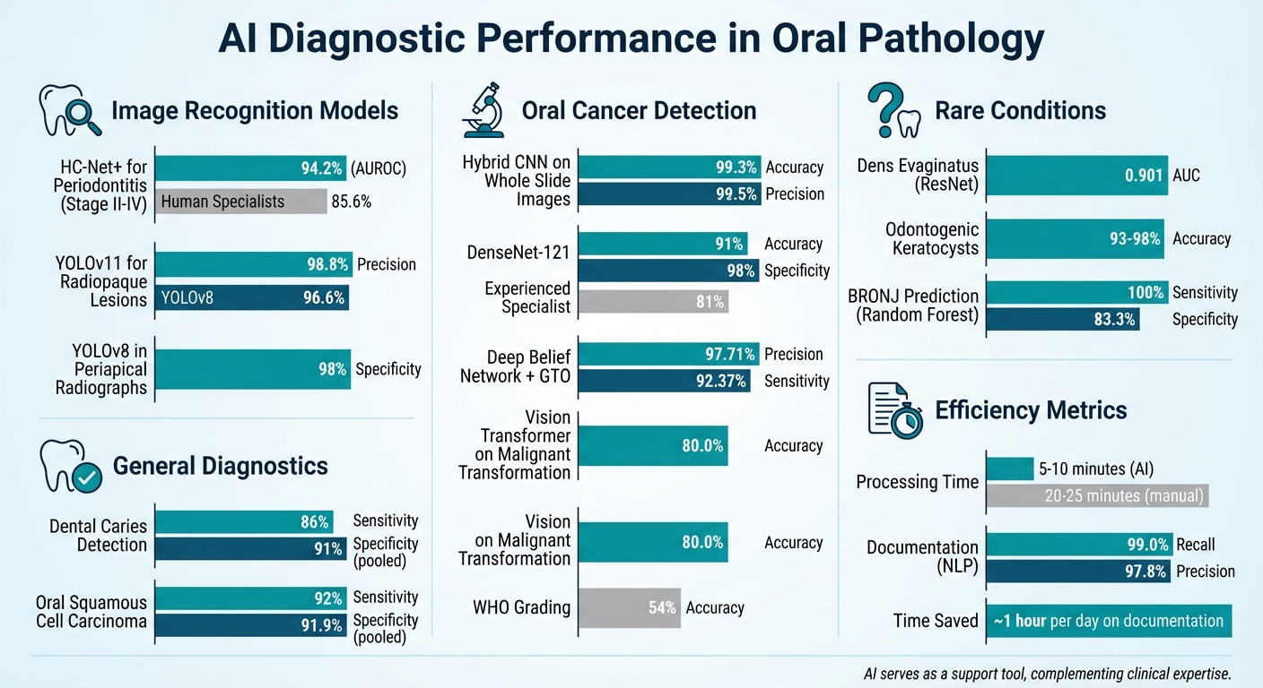
AI Diagnostic Accuracy in Oral Pathology: Performance Metrics Across Applications
AI in Image Recognition and Analysis
AI models are tackling some of the biggest challenges in diagnostics, such as variability and clinician fatigue, by delivering consistent and precise results. Take HC-Net+, for example. This model mirrors the way clinicians work – analysing individual teeth before assessing the entire mouth. It even generates probability scores for conditions like stage II–IV periodontitis, offering clear and actionable diagnostic insights [9].
Another standout is the YOLO (You Only Look Once) algorithm, known for its real-time object detection capabilities in dental imaging. The latest version, YOLOv11, achieved an impressive 98.8% precision in identifying radiopaque lesions like Idiopathic Osteosclerosis, surpassing YOLOv8‘s 96.6% [11]. This level of precision is vital for distinguishing between conditions that can look deceptively similar, even to seasoned clinicians.
These advanced image recognition techniques are the backbone of the diagnostic accuracy seen in modern clinical studies.
Accuracy Rates and Clinical Examples
AI’s diagnostic performance in oral pathology is nothing short of impressive. A 2025 multicentre study published in Nature showcased the capabilities of the HC-Net+ model. Tested across four international centres – Hong Kong, Shanghai, and Rome – on 760 radiographically labelled orthopantomograms, it achieved an AUROC of 94.2% for detecting stage II–IV periodontitis. This was a significant improvement over the 85.6% average achieved by periodontal specialists. Interestingly, when junior dentists used HC-Net+ for assistance, their performance matched that of experienced specialists [9].
In oral cancer detection, AI systems analysing whole slide images achieved an accuracy of 99.3% and a precision of 99.5%, thanks to hybrid CNN fusion features [5]. Similarly, a December 2025 study by the Kerman Faculty of Dentistry tested three deep learning models on 518 intraoral clinical images collected over 14 years. The DenseNet-121 model stood out, achieving 91% accuracy and 98% specificity – well above the 81% accuracy of an experienced oral medicine specialist [13].
For detecting dental caries across various imaging types, AI models demonstrated a pooled sensitivity of 86% and specificity of 91% [10]. These numbers highlight AI’s ability to consistently outperform or match human expertise, offering a powerful tool to reduce diagnostic errors in oral pathology. This technology also supports the dentist’s role in oral cancer prevention by identifying high-risk lesions during routine check-ups.
Applications of AI in Oral Disease Detection
Detecting Oral Cancer
AI has become a game-changer in identifying oral cancers, especially Oral Squamous Cell Carcinoma (OSCC) and other malignant disorders. By analysing clinical photographs, histopathological slides, and advanced imaging techniques like Optical Coherence Tomography (OCT), AI can detect subtle patterns that might be overlooked during routine examinations [8][14].
Recent advancements highlight its effectiveness. For instance, in January 2025, researchers Wang, Liu, and Wu introduced a hybrid AI system combining a Deep Belief Network with a Group Teaching Optimisation algorithm. This system, using standard clinical images, achieved an impressive 97.71% precision and 92.37% sensitivity in distinguishing cancerous from non-cancerous samples [2][5].
AI’s capabilities go beyond detection – it can predict the likelihood of malignant transformation. Early in 2025, Rutgers University developed a multiresolution Vision Transformer model that analysed 221 whole-slide images to predict oral lesions likely to turn cancerous within five years. This model achieved 80.0% accuracy, significantly outperforming the traditional WHO dysplasia grading system, which had a positive predictive value of just 54% [14]. Such predictive tools are vital for reducing missed diagnoses in potentially malignant disorders.
"AI algorithms have the potential to function as reliable tools for the early diagnosis of OPMDs and oral cancer, offering significant advantages, particularly in resource-constrained settings."
– Frontiers in Oral Health [6]
Periodontal Disease and Bone Loss Analysis
AI is also transforming how periodontitis is assessed, combining radiographic data with patient information to refine diagnostic accuracy. For example, AI can measure the distance between the cemento-enamel junction and the marginal alveolar bone in radiographs, enabling precise staging of periodontitis from stages I to IV [15][16].
Tools like HC-Net+ have proven to be highly effective. When junior dentists used HC-Net+ for decision support, their diagnostic accuracy matched that of experienced specialists. Furthermore, AI models like YOLOv8 have demonstrated 98% specificity in analysing periapical radiographs, processing data nearly 30% faster than earlier two-stage models [12][16]. By incorporating patient risk factors – such as smoking and diabetes – AI automates disease grading (A–C), reducing variability between clinicians and minimising interpretation errors [12][15].
Identifying Rare Conditions
AI is also making strides in detecting rare oral disorders, which are often missed due to their infrequent occurrence. By training on pooled datasets, AI systems can recognise patterns in conditions like Dens Evaginatus (DE), a rare anomaly affecting 0.5% to 4.3% of the population [7]. A ResNet-based model achieved an AUC of 0.901 in diagnosing DE, outperforming 14 endodontic specialists. This model excelled at identifying subtle diagnostic features, such as the projection of pulp in worn-down tubercles, which might be overlooked in periapical radiographs [7].
"AI can recognise it [Dens Evaginatus] if PA [periapical radiography] was taken, enabling clinicians to choose preventive treatment options to avoid tooth damage."
– Nature Scientific Reports [7]
AI has also shown exceptional accuracy – ranging from 93% to 98% – in distinguishing Odontogenic Keratocysts from other jaw cysts in microscopic images [4]. Additionally, a Random Forest model achieved 100% sensitivity and 83.3% specificity in predicting bisphosphonate-related osteonecrosis of the jaw (BRONJ) following tooth extractions, surpassing traditional clinical prediction methods [4]. This level of precision is crucial for minimising diagnostic errors in rare oral anomalies.
Benefits and Limitations of AI in Oral Pathology
Key Benefits of AI
AI has introduced a new level of efficiency in oral pathology diagnostics. It can analyse complex clinical images in just 5–10 minutes, compared to the 20–25 minutes typically required for manual analysis [21]. This time-saving capability allows pathologists to dedicate more attention to rare and complex cases that demand human expertise. By streamlining routine diagnostics, AI plays a role in reducing diagnostic errors and improving accuracy.
Another standout benefit is AI’s consistency. Unlike human practitioners, who may be affected by fatigue or cognitive biases, AI delivers reliable and impartial results regardless of workload [17][20]. Considering that diagnostic errors contribute to about 10% of patient deaths and 17% of adverse events in hospitals [18], AI’s ability to minimise errors linked to clinician fatigue or biases is invaluable. Dr. Kevin Sandeman, a Clinical Pathologist at Region Skåne pathology lab, highlights this advantage:
"If the AI system can take that 2% area [of a tumour] and present it to me to review first, the diagnosis is much faster. This benefits my productivity."
– Dr. Kevin Sandeman [17]
Beyond diagnostics, AI automates tedious tasks like cell counting and Ki-67 scoring [17][19]. Hybrid diagnostic systems have also shown impressive results, with models for oral squamous cell carcinoma achieving 99.3% accuracy and 98.35% specificity [5]. These advancements make AI a powerful tool in enhancing the diagnostic process.
Challenges in AI Implementation
Despite its advantages, implementing AI in oral pathology comes with a set of challenges. One major hurdle is the need for large, high-quality datasets. Rare conditions in oral pathology often lack sufficient data, limiting the effectiveness of AI models [5]. Additionally, inconsistencies in tissue preparation – such as fixation, staining, and cutting – can negatively impact model performance [5][1]. Even highly accurate systems are not immune to errors, with some "hallucinating" or misinterpreting clinical data [5].
The "black box" nature of many AI models is another issue. These systems often lack transparency, making it difficult for clinicians to understand how a diagnosis was reached [5][1]. This lack of interpretability can erode trust and slow clinical adoption, especially when practitioners cannot verify the AI’s reasoning. Moreover, AI models may struggle when applied to patient demographics or clinical environments that differ from their training data [5][8].
Financial and regulatory obstacles further complicate AI adoption. The costs of hardware, software, and ongoing maintenance can be prohibitive, while lengthy regulatory approval processes add another layer of difficulty [5]. For example, the EU AI Act classifies AI as "high-risk", requiring rigorous assessments and human oversight [21]. Legal challenges, including compliance with HIPAA/GDPR, patient privacy concerns, algorithmic bias, and liability issues, also persist [5][1][4].
Practical solutions are needed to address these challenges. Collaborative data sharing across institutions can help create larger, more diverse datasets to improve AI robustness [5]. Local calibration of AI models can account for variations in imaging equipment and patient demographics [5]. Most importantly, AI should act as a supportive tool rather than a replacement for clinical judgement. Pathologists must critically evaluate AI outputs to avoid "automation bias" and ensure that human expertise remains central to the diagnostic process [19][20]. These measures reinforce the idea that AI is a partner in reducing diagnostic errors, not a substitute for clinical decision-making.
Future Potential of AI in Oral Pathology
Improved AI Algorithms and Models
AI in oral pathology is evolving with significant advancements in algorithms and models. The shift from traditional Convolutional Neural Networks (CNNs) to Vision Transformers (ViTs) is noteworthy. ViTs excel at capturing global patterns in tissue samples, offering a fresh perspective on diagnostic accuracy. For instance, a multiresolution ViT model reached 80.0% accuracy in predicting the malignant transformation of oral lesions over a five-year span, providing a more consistent diagnostic approach.
Multimodal deep learning is another breakthrough. The OMMT-PredNet framework, developed using data from 649 histopathologically confirmed leukoplakia cases between 2003 and 2024, combines clinical photographs with medical records for cancer risk predictions. This system achieved impressive results, with an AUC of 0.9592 for cancer risk prediction and 0.9219 for identifying oral epithelial dysplasia – without relying on manual region annotations.
Explainable AI (XAI) tools like SHAP (SHapley Additive Explanations) are addressing the "black box" challenge by highlighting the features that influence diagnostic decisions. Federated learning is also making waves by enabling institutions to collaborate on AI model development without compromising patient privacy. Additionally, Generative Adversarial Networks (GANs) are being used to create synthetic training data, such as radiographs and CBCT slices, tackling the issue of limited datasets for rare conditions.
These advancements are laying the groundwork for smoother integration with clinical tools.
Integration with Clinical Tools
AI is becoming an integral part of clinical workflows, improving efficiency and accuracy. For example, tools like Impetus categorise histological slides into confidence levels – high, medium, and low – allowing pathologists to quickly verify high-confidence diagnoses while focusing on complex cases that need manual review. Open-source platforms like QuPath are also making an impact by standardising biomarker quantification, such as PD-L1 and p53, which reduces observer bias and enhances reproducibility.
Natural Language Processing (NLP) is transforming clinical documentation. AI scribes can now transcribe pathologists’ verbal observations in real time, achieving recall rates of 99.0% and precision rates of 97.8%. This eliminates the need for pathologists to alternate between microscopes and keyboards, saving about an hour of documentation time each day. Portable solutions like Cellscope are also enabling AI-assisted screenings in areas with limited resources.
These tools aren’t just refining diagnostics – they’re setting the stage for AI’s broader role in dental care.
Broader Applications in Dentistry
AI’s reach in dentistry is extending beyond diagnostics to preventive care and treatment planning. Digital twins, which integrate radiographs, genetic information, and treatment histories, are now being used to simulate patient responses and predict outcomes. This technology opens up possibilities for personalised treatment planning, allowing clinicians to virtually test different strategies before implementation.
Automated segmentation tools are also helping create 3D visualisations of cysts and tumours, enhancing surgical planning precision. By integrating imaging data with other clinical variables – like age, tobacco use, and genetic factors – AI is paving the way for more comprehensive risk assessments. These systems are not only improving diagnostic reliability but also supporting tailored dental care that considers the unique needs of each patient.
Conclusion
AI is reshaping oral pathology by detecting subtle cellular changes with remarkable consistency – changes that might otherwise go unnoticed during manual examination. For instance, AI achieves a pooled sensitivity of 92% and specificity of 91.9% in identifying oral squamous cell carcinoma [21]. By minimising inter-observer variability, AI ensures a level of objectivity that remains uniform across clinicians and pathologists.
However, AI’s true strength lies in its role as a support tool rather than a replacement for clinical expertise. As G. D’Albis and S. Capodiferro explain:
"AI should be seen as a helpful tool that complements, rather than replaces, the expert judgement of oral surgeons" [3].
This partnership between advanced algorithms and the nuanced judgement of dental professionals creates a powerful combination. While AI brings precision and efficiency, clinicians contribute critical thinking and contextual understanding.
The impact of these advancements is already evident in various clinical settings. AI demonstrates its adaptability in tasks like image recognition for radiographs, whole slide imaging, and natural language processing. For example, AI-driven dental charting achieves a recall rate of 99.0% [5], saving clinicians up to one hour per day on documentation. These tools not only improve efficiency but also enhance diagnostic accuracy. However, for AI to succeed in practice, high-quality data, proper clinician training, and transparent decision-making processes are essential.
In Australia, the future of AI in dentistry looks especially promising. AI-powered teledentistry is emerging as a critical solution for improving access to care in remote and underserved communities. At the same time, innovations like digital twins and multimodal data integration are enabling more personalised treatment plans. This shift from reactive to proactive care could allow conditions to be detected years earlier than traditional methods.
The key to unlocking AI’s full potential lies in its thoughtful integration into clinical workflows. Dental professionals must remain central to the process, using their expertise to critically evaluate AI-generated insights. Rather than viewing AI as a standalone solution, it should be embraced as a tool that extends their diagnostic capabilities. With the right training and implementation strategies, AI can significantly enhance patient outcomes while supporting clinicians in delivering more precise, efficient, and accessible care.
FAQs
Will AI replace oral pathologists?
AI is proving to be an impressive tool in oral pathology, helping to improve diagnostic precision and minimise errors. Some AI systems can reach over 98% accuracy in identifying conditions like oral cancer. However, it’s important to remember that they are meant to assist rather than replace human professionals. The expertise, clinical judgement, and comprehensive evaluations provided by oral pathologists are still crucial, particularly when dealing with more complex cases. Moving forward, AI is set to remain a supportive tool, complementing the care provided by professionals rather than replacing it.
How is AI checked for mistakes or bias?
AI is making strides in oral pathology, but its effectiveness hinges on clinician oversight and thorough testing. Tools like convolutional neural networks (CNNs) rely on extensive datasets and are assessed using metrics like sensitivity (how well it identifies true positives) and specificity (how well it avoids false positives).
To bolster these models, synthetic data generated by methods like Generative Adversarial Networks (GANs) plays a key role in refining accuracy and adaptability. However, AI doesn’t operate in isolation – clinicians meticulously review AI-generated results to catch errors and reduce bias.
Despite its potential, challenges remain. Issues like inconsistent data quality, privacy concerns, and the substantial costs of implementation need to be addressed to fully integrate AI into oral pathology workflows.
What does an AI result mean for my treatment?
AI in oral pathology plays a key role in improving diagnostic precision, especially for conditions like oral cancer or periodontitis. With its high sensitivity and specificity, AI helps dentists spot potential problems earlier, allowing for more personalised and timely treatment plans. However, while AI reduces the chances of diagnostic errors, it serves as a supportive tool. Your dentist will always evaluate all clinical factors to ensure the most accurate and comprehensive treatment decisions are made.
Related Blog Posts
- AI Research in Oral Disease Detection: Key Findings
- AI in Dentistry: Predicting Periodontal Disease
- AI in Oral Cancer Detection: How It Works
- AI-Powered Image Analysis: What Dentists Need to Know
Important Notice: Any surgical or invasive procedure carries risks. Before proceeding, you should seek a second opinion from an appropriately qualified health practitioner.
Individual results may vary. The information provided in this article is for educational purposes only and does not constitute medical advice.
Checkout Related Blogs




Get in touch with us
For more information, call us now to start feeling better. Or fill the form below to make appointment
The Latest News from Complete Smiles
How to Clean Clear Plastic Retainers
Checklist for Choosing Wearable Dental Devices
Checklist for Choosing Cloud AI Platforms in Dentistry

Complete Smiles Bella VistaAccepts All Major Health Funds, Including





