





Electrochemical Surface Modification for Implants
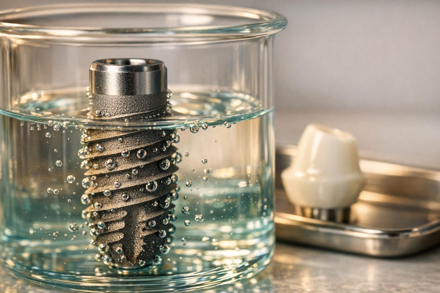
Electrochemical surface modification is transforming how dental implants connect with surrounding tissues. By creating bioactive coatings on titanium and zirconia surfaces, this technology improves implant integration, reduces inflammation risks, and enhances durability. Here’s what you need to know:
- What It Does: Modifies implant surfaces to mimic natural bone, promoting chemical bonding and faster healing.
- Why It Matters: Reduces peri-implantitis, which affects up to 20% of patients within five years.
- Key Techniques:
- Anodic Spark Deposition (ASD): Creates microporous layers with bioactive ions.
- Electrochemical Anodic Oxidation: Produces nano-scale structures for better hydrophilicity and cell attachment.
- Micro-Arc Oxidation (MAO): Forms thick, porous coatings resembling bone structure.
Modified implants show over 95% success rates in clinical studies. They also reduce bacterial colonisation and improve long-term stability, making them a game-changer for patients with systemic health challenges like diabetes or smoking-related complications. Whether you’re a practitioner or patient, understanding these advancements ensures better outcomes for dental implants.
The HAnano Surface Coating Process
sbb-itb-2be92ed
Electrochemical Surface Modification Methods
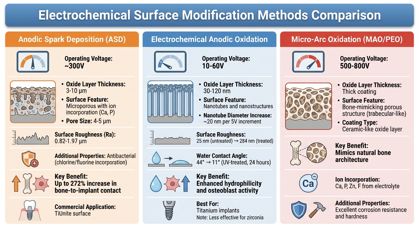
Comparison of Three Electrochemical Surface Modification Methods for Dental Implants
To support better osseointegration, three electrochemical techniques are commonly used to modify implant surfaces. These methods vary in voltage, the thickness of the oxide layer they produce, and their specific benefits for titanium and zirconia implants.
Anodic Spark Deposition (ASD)
ASD uses high voltage to create a thick oxide layer, usually between 3–10 μm, resulting in a microporous surface with pores measuring 4–5 μm [5]. This technique allows for the direct incorporation of ions like calcium and phosphorus from the electrolyte, which has been shown to increase bone-to-implant contact by up to 272% in rabbit studies compared to untreated controls [5]. A commercial application of this method, the TiUnite surface, has a moderately rough texture with an average roughness (Ra) of 0.82 to 1.97 μm [5]. Additionally, incorporating elements such as chlorine or fluorine can provide antibacterial benefits by disrupting bacterial cell walls and inhibiting pathogens like Porphyromonas gingivalis [5][6]. This combination of ion incorporation and antibacterial properties enhances the interaction between the implant and surrounding tissue.
Electrochemical Anodic Oxidation
This method operates at lower voltages, typically between 10–60 V, to create nanostructures and nanopores on titanium surfaces via electrode reactions and ion diffusion [6][9]. One notable outcome is a significant increase in surface energy and hydrophilicity. For instance, UV-treated anodised porous TiO₂ surfaces can reduce the water contact angle from 44° to 11° within 24 hours [5]. The process forms thin oxide layers of 30–120 nm, and the diameter of TiO₂ nanotubes increases by approximately 20 nm for every 5 V increment [9]. When performed in a 1 M H₃PO₄ solution with 2% HF, anodic oxidation increases surface roughness from 25 nm (untreated) to 284 nm [8]. These modified surfaces promote osteoblast activity, with significantly higher cell numbers observed within 24 hours compared to untreated surfaces [7].
"Anodic oxidation in H₂SO₄ solution combined with subsequent heat treatment was an effective method to prepare bioactive titanium." – Yang et al. [6]
While this method works well for titanium, it is less effective for zirconia implants. Zirconia is often chosen for soft-tissue applications due to its lower inflammatory response and smoother surface, which helps reduce biofilm formation [6]. This method enhances implant-tissue interaction by improving hydrophilicity and cellular response. For bone-mimicking porous structures, however, Micro-Arc Oxidation is a better alternative.
Micro-Arc Oxidation
Micro-Arc Oxidation (MAO), also called Plasma Electrolytic Oxidation (PEO), uses plasma discharge at high voltages (500–800 V) to create thick, porous coatings that resemble the trabecular structure of bone [4][10]. The resulting ceramic-like oxide layer features controlled porosity, providing a scaffold for mechanical interlocking and bone growth [6].
"The use of electrochemical treatment such as anodic oxidation or plasma electrolytic oxidation allows for the production of porous coating that mimics the bone structure and accelerates the osseointegration process." – Materials Today Communications [4]
MAO coatings are known for their excellent passivation and corrosion resistance, as indicated by a current plateau in the passive range [8]. These oxide films are chemically bonded to the substrate, offering better hardness and adhesion compared to many physical vapour deposition coatings [11]. Similar to ASD, MAO can incorporate bioactive elements like calcium, phosphorus, zinc, and fluoride directly from the electrolyte into the oxide layer [11]. Factors such as the electrolyte composition, processing time, duty cycle, and applied voltage can be adjusted to achieve specific coating properties [10]. This method enhances implant-tissue interaction by mimicking bone architecture.
| Method | Operating Voltage | Oxide Thickness | Primary Surface Feature |
|---|---|---|---|
| Anodic Spark Deposition | ~300 V | 3–10 μm | Microporous with ion incorporation (Ca, P) |
| Anodic Oxidation | 10–60 V | 30–120 nm | Nanotubes and nanostructures |
| Micro-Arc Oxidation (MAO) | 500–800 V | Thick coating | Bone-mimicking porous structure |
This table highlights the distinct conditions and outcomes of each electrochemical modification method.
The Electrochemical Surface Modification Process
The electrochemical surface modification process involves a series of carefully controlled steps to create bioactive surfaces on dental implants. These steps are key to achieving the enhanced properties highlighted earlier.
Preparation and Immersion
The process starts with mechanically preparing the implant surface using techniques like sandblasting, acid-etching, or polishing. These methods increase surface roughness and remove contaminants.
Next, the implant is immersed in an aqueous electrolytic solution within an electrolytic cell. For calcium phosphate coatings, the solution typically contains calcium nitrate tetrahydrate (Ca(NO₃)₂·4H₂O) and ammonium dihydrogen phosphate (NH₄(H₂PO₄)) [2]. If the goal is to create nanoporous oxide layers via anodisation, phosphoric acid (H₃PO₄) modified with calcium acetate or potassium acetate is used instead [12].
In this setup, the implant serves as the active electrode – acting as the cathode during electrodeposition and as the anode during anodisation [2][12]. Electrical energy is applied in different modes: direct current (DC) provides constant energy but may lead to dihydrogen bubble accumulation, while pulsed currents help disperse bubbles and maintain ion balance [2]. Mixed-mode techniques, which combine constant voltage and current, operate within specific limits, such as 150 V and 0.2 A [12].
During anodisation, the current may drop to zero within 12 seconds as the oxide layer forms and increases resistance [12]. The electrolyte temperature is kept near 35°C without active cooling [12]. Under these controlled conditions, redox reactions occur, either depositing calcium phosphate onto the surface or forming a nanoporous oxide layer. Once the desired surface layer is achieved, the process moves to post-modification treatments.
Post-Modification Steps
After electrochemical treatment, the implants are rinsed thoroughly and subjected to sonication to remove any loosely attached ions or debris [12]. These steps are essential for maintaining the bioactivity of the surface, which plays a crucial role in implant-tissue integration.
Thermal annealing is then used to improve the stability and functionality of the surface coating. Heating above 800°C converts amorphous coatings into stable crystalline forms like hydroxyapatite or β-tricalcium phosphate, enhancing osseointegration [2]. For anodised titanium implants, heat treatment at roughly 450°C for one hour optimises the crystalline phase, encouraging hydroxyapatite formation when exposed to body fluids [13]. This step can also significantly increase the thickness of the natural titanium dioxide layer from 17–200 nm to 600–1,000 nm [13].
However, these high-energy, modified surfaces are prone to contamination from the atmosphere [13]. Studies indicate that implants treated with alkali-heat methods can retain their stability for up to 52 weeks if stored in a vacuum [13]. This highlights the importance of controlled storage conditions to preserve the implant’s bioactivity until it is ready for clinical use.
Benefits of Electrochemical Surface Modification
Electrochemical surface modification brings a range of advantages to implants, enhancing their compatibility with biological systems, appearance, and resistance to bacterial colonisation.
Improved Biocompatibility
By mimicking the natural bone environment at the nanoscale, modified surfaces improve protein adsorption and cell adhesion. These surfaces often incorporate trace elements that promote biological activity. For instance, in 2019, Zhang and colleagues developed a layered micro/nanostructured coating known as MHTZn. This coating combined micro-arc oxidation, hydrothermal treatment, and thermal zinc deposition on a macroporous TiO₂ base. Testing revealed a 25% boost in osteogenesis, reflected in increased alkaline phosphatase activity and the upregulation of osteogenic markers compared to standard TiO₂ coatings [15]. Similarly, magnesium-doped surfaces demonstrated impressive results, showing a 4.5-fold increase in cell proliferation and a 38% rise in alkaline phosphatase activity, a critical indicator of bone mineralisation [15].
These modifications transition titanium implants from relying on physical bone bonds – which can weaken over time – to forming chemical osseous bonds, ensuring greater long-term stability [1]. As a bonus, these biocompatible changes also improve the implant’s appearance.
Better Aesthetic Results
Controlling the thickness and crystallinity of the titanium dioxide layer plays a key role in aesthetics. Adjusting these factors allows practitioners to refine the implant’s colour to a light grey tone, which blends more naturally in the transgingival area. This prevents the dark metallic shadow that often appears beneath thin gingival tissue when untreated implants are used. Such improvements are especially important in Australia, where patients often prioritise natural-looking results.
Porous coatings, such as those created via chemical vapor deposition for dental implants, replicate the structure of natural bone help implants integrate more effectively with surrounding tissues, leading to predictable soft tissue contours [19]. Additionally, coatings like TiO₂/ZnO composites improve hydrophilicity, enhancing protein adsorption and cell attachment, which supports healthier tissue development around the implant [15]. These aesthetic gains are complemented by the antibacterial benefits of modified surfaces.
Antibacterial Properties
Modified surfaces employ physical, chemical, and antibiotic-based strategies to combat bacterial colonisation. Nanopillars, for example, physically disrupt bacterial membranes, reducing Pseudomonas aeruginosa by 87% ± 2% and Staphylococcus aureus by 72.5% ± 13% within 24 hours [17]. Copper-doped surfaces also show over 98% antibacterial efficacy against common pathogens [15].
In 2016, researchers combined anodic oxidation and plasma immersion ion implantation to embed silver nanoparticles into TiO₂ nanotubes. This method successfully inhibited methicillin-resistant S. aureus (MRSA) ST239 in a rabbit model of periprosthetic joint infection. Remarkably, it achieved this without significant silver ion release, all while maintaining high biocompatibility [1].
Additionally, titanium oxide nanotubes act as reservoirs for localised antibiotic delivery, reducing systemic toxicity while improving efficiency [16]. With nearly 20% of patients at risk of developing peri-implantitis within five years, these antibacterial properties significantly enhance long-term implant success [1].
Clinical Considerations for Australian Dental Practitioners
Material-Specific Recommendations
When selecting dental implant materials, it’s essential to weigh factors like load distribution, compatibility with biological tissues, and overall appearance. Titanium remains the go-to material for electrochemical modification, with commercially pure titanium (cpTi) categorised into grades 1 to 4 based on its oxygen, carbon, and iron levels [3]. For cases requiring higher mechanical strength, Ti-6Al-4V alloys stand out, offering a minimum yield strength of 795 MPa and tensile strength of 860 MPa, making them ideal for high-stress applications [21].
Zirconia is gaining traction, particularly in aesthetic cases, thanks to its opaque white colour, which eliminates the dark metallic shadow sometimes seen with titanium [20]. However, zirconia modifications don’t yet have the extensive long-term clinical data available for electrochemically treated titanium surfaces [23].
"An effective dental biomaterial should osseointegrate, maintain structural integrity, resist corrosion and infection, and not cause systemic toxicity or cytotoxicity." – OK Semisch-Dieter, University of Technology Sydney [20]
Australian researchers from the University of Technology Sydney and the University of Sydney highlight the need for ongoing clinical studies, especially when evaluating newer titanium–zirconium alloys that combine strength and biological benefits [18][20]. These material choices also play a role in managing and preventing peri-implantitis, as discussed below.
Applications in Peri-Implantitis Management
Peri-implantitis affects about 10% of implants and 20% of patients within 5–10 years [21], making surface modifications a practical tool for both prevention and treatment. Techniques like anodisation create micro-pores and nanotube arrays, which can act as delivery systems for bioactive agents, such as bone morphogenetic proteins and antibiotics, to aid in re-establishing osseointegration in affected areas [24].
Surface roughness is another key factor. Surfaces with an Ra value greater than 0.8 μm can increase bacterial attachment by up to 25 times compared to surfaces with an Ra of 0.3 μm [21]. For patients with a higher risk of infection, selecting the right surface is critical. Hydrophilic-modified surfaces encourage faster early bone formation and higher expression of osteogenic markers, which can support early loading protocols within six weeks of insertion [23]. Additionally, fluoride-modified implants report survival rates of around 96% when loaded immediately, while SLA implants have shown a 0% failure rate in a five-year randomised controlled study under early loading conditions [23].
Conclusion
Electrochemical surface modification is reshaping the way osteoimmunology influences how dental implants interact with surrounding tissues. By enabling chemical bonds that promote rapid and stable osseointegration, while also incorporating antimicrobial properties, this approach directly addresses the risk of peri-implantitis [1][4]. This enhanced bioactivity is a key driver behind the impressive clinical outcomes seen in practice.
In Australia, research institutions like the University of Queensland‘s GATORs are leading the charge in developing advanced dental implants. These next-generation designs aim to deliver targeted bioactivity, significantly reducing implant failures. This progress is particularly impactful for patients with systemic health challenges, such as diabetes or smoking-related complications, where traditional osseointegration often falls short [22][25]. Such advancements align with the high success rates reported in clinical studies.
Commercially available surface-treated implants consistently demonstrate efficacy rates exceeding 95% over five years [3]. Clinical findings reveal that zinc-doped surfaces can enhance osteoblast proliferation by 25%, while copper-doped coatings achieve over 99% antibacterial effectiveness [15]. These results highlight the importance of practitioners understanding material-specific innovations and incorporating them into daily practice.
For Australian dental professionals, staying informed about emerging material modifications is crucial. While titanium remains the go-to material, new techniques involving zirconia and titanium–zirconium alloys are gaining attention. As more long-term clinical data emerges, these materials may redefine expectations for implant performance, especially in complex cases [14][26]. The shift from passive to therapeutic implants is setting a new standard for dental care in challenging scenarios.
FAQs
Are electrochemically modified implants safe long term?
Electrochemically modified implants are regarded as safe for extended use. Studies point to their robust electrochemical stability, which ensures reliable performance and strong cellular compatibility over time. These characteristics play a key role in their durability and ability to integrate well with biological systems.
Which surface method is best for my implant material (titanium or zirconia)?
When choosing a surface modification method, the implant material plays a key role. For titanium implants, techniques like acid etching and specialised coatings work well to encourage osseointegration. On the other hand, zirconia implants benefit from physical treatments, chemical processes, and surface coatings to boost bioactivity and ensure stability. These methods help implants integrate better with surrounding tissues, setting the stage for lasting success.
Do these coatings increase peri-implantitis risk or reduce it?
These coatings aim to minimise the risk of peri-implantitis by preventing bacterial biofilm from forming and boosting antimicrobial effectiveness. This reduces the chances of peri-implantitis occurring.
Related Blog Posts
- Electrochemical Deposition for Titanium Implants
- How Hybrid Coatings Improve Osseointegration
- Surface Modifications for Better Osseointegration
- Titanium Implant Surface Modifications for Osseointegration
Important Notice: Any surgical or invasive procedure carries risks. Before proceeding, you should seek a second opinion from an appropriately qualified health practitioner.
Individual results may vary. The information provided in this article is for educational purposes only and does not constitute medical advice.
Checkout Related Blogs




Get in touch with us
For more information, call us now to start feeling better. Or fill the form below to make appointment
The Latest News from Complete Smiles
How to Clean Clear Plastic Retainers
Checklist for Choosing Wearable Dental Devices
Checklist for Choosing Cloud AI Platforms in Dentistry

Complete Smiles Bella VistaAccepts All Major Health Funds, Including





