





Osteoimmunology in Implant Healing
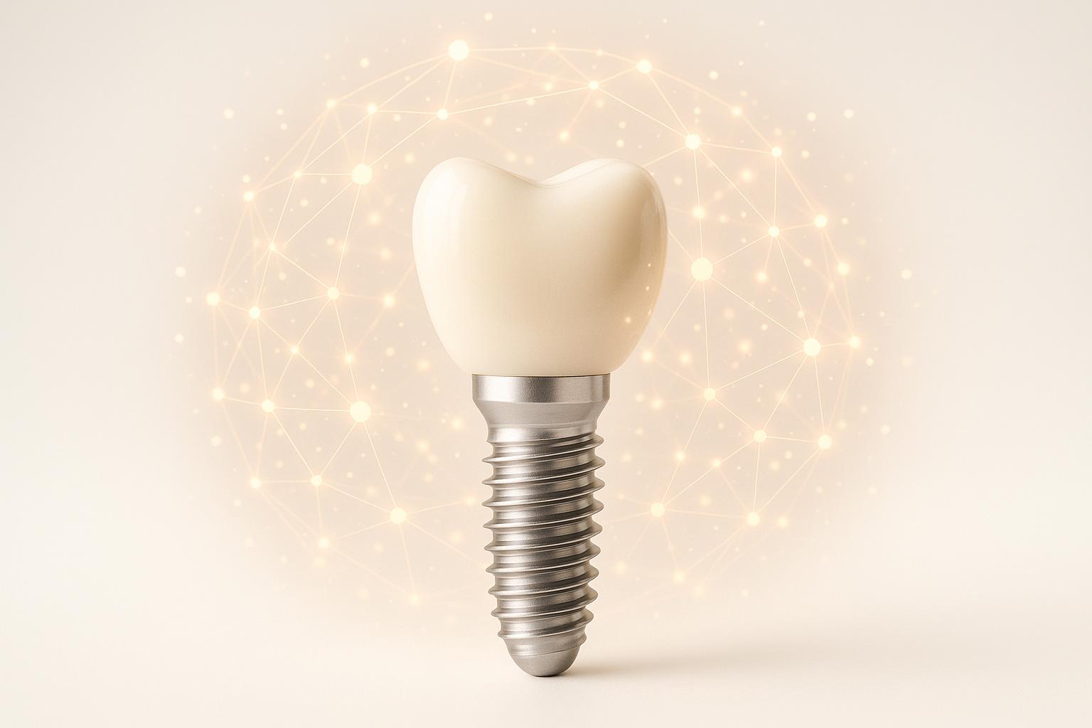
Dental implants aren’t just about bone mechanics anymore. The immune system plays a critical role in healing, influencing implant success or failure. This concept, called osteoimmunology, highlights how immune cells like macrophages drive bone integration and manage inflammation. Here’s what you need to know:
- Osseointegration is immune-driven: Healing involves inflammation, bone formation, and remodelling, regulated by immune cells.
- Complications like peri-implantitis are common: It affects ~19.5% of patients and can lead to implant failure.
- Material and surface matter: Advances like nanoscale titanium surfaces and ceramic implants reduce inflammation and improve outcomes.
- Personalised care is crucial: Diagnostics like trans-alveolar ultrasonography (TAU) and immune profiling help tailor treatments.
Understanding osteoimmunology helps dentists refine implant strategies, reduce complications, and improve long-term success rates.
Osteoimmunology and oxidative stress: the 2 missing pieces of the osseointegration puzzle
Immune Cell Activity in Osseointegration
The immune system’s response to dental implants is a key factor in determining whether they successfully integrate with surrounding bone tissue. By examining cellular activities – especially those involving macrophages – we can better understand why some implants achieve stable osseointegration while others face complications.
How Macrophages Aid Implant Healing
Macrophages play an essential role in implant healing due to their ability to adapt to different phases of the process. In the first 24 hours, inflammation peaks, with M1 macrophages dominating the scene. Regulated in part by NF-κB, these cells release cytokines that attract stem and progenitor cells, kickstarting bone regeneration and the formation of new blood vessels.
As healing advances, the macrophages undergo a key transformation from the inflammatory M1 type to the more reparative M2 type. This shift, supported by specialised osteal macrophages, is critical for tissue remodelling and the mineralisation of the bone matrix. These changes create the conditions needed for long-term implant stability. Research by Chu et al. highlighted that collagen membranes modified with epigallocatechin gallate (EGCG) promoted M2 macrophage recruitment in guided bone regeneration membranes and bone defects. This led to increased growth factor secretion and osteoblast differentiation, ultimately encouraging bone regeneration in vivo [3]. Without this balanced transition, the healing process can falter, potentially leading to the complications discussed below.
Consequences of Prolonged Inflammation
Maintaining a balanced immune response is central to osteoimmunology and crucial for avoiding extended inflammation. When macrophages fail to shift from the M1 to M2 phenotype, prolonged inflammation can jeopardise implant stability. As highlighted by Nasser M. Assery et al.:
"Peri-implantitis is an inflammatory condition induced by bacterial biofilm that affects the soft and hard tissues surrounding dental implants, compromising the success of implant therapy." [4]
Peri-implantitis is a significant clinical issue, affecting a notable percentage of patients and implant sites. The condition often develops over a period of about five years [5], presenting with symptoms such as bleeding during probing, pus formation, and progressive marginal bone loss. In severe cases, it can lead to implant removal.
This localised inflammation can also trigger systemic effects, as peri-implantitis increases levels of systemic pro-inflammatory cytokines [4]. Systemic health issues like uncontrolled diabetes can worsen these inflammatory responses. Moreover, peri-implant tissues are structurally different from natural teeth – they lack cementum, a periodontal ligament, and protective fibres, and they have fewer blood vessels. These differences make them more vulnerable to bacterial invasion and less capable of maintaining a robust immune defence [6].
Understanding how immune cells behave during osseointegration – and the potential complications that can arise – equips dental professionals to intervene early. By managing these processes effectively, they can help maintain the delicate balance needed for successful implant integration and long-term stability.
Recent Research and Immune Response Strategies
The study of osteoimmunology has made impressive strides in developing materials and methods that actively shape immune responses [7]. This shift highlights the importance of managing inflammation to ensure proper wound healing and tissue regeneration around dental implants. Instead of merely reacting to inflammation, these modern approaches work in harmony with the immune system to create an environment that supports healing.
New Biomaterials and Surface Treatments
Advancements in surface modification technologies have significantly influenced how immune cells interact with dental implants. These modifications can encourage helpful immune cell behaviours while minimising harmful inflammatory responses, ultimately improving the healing process.
Nanoscale Surface Modifications have shown great promise. Research indicates that titanium implants with nanoscale roughness reduce inflammatory responses more effectively than those with micro-roughened surfaces. This adjustment encourages the M2 macrophage phenotype, which plays a key role in healing [7].
A noteworthy development involves nano-octahedron ceria (nano-CeO2). When applied to titanium surfaces, this material has demonstrated strong anti-inflammatory properties, creating a healing-friendly environment with minimal inflammation [7].
Hydrophilic Surface Treatments also stand out for their ability to increase levels of IL-10, an anti-inflammatory cytokine, while reducing pro-inflammatory cytokines like TNF-α, IL-1α, IL-1β, and the chemokine CCL-2. This balance supports a more controlled immune response [7].
Chemical modifications offer additional benefits. For instance, a study by Shin et al. introduced a negatively charged polylactic acid (PLA) scaffold crosslinked with phosphate. This material captured inflammatory molecules such as TNF-α, IL-1β, and IL-6, effectively removing them from the healing environment. Similarly, ionic-treated surfaces have shown potential for immune modulation. High magnesium (Mg) concentrations can reduce pro-inflammatory cytokine secretion, while lithium chloride (LiCl) helps mitigate macrophage-driven inflammation [7] [2].
Graphene nanoparticle modifications on magnesium alloys address two critical challenges: reducing degradation by four times and enhancing antibacterial activity by five times. This dual benefit plays a vital role in improving implant healing [7].
Different surface properties also influence immune cells in distinct ways. For example, hydrophilic surfaces tend to promote immature dendritic cell phenotypes, which are less inflammatory. In contrast, hydrophobic surfaces encourage mature dendritic cell phenotypes, which can trigger stronger immune responses. Additionally, cationic surfaces typically provoke more inflammation compared to neutral or anionic surfaces [7] [9].
Implant Material Comparison
Beyond surface treatments, the choice of implant material itself plays a crucial role in determining immune response and healing outcomes. Understanding how different materials interact with the immune system helps clinicians select the most suitable options for their patients, influencing not just healing but also long-term stability and satisfaction.
| Comparison Criteria | Ceramic Implants (Zirconia) | Titanium Implants |
|---|---|---|
| Material Type | Ceramic (Zirconia) | Metal (Titanium) |
| Immune Response | Excellent biocompatibility; low inflammation response and bone integration comparable to titanium | Strong osseointegration with a 97% success rate over 10 years |
| Aesthetics | Natural appearance, ideal for front teeth restorations | Good appearance, though metal may show with gum recession |
| Strength & Durability | High strength but more prone to fracture in smaller sizes | High strength; titanium-zirconium alloy offers 30% more strength than pure titanium |
| Plaque Resistance | Lower tendency to attract plaque and bacteria | Standard plaque attraction typical of metal surfaces |
| Cost | Generally higher due to complex manufacturing | Roughly $300 less expensive than zirconia implants |
| Allergic Reactions | Metal-free, suitable for patients with metal allergies | Only 0.6% of patients show sensitivity to titanium |
| Long-Term Data | Limited due to recent clinical adoption; market expected to grow 50% annually | Extensive data with over 50 years of clinical success |
| Surgical Complexity | Higher technical demand, especially for one-piece designs | Easier placement; two-piece design offers more flexibility |
Titanium implants typically trigger an initial inflammatory phase, which transitions into healing-focused immune activity, leading to successful osseointegration. Zirconia implants, although newer, exhibit excellent biocompatibility and comparable inflammatory responses to titanium. They are especially suitable for patients with metal sensitivities or those prioritising aesthetics in visible areas.
Surface modifications can optimise both types of implants. Research shows that dental-grade implant materials with surface roughness between 0.5 and 3.5 micrometres enhance the anti-inflammatory activation of macrophages more effectively than smooth surfaces [7].
Emerging stimuli-responsive biomaterials represent the next step in implant technology. These materials can adjust their properties based on environmental changes, offering more precise control over immune responses during different healing stages [8].
sbb-itb-2be92ed
Clinical Applications for Dental Implant Practice in Australia
Incorporating osteoimmunology into dental implant practices has brought a new layer of precision and understanding to treatment planning and execution. Australian dentists now recognise that achieving successful implant outcomes goes beyond surgical expertise – it also requires a deep understanding of how the immune system interacts with bone metabolism during the healing process.
Pre-Implant Site Assessment
Pre-surgical planning for implants has evolved to include a focus on inflammation as a critical factor in ensuring long-term success. Evaluating inflammation levels is now a cornerstone of implant site assessment.
One tool making waves in diagnostics is trans-alveolar ultrasonography (TAU). This technique helps assess bone density and identify bone marrow defects in the jaw (BMDJ) before implant placement. Another game-changer is the titanium stimulation test (Ti-Stim), which evaluates immune reactivity to titanium. This test is particularly useful for identifying patients who might experience excessive inflammatory responses to titanium implants, enabling dentists to make more informed decisions about implant material selection [10].
It’s worth noting that around 23% of dental implants are impacted by peri-implantitis, a condition that highlights the need for thorough pre-implant evaluations. By assessing inflammation, immune health, and bone quality, dentists can tailor treatment plans to reduce complications and improve long-term results [10]. These advanced diagnostics pave the way for personalised care and better outcomes.
Customised Treatment Approaches
Every patient’s immune status and bone condition are unique, and these factors significantly influence the choice of implant material and approach. By aligning implant materials and surface treatments with a patient’s immune profile, clinicians can leverage osteoimmunology to enhance healing and integration.
For patients with higher levels of inflammation, ceramic implants are emerging as a promising option. They tend to cause less local inflammation and integrate more effectively with bone [10]. Research has shown that bone marrow defects around titanium implants exhibit a 20-fold increase in CCL5 (C-C motif chemokine 5) expression compared to ceramic implants. Additionally, TAU measurements for ceramic implants are more than double those for titanium, reinforcing their superior integration [10].
Customised strategies for implant selection and immune system modulation further enhance osseointegration. For instance, titanium oxide nanotube surface modifications can encourage macrophages to adopt a regenerative role, while magnesium-treated surfaces help reduce inflammation [2]. In cases where the immune response is compromised, treatments like IL-4 can shift macrophages from an inflammatory state (M1) to a regenerative one (M2). Similarly, bone marrow-derived mesenchymal stem cells (BMMSCs) can be employed to fine-tune immune responses [2].
Role of Full-Service Dental Care Providers
Comprehensive dental practices are uniquely positioned to integrate these advanced approaches into every stage of implant care. Full-service providers, such as Complete Smiles Bella Vista, are leading the way by incorporating osteoimmunology principles into their diagnostics and treatment protocols (https://completesmilesbv.com.au).
These practices combine cutting-edge diagnostics with continuous monitoring of immune and bone health to create tailored treatment plans that prioritise long-term success [11]. By utilising advanced surface modification technologies and selecting implant materials based on individual immune profiles, they’re setting a new standard in implant dentistry.
Coordinated care across multiple dental specialties ensures that osteoimmunology considerations are addressed at every stage – from initial assessment to long-term maintenance. This holistic approach has contributed to the impressive success rate of modern endosteal implants, which exceeds 95% when properly planned and executed [11].
Future Developments in Osteoimmunology and Implant Technology
The dental implant market is projected to reach A$9.62 billion by 2030 [12], driven by advancements in osteoimmunology that refine the complex interactions between implants, the immune system, and bone tissue.
Smart Bone Implants and Immune Modulation
Smart bone implants represent a leap forward, shifting from passive integration to active engagement with the body’s biological environment. These implants are designed to not only support bone repair but also to adapt to the surrounding tissue. By combining biomaterials, innovative structural designs, and functional features, they aim to regulate cellular responses and improve their compatibility with natural bone [13].
One major development is the use of osteoimmunomodulation – where material designs, like bioactive coatings and surface nanostructures, are engineered to influence the immune system positively [13]. For instance, smart coatings on implants can detect environmental changes such as shifts in temperature, pH levels, or light, and respond to support tissue healing [14]. These coatings also play a role in reducing inflammation by encouraging macrophages to transition from the pro-inflammatory M1 state to the regenerative M2 state, which is crucial for promoting bone growth [14].
A notable example comes from Zheng et al., who developed a programmable PEEK implant with a biodegradable hybrid coating. This coating sequentially releases IL‑4, alendronate, and calcium ions to shift macrophages toward a regenerative state [1]. Similarly, research by Chen et al. and Xie et al. highlights bioactive coatings that control immune responses by modulating reactive oxygen species and releasing IL‑10 and dexamethasone [1][13].
These advancements pave the way for more personalised and effective implant therapies in the near future.
Patient-Specific Therapies
Building on these material innovations, patient-specific therapies are emerging as a way to customise treatment protocols based on an individual’s unique immune profile. Since each person’s immune system reacts differently to implant materials, researchers are now focusing on tailoring treatments to specific biological markers and immune characteristics.
A key area of study is maintaining the delicate balance of the foreign body equilibrium, which is critical for the long-term success of implant-based prosthetics [2]. This involves understanding how regulatory T cells suppress inflammation to facilitate healing and recognising the risks posed by an imbalanced M1/M2 macrophage ratio, which can lead to implant failure [2].
Factors like genetics, smoking, alcohol consumption, and medication use significantly influence how a patient’s body responds to implants [2]. Personalised approaches aim to address these individual risk factors to minimise complications.
New implant-abutment designs, such as platform switching and transmucosal zirconia abutments, are also enhancing outcomes by improving soft tissue stability and reducing bone loss. Meanwhile, next-generation materials with immunomodulatory properties are being explored to direct macrophage behaviour and improve wound healing for individual patients.
Researchers are also developing advanced surface modification techniques to create multifunctional coatings that combine osteogenic, antibacterial, and immunomodulatory properties [15]. For example, zinc-doped coatings can boost osteoblast proliferation by 25% and improve cell adhesion by 40%, while copper-doped coatings have demonstrated 99.45% antibacterial effectiveness against Staphylococcus aureus [15].
The concept of smart orthopaedic implants tailored to an individual’s osteogenic environment is an exciting step forward. These implants consider factors such as immune response, bone density, and healing patterns to optimise implant design and surface treatments for each patient’s biological needs [13]. This personalised approach could significantly enhance bone regeneration and long-term implant success.
Conclusion
Osteoimmunology has reshaped how we view dental implant healing, showing that osseointegration is driven by immune processes rather than just the physical growth of bone around titanium. This perspective highlights the pivotal role of immune cell behaviour in successful implant integration.
The interplay between M1 and M2 macrophages is particularly important. When this balance tips toward chronic inflammation, implant stability can be jeopardised, potentially leading to complications like peri-implantitis.
Advances in implant surface technologies, such as specialised coatings, are helping to encourage M2 macrophage activity while minimising inflammation. These developments align well with the growing focus on tailoring treatments to individual patients.
Personalised care is becoming increasingly important, as each patient’s immune response and healing capacity differ. Tools like trans-alveolar ultrasonography are proving invaluable for assessing bone density and identifying risks before treatment begins.
For practitioners in Australia, incorporating osteoimmunology into pre-implant evaluations and adopting tailored immunomodulatory strategies can significantly enhance healing outcomes. By understanding the intricate relationship between the immune system and bone, clinicians can deliver more predictable results and elevate patient care through evidence-based practices.
FAQs
What role does the immune system play in dental implant healing?
The immune system plays an essential role in the healing and integration of dental implants, a process known as osteoimmunology. This process involves the interaction between the body’s immune response and the bone regeneration that occurs around the implant. When the immune system is well-regulated, it supports osseointegration, allowing the implant to bond securely with the surrounding bone.
Several factors influence this healing process, including immune cell activity, the design of the implant surface, and cellular mechanisms like autophagy. When the immune response functions as it should, it helps ensure the implant remains stable and performs well over the long term. On the other hand, disruptions to this balance can interfere with the implant’s integration and overall effectiveness. Maintaining good oral hygiene is key to fostering a supportive immune environment, which is crucial for proper healing and the long-term success of the implant.
How do macrophages support the healing process after dental implant placement?
Macrophages are central to the healing process after dental implant placement, guiding the body through three main stages: inflammation, bone formation, and remodelling. These immune cells play a key role in managing the body’s reaction to the implant while encouraging bone repair.
Among them, M2 macrophages are especially important. They help calm inflammation and draw stem cells to the area, facilitating tissue repair and the integration of the implant. Achieving this balance between immune activity and bone regeneration is essential for proper healing and ensuring the implant’s long-term stability.
What are the advantages of ceramic dental implants over titanium implants in terms of immune response and bone integration?
Research indicates that ceramic implants, like zirconia, tend to provoke a milder inflammatory response compared to titanium implants, which are known to cause acute inflammation during the initial stages. Ceramic materials also show better compatibility with the immune system, supporting a smoother process of osseointegration – the bonding of the implant with the jawbone.
Moreover, ceramic implants are less prone to triggering immune reactions or allergies, making them an excellent choice for people who have sensitivities to metals. These advantages, combined with their compatibility with human tissues, are driving their increasing popularity in dental implantology.
Related Blog Posts
- Smoking and Dental Implants: Impact on Healing
- Gene Therapy in Dental Implants: Overview
- 3D Bioprinting in Dental Implants: How It Works
- When to Place Implants After Bone Grafting
Important Notice: Any surgical or invasive procedure carries risks. Before proceeding, you should seek a second opinion from an appropriately qualified health practitioner.
Individual results may vary. The information provided in this article is for educational purposes only and does not constitute medical advice.
Checkout Related Blogs




Get in touch with us
For more information, call us now to start feeling better. Or fill the form below to make appointment
The Latest News from Complete Smiles
How to Clean Clear Plastic Retainers
Checklist for Choosing Wearable Dental Devices
Checklist for Choosing Cloud AI Platforms in Dentistry

Complete Smiles Bella VistaAccepts All Major Health Funds, Including





