





Radiopacity of Bioceramic Sealers in Dental Imaging
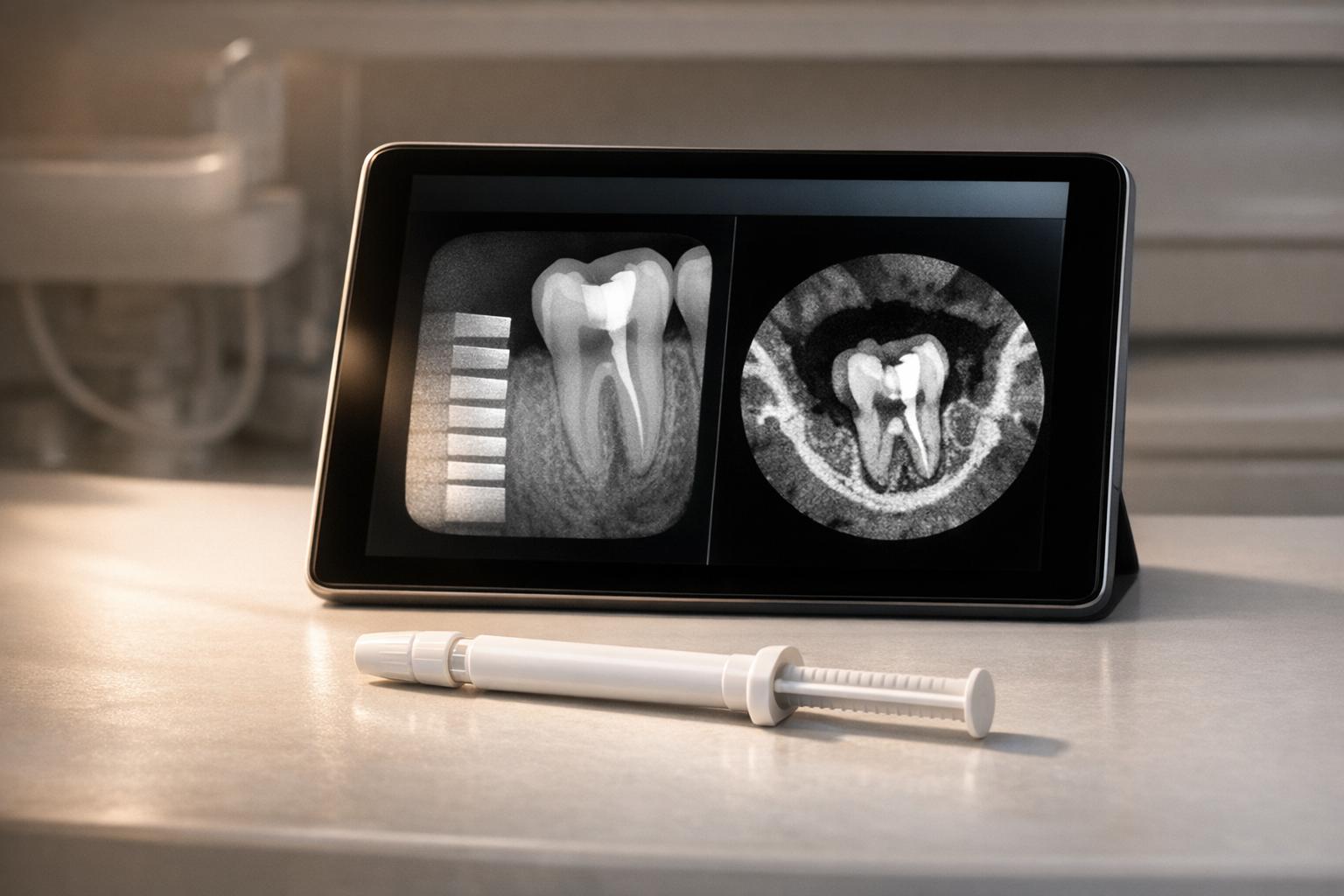
Bioceramic sealers in dentistry have lower radiopacity than traditional resin-based sealers, but they meet ISO standards and offer key benefits in imaging accuracy. Radiopacity ensures materials are visible on X-rays, allowing dentists to confirm proper root canal sealing. Here’s what you need to know:
- ISO Standards: Sealers must have a radiopacity of at least 3 mmAl to comply with ISO 6876 guidelines.
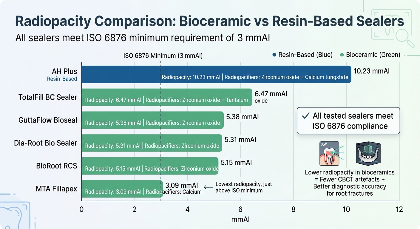
- Bioceramic vs Resin-Based Sealers: Bioceramic sealers like TotalFill BC (6.47 mmAl) and BioRoot RCS (5.15 mmAl) have lower radiopacity compared to resin-based AH Plus (10.23 mmAl).
- Imaging Benefits: Lower radiopacity in bioceramics reduces artefacts and distortion in CBCT scans, improving diagnostic accuracy for root fractures.
- Key Components: Zirconium oxide and tantalum oxide enhance radiopacity in bioceramic sealers, while resin-based sealers often use bismuth or calcium tungstate.
Bioceramic sealers provide sufficient visibility for clinical use while minimising imaging artefacts, making them a reliable choice for root canal treatments.
Research on Bioceramic Sealer Radiopacity Measurements
How Radiopacity is Tested
Radiopacity in bioceramic sealers is assessed using a standardised procedure. The sealer is prepared according to the manufacturer’s instructions and placed into moulds, typically 7.5 mm in diameter and 1 mm in height [1][3].
To establish a reference, an aluminium step-wedge (99.6% pure aluminium) is used. This wedge is graduated in 1 mm increments from 1 to 10 mm and is radiographed alongside the sealer samples [1][3]. Digital radiography systems capture these images using settings of approximately 70 kVp, 7–8 mA, with exposure times ranging from 0.07 to 0.17 seconds. Specialised software then converts the Mean Grey Values from the images into mmAl (millimetres of aluminium) using regression analysis [1][3].
Testing conditions can significantly impact radiopacity outcomes. For instance, BioRoot RCS showed a radiopacity of 8.32 mmAl in one study conducted at a 30 cm distance [3], while a different study using a 10 cm distance recorded a much lower value of 4.88 mmAl [1].
Measured Radiopacity Values from Studies
Using this standardised method, radiopacity values for bioceramic sealers vary depending on imaging conditions. While all tested sealers surpass the ISO minimum requirements, they generally exhibit lower radiopacity compared to AH Plus, a commonly used epoxy resin-based sealer. A study published by Bezmialem Science highlighted that:
All the tested sealers met the ISO requirements for radioopacity and flow [1].
Similarly, researchers at the University of Belgrade reported:
Radiopacities of all evaluated calcium silicate-based sealers were higher than minimal values recommended by standards [3].
For example, TotalFill BC Sealer achieves a radiopacity of 6.47 mmAl, attributed to its dual radiopacifiers – zirconium oxide and tantalum oxide [1]. BioRoot RCS displays a range of 4.51 to 8.32 mmAl, depending on the testing parameters [1][3]. GuttaFlow Bioseal measures between 4.83 and 7.64 mmAl [1][3]. On the other hand, MTA Fillapex registers 3.09 mmAl in recent studies, a drop from earlier formulations containing bismuth trioxide, which reached up to 8.9 mmAl [1]. In contrast, AH Plus consistently exceeds 10 mmAl [1][3].
These findings highlight the importance of consistent imaging protocols to ensure reliable comparisons in clinical settings.
sbb-itb-2be92ed
Radiopacity Comparison of Different Bioceramic Sealers
Radiopacity Comparison of Bioceramic vs Resin-Based Dental Sealers
Radiopacity Results for Common Sealers
Using established testing methods and ISO standards, let’s examine how the radiopacity of various bioceramic sealers stacks up.
AH Plus, a resin-based sealer, stands out with a radiopacity of 10.23 mmAl, making it a reliable high-radiopacity control. This is thanks to its formulation, which includes calcium tungstate and zirconium oxide, giving it a clear edge over bioceramic sealers [1].
Within the bioceramic category, TotalFill BC Sealer leads the pack with a radiopacity of 6.47 mmAl. This is attributed to its use of two radiopacifiers – zirconium oxide and tantalum oxide. On the other hand, GuttaFlow Bioseal, Dia‐Root Bio Sealer, and BioRoot RCS rely solely on zirconium oxide for radiopacity. Meanwhile, MTA Fillapex has the lowest radiopacity at 3.09 mmAl, which only slightly surpasses the ISO 6876 minimum requirement. This lower value is due to a formulation adjustment that now incorporates calcium tungstate [1].
The table below provides a clear comparison of radiopacity values and the main radiopacifiers used in each sealer.
Sealer Radiopacity Comparison Table
| Sealer Name | Radiopacity (mmAl) | ISO 6876 Compliance | Primary Radiopacifier(s) |
|---|---|---|---|
| AH Plus (Control) | 10.23 | Yes | Zirconium oxide, Calcium tungstate |
| TotalFill BC Sealer | 6.47 | Yes | Zirconium oxide, Tantalum oxide |
| GuttaFlow Bioseal | 5.38 | Yes | Zirconium oxide |
| Dia-Root Bio Sealer | 5.31 | Yes | Zirconium oxide |
| BioRoot RCS | 5.15 | Yes | Zirconium oxide |
| MTA Fillapex | 3.09 | Yes | Calcium tungstate |
Deniz Burcu Mert and Nimet Gençoğlu from Marmara University Faculty of Dentistry highlight:
All the tested sealers met the ISO requirements for radioopacity and flow [1].
This compliance assures that, while bioceramic sealers are generally less radiopaque than resin-based alternatives, they still offer sufficient visibility for effective use in root canal procedures.
What Affects Sealer Visibility in CBCT and X-rays
Imaging Settings and Methods
The visibility of sealers on CBCT and X-rays largely depends on imaging parameters like kVp, mA, and exposure time, which influence the contrast and clarity of the images [2][3].
For endodontic imaging, CBCT protocols often use settings such as 89 kVp and 10 mA with a voxel size of 0.085 mm [2]. These smaller voxel sizes offer the detailed resolution necessary for examining root canal fillings, though they can also exaggerate volumetric distortion – where dense materials appear larger than they actually are [2]. Digital radiography systems, being more sensitive than conventional film, allow for lower radiation doses without sacrificing diagnostic quality [3].
The field of view (FOV) is another critical factor. Smaller FOVs, like 6 x 4 cm, are preferred in endodontics because they reduce radiation exposure while enhancing local resolution [2]. However, variations in imaging settings can affect radiopacity measurements, making standardisation essential for consistent comparisons [2]. Beyond imaging parameters, the material composition of the sealer also plays a significant role in its radiographic visibility.
Material Components and Radiopacifiers
The composition of a sealer, particularly its radiopacifiers, is a key factor in determining its radiographic profile. Zirconium oxide (atomic number 40) is widely used in modern bioceramic sealers such as Bio-C Sealer, BioRoot RCS, and Dia-Root Bio Sealer [1][2]. Some products, like TotalFill BC, also include tantalum oxide to further enhance radiopacity [1].
Interestingly, lower radiopacity in bioceramic materials can be advantageous for CBCT imaging. Research led by Alice Corrêa Silva-Sousa at the University of São Paulo revealed that:
Bioceramic materials, which had lower radiopacity, generated fewer artefacts and less volumetric distortion compared to conventional gutta-percha cones and AH Plus sealer [2].
Materials with higher atomic numbers, such as bismuth (83) or barium (56), tend to cause more beam hardening and blooming effects, which can obscure critical clinical details [2]. To address these issues, newer formulations like MTA Fillapex now use calcium tungstate, though this adjustment results in reduced radiopacity [1].
Testing Conditions and Clinical Application
While standardised imaging settings provide reliable laboratory data, clinical conditions introduce variables that can affect sealer performance. Unlike controlled ISO tests, clinical environments involve factors like moisture and tissue interactions that influence the sealer’s setting and imaging clarity [6].
To replicate these conditions, researchers often use MixD – a blend of paraffin, polyethylene, magnesium oxide, and titanium dioxide – to mimic the photon absorption of human soft tissue during CBCT scans [2]. This simulation is crucial because clinical imaging includes photon interactions with surrounding tissues, which are absent in laboratory setups.
Moisture in the clinical environment also plays a vital role. Bioceramic sealers rely on tissue fluids to set properly and develop their bioactive properties [6]. This interaction forms an interfacial layer between the sealer and dentine, which doesn’t occur in dry laboratory conditions. Such differences explain why bioceramic materials often perform better clinically than their laboratory radiopacity values suggest – especially when diagnosing issues like vertical root fractures [7].
How Radiopacity Affects Root Canal Treatment
Benefits and Drawbacks of Bioceramic Sealers
The radiopacity of a sealer plays a crucial role in how effectively clinicians can evaluate root canal treatments and identify potential complications. Bioceramic sealers, in particular, offer some advantages when it comes to post-treatment imaging, especially with CBCT technology. Their lower radiopacity, compared to traditional resin-based sealers, helps to minimise imaging artefacts, leading to better diagnostic accuracy.
A study conducted at Hamadan University of Medical Sciences between March and August 2019 examined 100 mandibular premolars using a Cranex 3D CBCT device with a 200 µm voxel size. The results showed that bioceramic root filling materials achieved a 94% accuracy rate in detecting vertical root fractures. This was notably higher than the 81% accuracy observed with AH26 and gutta-percha. Sensitivity increased from 84% to 93%, while specificity improved from 78% to 95% [7].
However, not all bioceramic materials perform equally in radiographic evaluations. For instance, Biodentine poses a challenge due to its radiodensity (113.22 HU), which is nearly identical to natural dentine (115.44 HU, P = 0.649) [9]. Cirstea Corneliu-Aurelian from the University of Medicine and Pharmacy of Craiova explains:
Biodentine-based fillers have a radiodensity close to that of dentin, which makes it difficult for the physician to radiologically assess the correctness of the canal filling [9].
On the other hand, highly radiopaque sealers like AH Plus (10.23 mmAl) can create their own challenges. They may mask internal voids or air bubbles within the filling, leading to diagnostic errors. Excessive radiopacity can also produce blooming effects, where noise levels obscure clinical details or even mimic vertical root fractures, adding to diagnostic confusion [2][10].
Better Results Through Consistent Imaging Protocols
While varying radiopacity levels can complicate image interpretation, following standardised imaging protocols can help address these challenges. Consistency in imaging parameters is key to improving diagnostic reliability.
Using consistent CBCT settings – such as specific kVp, mA, and field of view parameters – reduces artefacts and allows for reliable comparisons across different filling materials [2][7]. Techniques like parallelism in digital radiography produce reproducible and undistorted images, enabling objective measurement of radiopacity using Hounsfield units or grey values [9]. Measuring radiodensity at standardised intervals (such as 3 mm, 6 mm, and 9 mm from the radiological apex) provides a structured approach to evaluate canal filling homogeneity and detect voids or interfacial gaps [9].
For materials with lower radiopacity, such as Biodentine, preoperative radiographs are particularly important. These help pinpoint the canal or perforation site before treatment, as the material may become indistinguishable from dentine postoperatively [9]. Conversely, streak artefacts caused by high-density sealers in CBCT imaging can result in false-negative or false-positive diagnoses of root fractures [10].
Research from the University of São Paulo in May 2025 highlighted that Bio-C Sealer’s lower radiopacity (4.9 mmAl) compared to AH Plus (9.17 mmAl) significantly reduced volumetric distortion, enabling more precise measurements of filling volumes in CBCT scans [2]. Despite these imaging differences, a retrospective study of 248 endodontically treated teeth revealed an overall success rate of approximately 96%, regardless of whether bioceramic or resin-based sealers were used [8].
Conclusion
Bioceramic sealers comply with ISO 6876 standards but exhibit lower radiopacity compared to epoxy resin-based sealers like AH Plus, which typically measure around 9–10 mmAl. For instance, Bio-C Sealer and TotalFill BC Sealer have radiopacity values of approximately 5 mmAl and 6.5 mmAl, respectively, ensuring adequate visibility during clinical imaging procedures [1][2].
This reduced radiopacity offers distinct advantages in CBCT imaging by minimising beam hardening artefacts, image noise, and volumetric distortion. These properties enhance diagnostic accuracy, particularly when identifying complications like vertical root fractures. In May 2025, Prof. Hugo Gaêta-Araujo from the University of São Paulo highlighted that bioceramic materials generated fewer artefacts and less distortion than conventional materials, achieving a sensitivity of 93% and a specificity of 95% for fracture detection. This significantly outperformed conventional gutta-percha, which showed 84% sensitivity and 78% specificity [2][7].
Optimal radiopacity is not just about meeting technical benchmarks – it plays a crucial role in ensuring successful root canal treatments. The choice of radiopacifiers, such as zirconium oxide or tantalum oxide, directly impacts radiographic clarity and clinical outcomes, avoiding the tooth discolouration often associated with bismuth oxide [4]. Furthermore, bioceramic sealers have shown superior biological performance, with periapical healing rates around 90% compared to 75% for conventional sealers over a six-month period [11].
Retrospective data reveal that both bioceramic and resin-based sealers can achieve success rates of approximately 96% when appropriate imaging protocols are used [5]. Understanding the unique properties of each material is essential for selecting the right sealer, particularly in cases where post-operative CBCT imaging is necessary. By carefully considering radiopacity, clinicians can optimise treatment outcomes and enhance patient care.
FAQs
Does lower radiopacity make a bioceramic sealer harder to see on an X-ray?
Bioceramic sealers with lower radiopacity may appear less distinct on an X-ray. However, these sealers are formulated to comply with the required minimum radiopacity standards (≥3 mm Al) outlined in ISO 6876:2012. This ensures they remain adequately visible for radiographic assessments.
Why does a more radiopaque sealer create artefacts on CBCT scans?
A sealer with high radiopacity can lead to artefacts on CBCT scans because of its ability to significantly attenuate X-rays. This can create streaks or distortions in the images, which may obscure nearby structures and complicate accurate interpretation of the scan.
What CBCT settings most affect how a sealer looks in scans?
The visibility of a sealer in CBCT scans is largely determined by its radiopacity, which is influenced by the material’s composition. Additional factors, such as exposure parameters like voltage, current, and image acquisition settings, also significantly affect how well sealers appear in scans. Although precise settings aren’t specified, these elements are critical in shaping the sealer’s detectability.
Related Blog Posts
- 3D Bioprinting Materials for Endodontic Repair
- Bioceramic vs. Resin-Based Sealers: Comparison
- How Heat Affects Bioceramic Sealers
- CBCT in Detecting Root Fractures
Important Notice: Any surgical or invasive procedure carries risks. Before proceeding, you should seek a second opinion from an appropriately qualified health practitioner.
Individual results may vary. The information provided in this article is for educational purposes only and does not constitute medical advice.
Checkout Related Blogs




Get in touch with us
For more information, call us now to start feeling better. Or fill the form below to make appointment
The Latest News from Complete Smiles
How to Clean Clear Plastic Retainers
Checklist for Choosing Wearable Dental Devices
Checklist for Choosing Cloud AI Platforms in Dentistry

Complete Smiles Bella VistaAccepts All Major Health Funds, Including





