





3D Bioprinting Materials for Endodontic Repair

3D bioprinting is reshaping how we approach endodontic repair by aiming to regenerate living, functional tissue instead of using traditional inert materials like gutta-percha. This technology uses patient-specific data (e.g., CT or MRI scans) to create precise, layer-by-layer structures that mimic the complex architecture of teeth. Key materials include:
- Polymer-based bioinks: Natural polymers (e.g., collagen, alginate) support cell growth but lack strength, while synthetic ones (e.g., PCL, PLA) offer durability but may need surface modifications for better cell attachment.
- Composite/hybrid formulations: Combine natural and synthetic materials for better balance between bioactivity and strength; useful for both soft pulp and hard dentin regeneration.
- Resin-based materials: Provide high precision and mechanical strength, ideal for detailed structures, though cytotoxicity risks require careful post-processing.
- Bioactive glass/mineral-based options: Mimic natural tooth chemistry, promoting mineralisation and cell differentiation, but can be brittle and challenging to print.
Each material has its strengths and limitations, from biocompatibility and mechanical properties to degradation rates. While progress is promising, challenges like improving mechanical strength, controlled degradation, and clinical adoption remain. These advancements could transform dental care, offering new ways to repair and regenerate damaged teeth.
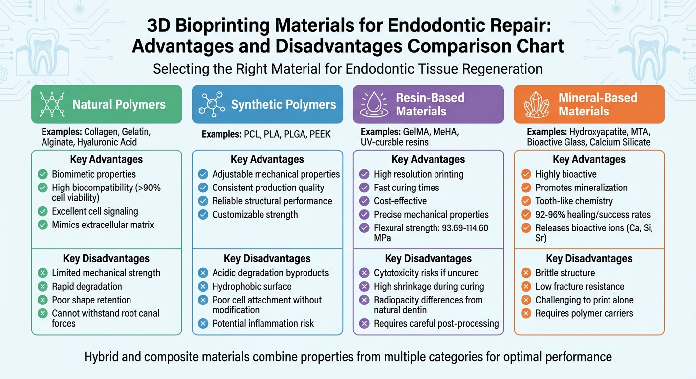
Comparison of 3D Bioprinting Materials for Endodontic Repair
What Is 3D Bioprinting? – The Pro Dentist
1. Polymer-Based Bioprinting Materials
Polymer-based bioinks play a key role in endodontic bioprinting. They are generally categorised into two groups: natural polymers like collagen, gelatin, alginate, and hyaluronic acid, and synthetic polymers such as polycaprolactone (PCL), polylactic acid (PLA), and poly(lactic-co-glycolic acid) (PLGA). Each type has its own strengths. Let’s explore their key aspects: biocompatibility, mechanical properties, degradation profiles, and clinical applications.
Biocompatibility
Supporting cellular activity is essential for regenerating functional dental tissues. Natural polymers closely imitate the extracellular matrix (ECM), providing biochemical cues that help dental pulp stem cells (DPSCs) adhere, grow, and transform into odontoblast-like cells. Studies show that cell viability with these materials consistently exceeds 90% [2][8][9]. As Sam P. Tarassoli and colleagues explain:
Natural bioinks differ to synthetic bioinks in their ability to mimic the native cellular microenvironment, offering support to growing cells and thereby increasing the likelihood of cell adhesion and secretion of matrix [9].
Synthetic polymers, like PCL, are also biocompatible and less likely to cause inflammation compared to some materials. However, they often lack natural cell-binding sites, which means surface modifications may be needed to improve cell attachment [5][8].
Mechanical Properties
While biological performance is critical, mechanical stability is just as important for endodontic applications. Natural polymers are excellent for cell signalling but tend to be mechanically weak, making it difficult for them to retain shape or withstand forces within the root canal [2][7]. Synthetic polymers, on the other hand, provide customisable mechanical strength and structural integrity. For example, PCL has a low melting point (59–64 °C), making it ideal for extrusion-based printing [5], and materials like PEEK offer exceptional load-bearing capabilities [10].
To address these challenges, hybrid bioinks that combine natural and synthetic polymers have been developed. These blends strike a balance between biological signalling and the mechanical support required for dental applications [2][8].
Degradation Profile
For successful tissue repair, the scaffold’s degradation must align with tissue formation. Natural hydrogels like GelMA degrade enzymatically through matrix metalloproteinase (MMP) pathways, allowing cells to remodel the scaffold into native tissue. This feature is particularly crucial for regenerating vascularised pulp [1][2].
In contrast, synthetic polymers such as PLA and PLGA degrade through hydrolysis. While their degradation rates can be controlled, they release acidic byproducts that may lead to inflammation in the root canal [1][5]. A unique challenge with alginate was highlighted by Fengxiao Zhao:
The enzyme that cleave alginate chain is absent in human body, leading to the uncontrollable biodegradation in vivo [1].
PCL, known for its slow degradation, is better suited for applications requiring long-term structural support, such as hard tissue repairs, rather than environments needing rapid tissue turnover [1][10].
Clinical Applications
The choice of material significantly impacts clinical outcomes. Alginate is a popular option in bioprinting, featuring in 30% to 41% of studies due to its affordability [9]. GelMA has gained attention for pulp regeneration, combining gelatin’s bioactivity with photo-crosslinkable stability [1]. For high-load applications, materials like PCL and PEEK are preferred, though surface coatings are often used to enhance their bioactivity [1][10].
2. Composite and Hybrid Bioprinting Materials
Composite and hybrid materials bring together natural hydrogels and synthetic polymers or bioactive minerals, offering a blend of biological signalling and mechanical strength. This combination is especially useful in endodontics, where regenerating both soft pulp tissue and hard dentine is critical. Let’s dive into how these materials balance bioactivity with structural integrity.
Biocompatibility
For composite materials to succeed, they must support cell survival during the bioprinting process. Factors like extrusion pressure, shear stress, and UV exposure during photocrosslinking can affect cell viability [2]. Research on collagen-chitosan-hydroxyapatite composites has shown promising results, with cell viability exceeding 95% just one week post-printing [2].
What makes composites stand out is their ability to replicate the extracellular matrix while also providing structural support. Inorganic additives like calcium silicate, hydroxyapatite, or tricalcium phosphate enhance mineralisation, encouraging the formation of dentine-like tissue [5][2]. For instance, combining calcium silicate with GelMA has been shown to stimulate dentine regeneration and odontogenic differentiation in dental pulp stem cells [2].
Mechanical Properties
Natural hydrogels are excellent for supporting cells but lack mechanical strength. On the other hand, pure bioceramics are strong but can be brittle [11]. Hybrid materials address this issue by balancing inorganic fillers with organic components. For example, adding mineral fillers like CaCO₃ or SrCO₃ to a PLGA matrix improves both degradation behaviour and mechanical stability [11].
Studies comparing hybrid hydrogels have found that CS/GelMA composites enhance odontogenic differentiation in human dental pulp stem cells more effectively than pure GelMA [11]. However, it’s worth noting that 3D-printed materials often have lower structural integrity compared to milled materials like zirconia. This is due to the layer-by-layer nature of additive manufacturing, which can create weak points between layers [12][13].
Degradation Profile
One of the key challenges in bioprinting is ensuring that scaffold degradation aligns with tissue regeneration. Hybrid materials offer adjustable degradation rates to address this issue. Natural hydrogels like GelMA include matrix metalloproteinase-degradable motifs, allowing cells to remodel the scaffold as they grow [1]. Meanwhile, synthetic components like PLGA degrade hydrolytically, though their acidic byproducts can sometimes trigger inflammation [1][5]. As Serge Ostrovidov from Tokyo Medical and Dental University explains:
The bioink should be biodegradable, ideally at a rate that fits the growth of the biological tissue, to be naturally replaced by the ECM components secreted by cells [3].
Mineral fillers can further fine-tune degradation. Strontium-doped calcium silicate scaffolds, for example, can release bioactive ions for over six months while maintaining structural support for bone regeneration [11]. Similarly, BMP-mimicking peptides tethered to GelMA-based hybrid bioinks have been shown to retain more than 50% of their initial volume after three weeks [11].
Clinical Applications
These properties have paved the way for practical uses in dentistry, particularly for complex structures like the dentine-pulp complex [11][12]. For dentine regeneration, PCL/bioglass composites are highly effective due to their support for mineralisation. Meanwhile, PCL/hyaluronic acid composites are better suited for regenerating soft dental pulp tissue [3]. For extrusion-based bioprinting of dental composites, the optimal viscosity typically ranges between 30 and 60 × 10⁷ mPa/s [2].
3. Resin-Based Bioprinting Materials
Resin-based materials, commonly utilised in SLA and DLP printing, are prized for their ability to achieve high-resolution structures with precise mechanical properties. These materials include UV-curable resins, gelatin methacryloyl (GelMA), and methacrylated hyaluronic acid (MeHA). Unlike natural hydrogels, which often lack fine detail, resin-based materials excel in applications requiring both precision and durability, making them highly relevant for advanced clinical uses.
Biocompatibility
Among resin-based options, GelMA stands out due to its biological origin. As a derivative of gelatin, it retains arginine-glycine-aspartic acid (RGD) motifs, which promote cell attachment and spreading, as well as matrix metalloproteinase (MMP)-degradable motifs, which allow cells to remodel the scaffold as they grow [1]. In a 2023 study led by Qian et al., researchers used a DLP printer to create GelMA-based microspheres loaded with dental pulp stem cells (DPSCs). These microspheres supported angiogenic, neurogenic, and odontogenic differentiation, leading to the successful regeneration of vascularised, pulp-like tissue in swine models [1].
Further studies demonstrated that human DPSCs encapsulated in GelMA constructs maintained a viability rate of over 90% throughout all experimental phases [2]. Similarly, periodontal ligament stem cells exhibited approximately 90% viability in composite hydrogels when the GelMA-to-PEGDA ratio ranged from 2:3 to 4:1 [2]. However, traditional resins such as Bis-GMA and UDMA can compromise cell viability if curing is incomplete, highlighting the importance of proper post-processing.
Mechanical Properties
In addition to biocompatibility, resin-based materials are valued for their mechanical strength. For example, 3D-printed dental resin has a flexural strength of around 93.69 MPa, which can be enhanced to 112.80–114.60 MPa with the addition of 10–20% zirconia or 5–10% glass silica nanoparticles [14]. As Abdullah Alshamrani from The University of Sydney explains:
Dental restorations demand materials that can endure chewing forces while maintaining slow biodegradation rates [14].
The mechanical properties of these materials can be fine-tuned by adjusting photoinitiator concentrations, crosslinker levels, and exposure times during the printing process [10]. However, increasing filler content can raise resin viscosity, potentially causing printing issues like clogging or uneven flow, which might weaken the final structure [14]. Tiago Reis from the University of Santiago de Compostela also notes:
One challenge is the mismatch in radiopacity and hardness between resin-based materials and natural dentine [15].
Degradation Profile
The degradation characteristics of resin-based materials vary depending on their composition. Synthetic resins like PLA and PLGA degrade through hydrolysis, breaking down in water. By adjusting the lactic-to-glycolic acid ratio, degradation rates can be controlled, though the acidic byproducts may cause inflammation in some cases [1][5].
GelMA, on the other hand, degrades via MMP mechanisms, allowing cells to remodel their environment, while PCL provides long-term stability with adjustable degradability [1][5]. Importantly, cell viability remains high with SLA printing (>85%) and laser-assisted methods (>95%) [5].
Clinical Applications
Resin-based materials are particularly advantageous for applications requiring precision and mechanical strength. In 2020, Duarte Campos et al. used a handheld bioprinter to deliver a collagen-based bioink modified with agarose into prepared root canals. The bioink demonstrated successful vascularisation without significant shrinkage, as confirmed by immunofluorescence imaging [1]. Beyond tissue regeneration, these materials are also widely used for creating surgical guides, temporary crowns, and highly detailed endodontic training models. For instance, PolyJet technology can replicate root canal anatomy with layer thicknesses as fine as 16 μm [15].
Proper post-processing is essential to ensure both biocompatibility and mechanical reliability. Steps like cleaning with isopropyl alcohol and a 10-minute light curing process (400–550 nm) help remove unpolymerised monomers that could otherwise compromise the material [14]. Testing of reinforced resins shows cell viability rates exceeding 80% across all groups, confirming their safety for clinical use [14].
sbb-itb-2be92ed
4. Bioactive Glass and Mineral-Based Materials
In addition to polymer-based and resin-based bioinks, mineral-based materials provide a strong foundation for regenerating hard tissues, especially in endodontic repair. Materials like Mineral Trioxide Aggregate (MTA), calcium silicate-based cements (CSCs), and hydroxyapatite closely resemble the inorganic makeup of natural teeth, making them particularly effective for this purpose. Among these, MTA stands out as the most extensively studied material and is often referred to as the "gold standard" [16].
Clinical results back this up. For instance, when MTA is used as an apical barrier in treating nonvital immature teeth, it achieves a remarkable healing rate of 96%. Similarly, bioceramics employed as root-end filling materials in endodontic microsurgery show success rates between 86.4% and 95.6% over one to five years [16]. A retrospective study spanning up to nine years highlighted that ProRoot MTA achieved a 92.1% success rate, while BC Putty, a premixed calcium silicate/phosphate material, reached 92.4% [16].
Biocompatibility
Mineral-based materials excel at supporting the attachment, survival, and differentiation of dental stem cells. These bioceramics release bioactive ions – such as calcium (Ca), silicon (Si), and strontium (Sr) – which play a key role in cell differentiation. For example, Biodentine, a tricalcium silicate-based material, promotes odontogenic and osteogenic differentiation through the MAPK and CaMKII pathways [16]. Strontium-doped calcium silicate scaffolds can release Sr and Si ions consistently for at least six months, ensuring sustained bioactivity to aid tissue regeneration [1].
That said, biocompatibility depends on the material’s composition and the type of cells involved. Biodentine, for instance, offers faster setting times (around 12 minutes) and better mechanical strength compared to traditional MTA, which takes several hours to cure and can cause tooth discolouration [16]. For cases where discolouration is a concern, aluminium-free alternatives like BioAggregate and ERRM are often recommended [16].
Mechanical Properties
While bioceramics are excellent at providing mineral cues for tissue repair, they come with mechanical challenges. These materials generally have low fracture resistance and low flexural resistance, which can limit their durability in load-bearing situations [1]. For context, natural dentine has a Young’s modulus of 17–42 GPa, a benchmark that bioceramics aim to meet [4].
To overcome these limitations, researchers often combine bioceramics with polymer matrices like GelMA or PCL. This hybrid strategy enhances the mechanical properties, such as compression modulus and equilibrium swelling rates, of the resulting bioinks [1][4]. For example, integrating mesoporous bioactive glass nanoparticles into bioinks improves both shape fidelity and surface roughness in 3D-printed constructs [1]. However, while BioAggregate is aluminium-free and has bond strength comparable to white MTA, its mechanical properties are generally weaker [16]. These shortcomings highlight the importance of controlled degradation to ensure full integration with tissue.
Degradation Profile
The degradation rate of these materials must align closely with the pace of tissue regeneration [3][17]. Bioceramics are classified either as bioactive (bonding directly with tissue) or biodegradable (resorbed and replaced by natural tissue) based on their reactivity [16]. Ideally, their degradation should match the rate at which cells synthesise new extracellular matrix, ensuring a seamless replacement of functional tissue [3][17].
As these materials degrade, they release bioactive ions that encourage odontoblastic differentiation and mineralisation [3][16]. For example, in bioprinted constructs using "NICE" bioink (GelMA mixed with nanosilicates), calcium levels steadily increased over a 60-day period [2]. Additionally, cell viability in bioceramic-reinforced bioinks typically exceeds 90% post-bioprinting [2][3].
One challenge, however, is the high solubility of some bioceramics. Reducing solubility while maintaining their bioactive properties remains a critical focus for improving their clinical performance in endodontics [16]. Achieving controlled degradation, paired with effective ion release, is key to their success.
Clinical Applications
Mineral-based materials play a variety of roles in endodontic treatments. For procedures like endodontic microsurgery, where a quick initial set is needed, fast-setting CSCs such as Biodentine or BC Putty are preferred over traditional MTA [16]. A prospective study reported that both MTA and BC Putty achieved one-year success rates exceeding 93%, highlighting their reliability [16].
In intentional replantation cases, fast-setting bioceramics help limit extraoral time to under 15 minutes, reducing the likelihood of ankylosis [16]. When used as apical barriers, materials like MTA, Biodentine, or CEM (calcium-enriched mixture) improve the fracture resistance of immature teeth, contributing to their long-term durability [16]. Additionally, in 3D bioprinting, bioceramics are often combined with natural hydrogels to strike a balance between printability and bioactivity, further supporting their use in comprehensive endodontic repair [1][4].
Advantages and Disadvantages
After examining the unique characteristics of each material category, it’s time to weigh their overall strengths and limitations.
Each type of material brings its own set of benefits and challenges. Natural polymers, such as collagen and alginate, closely mimic the extracellular matrix, offering excellent biocompatibility. However, their mechanical strength is limited, and they degrade quickly, making them unsuitable for applications requiring load-bearing support [2][7].
Synthetic polymers like PCL and PLA stand out for their adjustable mechanical properties and consistent production quality, which ensure reliable structural performance [5][2]. On the downside, their hydrophobic surfaces make cell attachment difficult, and their degradation releases acidic byproducts that may cause local inflammation [1][7]. PCL, with its lower melting point, is less likely to provoke inflammatory reactions [5].
Resin-based materials are known for their ability to achieve high-resolution printing at a relatively low cost, with rapid curing times that align well with clinical workflows [5][10]. Despite these advantages, they face notable drawbacks. Tiago Reis from the University of Santiago de Compostela highlights one key issue:
The major criticism of 3D-printed teeth is the difference in radiopacity and hardness between resin and human dentine [15].
Additionally, high shrinkage rates during curing can compromise dimensional accuracy [10][15].
Mineral-based materials, such as hydroxyapatite and bioactive glass, are highly bioactive and chemically resemble natural tooth structures, encouraging odontogenic differentiation [5][1]. However, their brittleness and low fracture resistance pose significant challenges. To improve their usability, they are often combined with polymer carriers, which enhance printability [5][1].
Lastly, composite and hybrid materials aim to balance mechanical stability with biological signalling. While promising, optimising these materials for both cell viability and printability remains a complex task [5][2][1]. Ultimately, the choice of material plays a critical role in achieving both tissue regeneration and structural integrity in endodontic repair.
| Material Category | Key Advantages | Key Disadvantages |
|---|---|---|
| Natural Polymers | Biomimetic, high biocompatibility (>90% cell viability) [6][1][2] | Limited mechanical strength, rapid degradation [2][7] |
| Synthetic Polymers | Adjustable properties, consistent production, reliable structure [5][2] | Acidic degradation products, hydrophobic surface [1][7] |
| Resin-Based | High resolution, fast curing, affordable [5][10] | Cytotoxicity risks, shrinkage issues, radiopacity differences [10][15] |
| Mineral-Based | Bioactive, promotes mineralisation, tooth-like chemistry [5][1] | Brittle, low fracture resistance, challenging to print alone [5][1] |
Conclusion
Selecting the right material for endodontic repair depends heavily on the specific clinical needs. Hybrid and composite materials strike a balance between natural bioactivity and synthetic durability, making them versatile choices for various applications [1][4]. For pulp regeneration, soft hydrogels like GelMA are often the go-to option, while dentin repair benefits from composite inks containing Treated Dentin Matrix or bioceramics, which provide critical mineral support [1][4].
While these materials show great potential, challenges still exist. Despite rapid advancements, significant gaps remain. The dental 3D printing market is expected to hit A$930 million by 2025, growing at an impressive 17% annually [18]. However, clinical adoption remains a hurdle. For instance, current resins often fall short of mimicking human dentine in terms of hardness and radiopacity, which complicates diagnostic imaging [15]. Brendan M. Leung from Dalhousie University highlights one promising aspect:
The fluidity of injectable scaffolds offers a number of advantages… including their critical ability to occupy and adapt to the irregular topology of the root canal space [7].
Looking ahead, research must focus on innovations like 4D bioprinting to create dynamic scaffolds and enhance vascularisation for sustaining bioprinted pulp tissue [4][5]. Additionally, developing standardised protocols for chairside bioprinting and cost-effective production methods will be crucial to transitioning these materials from the lab to everyday clinical use [1][7].
For now, dental practices, such as Complete Smiles Bella Vista (https://completesmilesbv.com.au), continue to rely on established root canal therapy techniques while keeping an eye on these emerging advancements. Moving forward, collaboration among researchers, clinicians, and regulatory bodies will be essential to create evidence-based guidelines that prioritise both innovation and patient safety.
FAQs
What are the benefits of using 3D bioprinting for endodontic tissue repair compared to traditional methods?
3D bioprinting is transforming endodontic tissue repair by enabling the creation of structures tailored to each patient, closely mimicking the natural design of a tooth. Unlike traditional approaches such as root canal therapy or resin fillings – which restore functionality without regenerating tissue – this technology supports genuine tissue regeneration.
By using advanced bio-inks loaded with stem cells and biomaterials, 3D bioprinting builds scaffolds layer by layer with remarkable precision. These customised scaffolds encourage better blood vessel formation, cell growth, and mineralisation, which are essential for regenerating pulp and dentine. The benefits? A lower chance of reinfection, improved long-term tooth health, and more reliable outcomes compared to conventional methods.
What challenges are involved in using 3D bioprinting materials for dental treatments?
Replicating the intricate structure of natural teeth is one of the biggest hurdles in using 3D bioprinting for dental treatments. Bio-inks currently fall short when it comes to mimicking the layered makeup of dentine, pulp, and enamel, all while maintaining the strength needed to endure chewing. On top of that, many materials either degrade too quickly or too slowly, making it tough to match the natural pace of tissue regeneration.
Another significant challenge lies in keeping cells alive during the printing process. Steps like UV curing and the high shear forces involved can harm stem cells and weaken the effectiveness of growth factors. Beyond the technical barriers, broader adoption faces issues like the steep cost of equipment, the need for specialised training, and unclear safety and regulatory guidelines in Australia. Overcoming these obstacles is crucial for 3D bioprinting to transition from experimental research into everyday dental practice.
What makes composite and hybrid bioprinting materials effective for dental tissue repair?
Composite and hybrid bioprinting materials are crafted to strike a balance between bioactivity and mechanical strength, making them ideal for dental uses like repairing endodontic tissues. These materials blend soft, cell-supportive hydrogels – such as gelatin-methacrylate or alginate – with rigid bioceramic particles like calcium phosphate or bio-glass. The hydrogel creates a moist, nurturing environment for cell growth and tissue repair, while the ceramic particles add strength, ensuring the material can handle the physical demands of dental applications.
Hybrid materials take this concept a step further by including additional functional elements, like nano-carriers for growth factors or interpenetrating polymer networks. These components can release bioactive cues to encourage dentin-pulp regeneration, all while the polymer-ceramic structure maintains the required mechanical stability (typically in the range of 10–20 MPa under compression). By fine-tuning the composition, researchers can design materials that meet both the biological and structural needs for effective dental tissue repair.
Related Blog Posts
- 3D Bioprinting in Dental Implants: How It Works
- Nanocomposites for Pulp Regeneration: Key Features
- Recent Advances in Biomaterials for Periodontal Regeneration
- How 3D Bioprinting Creates Periodontal Scaffolds
Important Notice: Any surgical or invasive procedure carries risks. Before proceeding, you should seek a second opinion from an appropriately qualified health practitioner.
Individual results may vary. The information provided in this article is for educational purposes only and does not constitute medical advice.
Checkout Related Blogs




Get in touch with us
For more information, call us now to start feeling better. Or fill the form below to make appointment
The Latest News from Complete Smiles
How to Clean Clear Plastic Retainers
Checklist for Choosing Wearable Dental Devices
Checklist for Choosing Cloud AI Platforms in Dentistry

Complete Smiles Bella VistaAccepts All Major Health Funds, Including





