





How 3D Bioprinting Creates Periodontal Scaffolds

3D bioprinting is transforming dental care by enabling precise reconstruction of periodontal tissues damaged by disease. Unlike conventional treatments, this technology uses bio-inks – materials containing living cells and growth factors – to build scaffolds that guide tissue regeneration. These scaffolds mimic the natural structure of gums, ligaments, and bone, supporting simultaneous healing of all components. With imaging data like CT scans, bioprinting creates patient-specific designs that fit defects perfectly, ensuring better integration and functionality.
Key points:
- What it solves: Periodontal disease causes complex tissue damage that traditional methods can’t fully repair.
- How it works: Bioprinters use bio-inks to create scaffolds layer by layer, embedding cells for targeted tissue growth.
- Benefits: Precise cell placement, custom designs, and improved tissue regeneration outcomes.
- Challenges: High costs, limited clinical trials, material limitations, and issues like vascularisation in small spaces.
This cutting-edge approach offers a tailored, cell-based solution to periodontal regeneration, addressing limitations of older methods.
Periodontal Tissue Regeneration Basics
Difficulties in Regenerating Periodontal Tissues
Regenerating the periodontium – comprising the gingiva, periodontal ligament (PDL), cementum, and alveolar bone – is a complex task because all four tissues need to regenerate at the same time. This is no small feat. For instance, the PDL itself is incredibly thin, measuring between 100 and 400 μm [1][7]. Achieving such precision with conventional surgical methods is nearly impossible.
Adding to the challenge, the tooth root surface lacks blood vessels, which restricts cell attachment and nutrient delivery. Meanwhile, the oral environment doesn’t make things any easier. Constant exposure to bacteria and the mechanical forces from chewing further complicate the healing process [1][7]. As Simon Raveau and Fabienne Jordana from the University of Nantes explain:
Conventional periodontal treatments, surgical or non-surgical, do not allow a complete periodontal regeneration. They can only obtain a partial localised regeneration in the apical part of the bone defect [2].
These hurdles highlight the need for more advanced solutions, which is where 3D bioprinting comes into play.
How 3D Bioprinting Solves These Problems
3D bioprinting offers a way to tackle these challenges head-on. By using CT or CBCT scans tailored to the patient, this technology creates scaffolds that perfectly match the shape and size of the defect [2][4]. Unlike one-size-fits-all treatments, bioprinted scaffolds are designed with specific compartments for different tissues. For example, a mineralised section can support bone growth, while a fibrous section is aligned to encourage PDL regeneration [1][4].
One of the standout features of bioprinting is its ability to precisely place cells. Traditional methods often struggle with uneven cell distribution and poor penetration into scaffolds. In contrast, 3D bioprinting embeds live cells layer by layer, ensuring they are evenly distributed and properly aligned [9][5]. Techniques like Melt Electrowriting produce fibres at micron-to-nanometre scales, mimicking the natural structure of collagen and providing directional cues that guide cell growth [1][6].
The results are promising. Studies show that bioprinted constructs can maintain cell viability rates of over 90% for more than seven days [8][5]. This creates an environment where different tissue types can grow simultaneously, while the scaffold’s precise design ensures functional restoration. By overcoming the limitations of traditional methods, 3D bioprinting opens the door to targeted, tissue-specific regeneration with extraordinary precision.
3D-printed scaffold enables controlled release of biomolecules into body
The Bioprinting Process for Periodontal Scaffolds
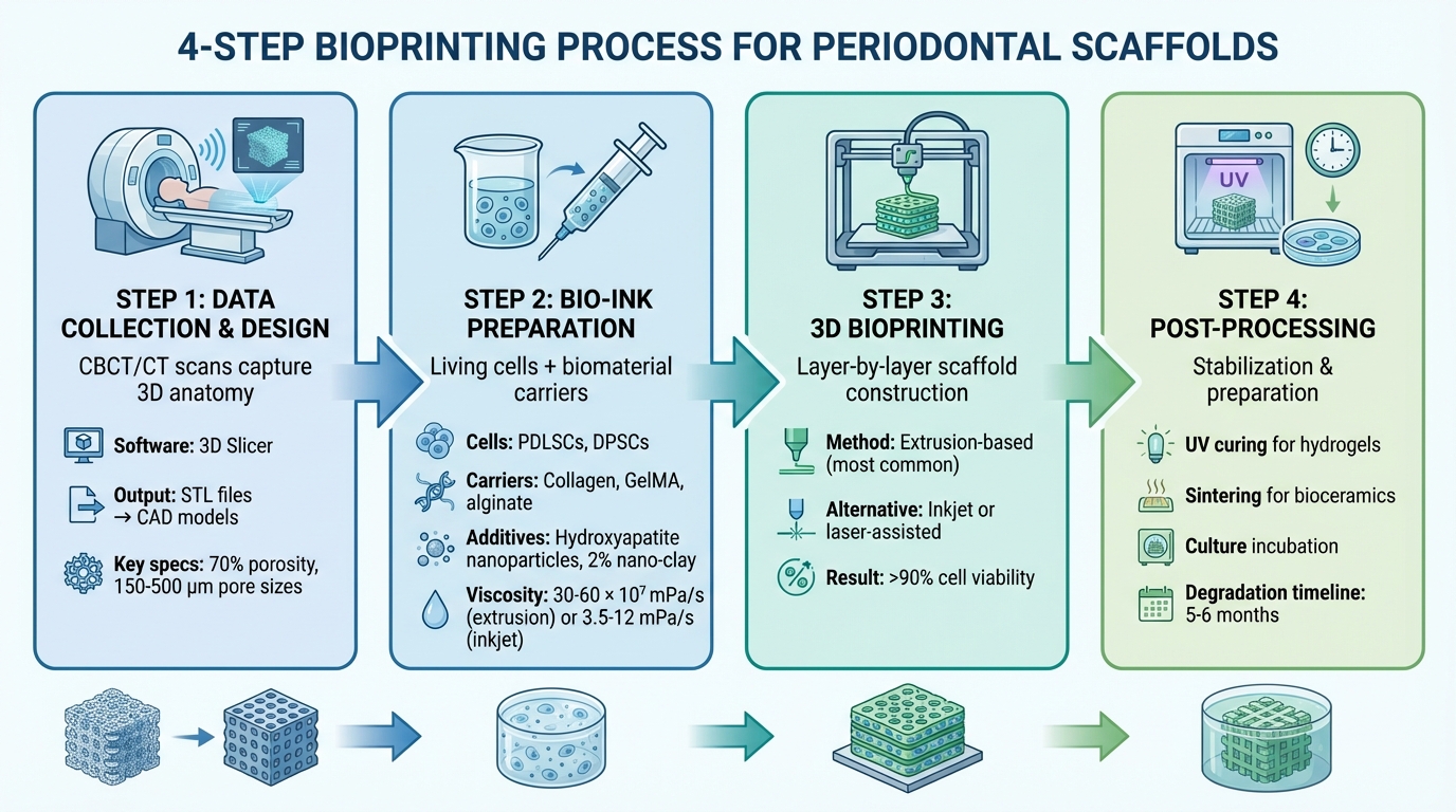
4-Step 3D Bioprinting Process for Periodontal Scaffolds
Data Collection and Scaffold Design
The creation of a periodontal scaffold begins with gathering detailed anatomical data of the affected area. Clinicians rely on Cone Beam Computed Tomography (CBCT) or standard CT scans to produce three-dimensional images of the patient’s periodontal tissues. Unlike traditional 2D X-rays, these scans provide a comprehensive view of the tooth root, alveolar bone, and surrounding structures, capturing their intricate geometry [11][2].
This imaging data is then processed using specialised medical software like 3D Slicer. The software segments the scans into categories such as teeth, bone, and soft tissues. These segmented images are converted into STL (Standard Tessellation Language) files, which serve as the foundation for Computer-Aided Design (CAD) models [11]. The scaffold design is tailored to match the defect site precisely. The internal surface is shaped to fit the tooth root, while the external surface mirrors the original bone structure. Designers adjust the scaffold’s thickness to reflect natural bone distribution – thicker at the base and thinner near the gingival margin. Key features like porosity (around 70% for alveolar bone) and pore sizes (150–500 μm) are incorporated to ensure proper vascularisation and cell infiltration [11][2].
Preparing Bio-Inks
Bio-inks are a blend of living cells, biomaterial carriers, and bioactive components. Commonly used cell types include periodontal ligament stem cells (PDLSCs) and dental pulp stem cells (DPSCs), which are suspended in hydrogel carriers made from materials such as collagen, gelatin methacryloyl (GelMA), or alginate [12][5]. These natural polymers closely mimic the body’s extracellular matrix, offering excellent biocompatibility, though they often lack mechanical strength.
To overcome this limitation, researchers have developed hybrid bio-inks. In 2023, Gao and colleagues demonstrated that a combination of polyethylene glycol diacrylate, Pluronic F127 diacrylate, and GelMA created superior porous structures compared to purely synthetic materials [12]. Another study by Cao et al. in the same year revealed that incorporating 2% nano-clay (laponite) into methacrylated gelatin and alginate enhanced viscosity and improved physical and biomedical properties [12]. Additionally, nanomaterials like hydroxyapatite nanoparticles are often added to strengthen the scaffold and boost its bone-forming capabilities. Proper viscosity is crucial – extrusion-based printing requires bio-inks with viscosities of 30–60 × 10⁷ mPa/s, while inkjet printing demands much lower viscosities of 3.5–12 mPa/s [5].
3D Bioprinting Methods
With a refined design and optimised bio-inks, the next step is selecting the appropriate printing method to balance resolution and cell viability. Extrusion-based bioprinting is the most commonly used technique for periodontal scaffolds. It uses pneumatic or mechanical pressure to push bio-ink through a nozzle, depositing material layer by layer. This method works well with high-viscosity materials and supports high cell densities, although it offers lower resolution and can expose cells to shear stress [5].
Inkjet bioprinting, on the other hand, deposits tiny droplets of bio-ink (1–100 picolitres) using thermal or piezoelectric pulses. It is fast, cost-effective, and provides high resolution, but nozzle clogging and the requirement for low cell concentrations (less than 5 × 10⁶ cells/mL) limit its application [5]. Laser-assisted bioprinting (LAB) is the most advanced option, using laser pulses to propel bio-ink onto a substrate without a nozzle. It achieves exceptional resolution and supports high cell densities (up to 1 × 10⁸ cells/mL), but it is slow and expensive, making it less practical for larger constructs [5].
Post-Processing and Stabilisation
After printing, scaffolds undergo additional treatments to ensure they are structurally sound and biologically functional. For photo-crosslinkable hydrogels like GelMA, UV curing is used to solidify the scaffold by forming chemical bonds under ultraviolet light. Bioceramic scaffolds made from materials such as hydroxyapatite and tricalcium beta-phosphate (β-TCP) are subjected to sintering, a high-temperature process that fuses the particles together. In one study, an extrusion-printed scaffold of hydroxyapatite and β-TCP implanted into sheep calvarial defects encouraged significant bone growth within eight weeks [4].
Some scaffolds are incubated in culture media before implantation, allowing cells to proliferate and begin producing their own extracellular matrix. Synthetic polymer scaffolds, such as those made from polycaprolactone, may undergo oxygen plasma treatment or be coated with graphene oxide to improve cell adhesion by reducing hydrophobicity [1]. The degradation rate of the scaffold is also carefully managed – for alveolar bone scaffolds, the ideal timeframe is five to six months, aligning with the natural pace of tissue regeneration [3]. These post-processing steps are essential for preparing the scaffolds for further laboratory and clinical testing.
sbb-itb-2be92ed
Testing and Clinical Use of Periodontal Scaffolds
Laboratory Testing
Before periodontal scaffolds can be used in real-world applications, they undergo extensive laboratory testing to ensure they can support cells and withstand the oral environment. Researchers use various methods to test cell viability, such as Live/Dead assays and confocal microscopy. Results from studies on 3D-bioprinted dental constructs often show viability rates exceeding 90% [5]. Beyond simply ensuring cells survive, tests like the CCK-8 assay monitor how well cells proliferate, while Alizarin Red S staining looks for calcium deposits, an important marker of bone formation. To further confirm differentiation, quantitative PCR analysis detects markers like RUNX2, linked to bone development, and CEMP1, associated with cementum formation [5][9].
A notable study from 2021 using a 3DX Printer showed that bioprinted scaffolds significantly improved marker expression and cell organisation compared to other methods [9].
Mechanical properties are also critical. Compression and tensile tests measure the scaffold’s strength and elasticity, while micro-CT imaging examines its internal structure. Together, these tests provide a solid foundation for moving on to in vivo evaluations.
Animal Testing and Patient Applications
Once laboratory tests are complete, scaffolds are tested in living models. Researchers use ectopic models, such as subcutaneous implants in mice, and orthotopic models, like periodontal defects in rats, dogs, or minipigs, to mimic the challenges of the oral environment [2][7].
In vivo testing and early human trials offer valuable insights into scaffold performance. While human clinical trials are still somewhat limited, they are steadily growing. One landmark case occurred in 2015 when Giulio Rasperini and his team applied a 3D-printed scaffold to a large periodontal defect in a human patient. The scaffold, made from polycaprolactone (PCL) and enriched with platelet-derived growth factors, initially showed promise. However, it had to be removed after 13 months due to exposure and bacterial contamination, highlighting ongoing issues with scaffold resorbability and interconnectivity [1][4]. Another important study in 2016, led by F. M. Chen, used a combination of autologous periodontal ligament stem cells and bovine-derived bone mineral to treat periodontal bone defects. While the approach was deemed safe and feasible, there were no statistically significant differences in regenerative outcomes compared to the control group [1].
Regulatory factors also play a key role in clinical applications. For example, the ex vivo expansion of periodontal ligament stem cells must comply with strict Good Manufacturing Practice (GMP) guidelines to ensure both safety and quality before transplantation [1]. As Ilaria Roato from the University of Turin explains:
Periodontal tissue engineering represents a proof of concept with high potential for innovative therapies in the near future [1].
Benefits and Drawbacks of 3D Bioprinting in Dentistry
Benefits of 3D Bioprinting
The accuracy of 3D bioprinting brings notable advantages to periodontal regeneration. By using CT or CBCT scans, scaffolds can be tailored precisely to fit a patient’s specific defect, ensuring they integrate seamlessly within the oral cavity [2][4].
One of the standout features of bioprinting is its ability to create complex, layered structures that mimic the natural transitions between alveolar bone, ligament, and cementum [2][1]. Researchers have highlighted this capability:
"Bioprinting techniques bring a biological functionality to a conventional 3D printed scaffold, as it mimics a cell-to-cell and cell-to-matrix interaction in construction" [4].
This layer-by-layer method ensures precise cell placement throughout the scaffold, addressing the common issue of uneven cell distribution seen in traditional seeding methods [5].
Another major advantage lies in its ability to support biological functionality. Scaffolds can incorporate living cells, growth factors like BMP-2 and BMP-7, and bioactive molecules, which help accelerate blood vessel development and tissue growth [2][5]. For instance, in May 2021, a team led by Pill-Hoon Choung at Seoul National University demonstrated this by bioprinting human periodontal ligament stem cells onto 3D-printed titanium scaffolds. When tested in a rat calvarial defect model, the bioprinted scaffolds achieved a 91.2% soft tissue-implant interface contact, compared to just 9.4% in traditional seeding methods [14].
Additionally, these scaffolds can replicate physiological functions, such as resisting infections and responding to mechanical forces during chewing – qualities that traditional dental implants often lack [14]. Despite these promising developments, several technical and clinical challenges still need to be addressed.
Current Limitations
While 3D bioprinting offers impressive precision and customisation, it comes with its own set of challenges. High costs and technical complexities, such as managing diverse material properties and ensuring the viability of multiple cell types, make clinical application difficult [5][1].
Material selection remains a key issue. Natural polymers like collagen are highly biocompatible but lack the mechanical strength needed for load-bearing dental applications [2][4]. On the other hand, synthetic polymers such as polycaprolactone (PCL) provide better structural support but are hydrophobic, which can hinder cell adhesion [1][2]. Past efforts have also faced problems with scaffold resorption and contamination.
A significant challenge lies in achieving vascularisation within the thin periodontal ligament space (100–400 μm) [1][13]. Establishing a functional blood supply in such a confined area remains a hurdle. Moreover, extrusion-based methods can expose cells to high shear stress and dispensing pressure, which may reduce their viability. Nozzle clogging is also a frequent issue when working with high-viscosity bioinks [5].
The lack of large-scale clinical trials is another major limitation. Most research is still confined to labs or animal studies, and meeting regulatory standards like Good Manufacturing Practice (GMP) adds further complexity to bringing these innovations into clinical settings [1][5].
Comparing Bioprinting Methods
Different bioprinting methods cater to various clinical needs, each with its own strengths and limitations. Here’s a breakdown of the primary techniques used for periodontal scaffolds:
| Method | Resolution | Cost | Periodontal Suitability |
|---|---|---|---|
| Extrusion-based | Moderate (Filaments) | Low | High; supports high-viscosity materials and high cell densities [5]. |
| Inkjet Printing | High (Droplets) | Low | Moderate; limited by low-viscosity requirements and potential nozzle clogging [5][4]. |
| Laser-assisted | Very High (Nano-scale) | High | High; nozzle-free, avoids shear stress on cells, but is slow and expensive [5][4]. |
Extrusion-based bioprinting is the most commonly used method in dentistry due to its versatility and affordability [5][12]. It works well with various materials and can maintain cell viability above 90% under optimised conditions [5]. Inkjet printing, while cost-effective and precise, struggles with structural stability for larger scaffolds because of its reliance on low-viscosity materials [5]. Laser-assisted bioprinting offers unmatched resolution and can handle extremely high cell densities (up to 1 × 10⁸ cells/mL), far exceeding inkjet’s capacity of less than 5 × 10⁶ cells/mL. However, its slow speed and high costs limit its broader application [5].
Emerging techniques like melt electrowriting (MEW) are showing potential. By combining thermal extrusion and electrospinning, MEW creates filaments as thin as 2 to 30 μm, closely resembling native collagen fibres [4]. This method could provide a more accurate way to replicate the intricate structure of the periodontal ligament.
Conclusion
3D bioprinting is transforming the way periodontal scaffolds are designed, shifting the focus from simple repair to the regeneration of the intricate bone-ligament-cementum interface [2][4]. Unlike traditional treatments, which often result in a long junctional epithelium rather than restoring the natural periodontal structure, bioprinting creates multi-layered scaffolds that closely replicate the native periodontium.
This advancement builds on the technical groundwork previously discussed. By integrating living cells, growth factors, and bioactive molecules directly into scaffolds during their fabrication, bioprinting ensures precise placement and distribution of cells [5][4]. When combined with patient-specific designs, these scaffolds can be tailored to match individual defects, guiding tissue growth in the right direction [2][10].
As Chan Ho Park from Kyungpook National University highlights:
"3D printing technology can be a key player in designing architectures for guiding tissue infiltration, forming distinct tissue interfaces, and promoting functioning PDL bundles."
– Chan Ho Park, Department of Dental Biomaterials [10]
The applications of 3D bioprinting extend far beyond periodontics. Emerging uses include vascularised pulp regeneration in endodontics, alveolar ridge augmentation for better implant support, and even the replacement of entire tooth roots [13][5]. With advancements in materials science and techniques like melt electrowriting improving fibre resolution to the nanometre scale, the gap between laboratory innovation and clinical application continues to shrink [4].
Key Takeaways
The development of multiphasic scaffolds targeting alveolar bone, periodontal ligament, and cementum marks a major step forward in periodontal tissue engineering [1][4]. Research shows that bioprinted constructs consistently achieve cell viability rates exceeding 90%, addressing a critical clinical need [1][5]. However, challenges such as vascularisation within the thin 100–400 μm periodontal ligament space still need to be addressed. The move towards personalised, biomimetic scaffolds that can adapt to mechanical forces offers an exciting future for dental regenerative medicine. These breakthroughs promise not only to revolutionise periodontal regeneration but also to open doors for broader dental therapies [13].
FAQs
What are the benefits of using 3D bioprinting for regenerating periodontal tissues?
3D bioprinting brings a new level of precision and personalisation to periodontal tissue regeneration. With this technology, customised scaffolds can be designed to match each patient’s unique anatomy, ensuring they fit perfectly and provide the ideal environment for tissue growth. What’s more, it allows for detailed architectural control, replicating the intricate structure of the periodontal ligament with remarkable accuracy.
These scaffolds can be further enhanced by incorporating stem cells or growth factors, which significantly improve their biological activity and support more effective tissue regeneration. By closely imitating the natural structure of tissues, 3D bioprinting opens the door to more successful and durable solutions for periodontal treatments.
How does 3D bioprinting help regenerate complex periodontal tissues?
3D bioprinting offers an innovative solution for regenerating periodontal tissues by creating scaffolds that closely resemble the natural structure of the periodontium. Unlike traditional approaches, this technology can produce multi-layered structures, each designed with specific materials and cell types to replicate the alveolar bone, cementum, and periodontal ligament (PDL). This level of precision supports better integration and functionality of the regenerated tissue.
One of the standout features of bioprinting is its ability to precisely control scaffold design. Factors like pore size and fibre orientation can be tailored to guide PDL fibre alignment and ensure mechanical stimulation during tissue development. Additionally, using digital workflows alongside CBCT imaging, patient-specific scaffolds can be crafted to fit perfectly, minimising the need for surgical adjustments.
Bioprinting also incorporates bio-inks enriched with stem cells, growth factors, or nanoparticles, delivering the biological signals needed to regenerate bone, cementum, and ligament simultaneously. With ongoing advancements in bioreactor systems and multicellular models, this technology is shaping the future of periodontal treatments. It holds the potential to provide more predictable and personalised care, improving outcomes for patients in clinics across Australia.
What challenges are preventing the widespread clinical use of 3D bioprinted scaffolds for periodontal regeneration?
The use of 3D bioprinted scaffolds for periodontal regeneration comes with its fair share of challenges. One of the biggest obstacles is recreating the intricate, multilayered structure of the periodontium while ensuring the scaffold is strong enough to maintain its shape and function. On top of that, ensuring proper vascularisation and allowing nutrients to diffuse effectively within the printed structure is another significant hurdle.
Another pressing issue lies in the development of bioinks. These specialised materials need to strike the right balance between being easy to print, supporting cell survival, and promoting tissue growth. At the same time, they must be durable enough to handle the mechanical loading necessary for healthy tissue development. Tackling these challenges is essential for pushing the boundaries of what 3D bioprinting can achieve in periodontal care.
Related Blog Posts
- Biodegradable Materials in Guided Tissue Regeneration
- Stem Cells in Periodontal Regeneration: Current Trials
- Recent Advances in Biomaterials for Periodontal Regeneration
- 3D Bioprinting for Oral and Facial Tissue Repair
Important Notice: Any surgical or invasive procedure carries risks. Before proceeding, you should seek a second opinion from an appropriately qualified health practitioner.
Individual results may vary. The information provided in this article is for educational purposes only and does not constitute medical advice.
Checkout Related Blogs




Get in touch with us
For more information, call us now to start feeling better. Or fill the form below to make appointment
The Latest News from Complete Smiles
How to Clean Clear Plastic Retainers
Checklist for Choosing Wearable Dental Devices
Checklist for Choosing Cloud AI Platforms in Dentistry

Complete Smiles Bella VistaAccepts All Major Health Funds, Including





