


3D Bioprinting for Oral and Facial Tissue Repair
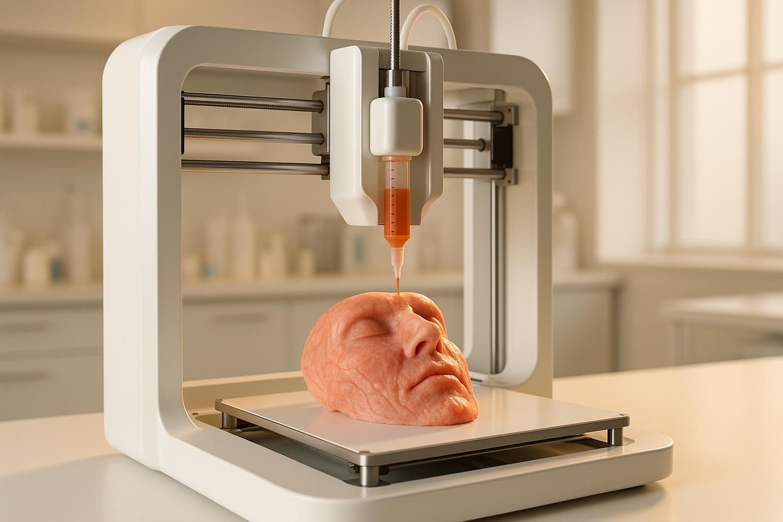
3D bioprinting is reshaping oral and facial reconstruction. It uses bioinks – combinations of cells, biomaterials, and growth factors – to create custom scaffolds that match a patient’s anatomy. Guided by imaging like CT or MRI, this method offers precision and control over tissue structure, promising better healing and recovery. While it’s still in experimental stages, 3D bioprinting shows potential in regenerating alveolar bone, periodontal structures, dental pulp, gingiva, and cartilage.
Here’s how it compares to other reconstruction methods:
- 3D Bioprinting: Customised, cell-laden scaffolds with bioactive properties. Minimal surgical morbidity but limited clinical use due to ongoing development.
- Autologous Bone Grafts: Reliable and widely used but involves donor-site surgery, which can cause complications.
- Microvascular Free Flaps: Vascularised tissue transfer for complex cases, requiring advanced surgical skills and long procedures.
- Alloplastic Implants: Synthetic materials for reconstruction with no donor-site surgery but higher infection risks.
Quick Comparison
| Aspect | 3D Bioprinting | Autologous Grafts | Free Flaps | Alloplastic Implants |
|---|---|---|---|---|
| Precision | High | Surgeon-dependent | Surgeon-dependent | High |
| Surgical Morbidity | Minimal | Donor-site complications | Donor-site complications | Minimal |
| Integration | Bioactive | Excellent | Excellent | Variable |
| Cost | High | Moderate | High | Moderate |
| Clinical Use | Experimental | Established | Established | Established |
While bioprinting holds promise for personalised tissue repair, challenges like regulatory approvals, bioink standardisation, and mechanical strength for load-bearing areas remain. Established methods, like bone grafts and implants, continue to dominate clinical practice in Australia. However, as bioprinting evolves, it could complement these techniques, particularly in complex cases requiring precise reconstruction.
CMFlex™ – Innovative bio-material for bone graft from Dimension Inx used on 3D-Bioplotter® printer

1. 3D Bioprinting
3D bioprinting is revolutionising the way oral and facial defects are addressed, offering precise, patient-specific solutions. By converting CT or CBCT imaging data into digital designs, this technology creates scaffolds and cell-laden structures that perfectly match the complex geometry of maxillofacial defects.
Precision in Reconstruction
One of the standout features of 3D bioprinting is its ability to precisely control both the external shape and internal structure of tissue constructs. Unlike traditional methods, where surgeons manually shape grafts during surgery, bioprinting allows for pre-designed scaffolds that influence cell behaviour and mechanical properties even before they reach the operating room.
This precision extends to the microscopic level. For example, research has shown that microgroove patterns at 25.40 µm intervals can align periodontal ligament cells, replicating the natural fibre orientations that support teeth[1].
Different bioprinting platforms cater to varying needs. Laser-assisted bioprinting offers exceptional precision with cell viability rates exceeding 95%, making it ideal for intricate craniofacial reconstructions[2]. On the other hand, extrusion bioprinters, though less precise, excel at creating larger constructs and handling multiple materials simultaneously. This makes them particularly useful for reconstructing composite tissues like bone, ligaments, and soft tissues in a single structure.
Multi-nozzle systems further enhance the technology’s capabilities, enabling the creation of compartmentalised, multi-material constructs that mimic the natural organisation of dental tissues. For instance, dental pulp stem cell-loaded GelMA microspheres have successfully regenerated vascularised and innervated pulp-like tissue in animal models, showcasing the potential for biologically "live" reconstructions[1].
This level of precision – both macroscopic and microscopic – is critical for achieving optimal tissue regeneration outcomes.
Recovery and Healing Outcomes
Preclinical studies highlight encouraging results in tissue healing across various applications. In periodontal regeneration, bioprinted cell-infused collagen has demonstrated more organised ligament-like soft tissue on titanium implants compared to traditional cell-seeding techniques[1]. This improved organisation enhances functional integration and may lead to longer-lasting results.
For bone regeneration, FDM bioprinted molar scaffolds loaded with stromal-derived factor-1 and bone morphogenetic protein-7 have shown increased cell recruitment and angiogenesis in rat models[2]. By combining precise scaffold geometry with bioactive growth factors, these constructs actively promote healing rather than simply providing a passive framework for tissue growth.
Gingival regeneration studies in beagle models have also been promising. Bioprinted hydrogel composites with growth factors significantly increased keratinised gingival width and collagen deposition compared to controls without growth factors[1]. These results suggest that bioprinted constructs can improve both the speed and quality of soft tissue healing around dental implants.
Stem cell integration offers another major advantage. Dental pulp stem cell-loaded GelMA microspheres have shown enhanced differentiation potential – angiogenic, neurogenic, and odontogenic – resulting in the successful regeneration of vascularised, innervated pulp-like tissue in animal models, including swine[1]. This represents a major leap over acellular scaffolds, which rely solely on the body’s ability to populate the construct with cells.
Integration with Digital Workflows
3D bioprinting fits seamlessly into the digital workflows already in place in many Australian dental practices. Building on tools like CBCT imaging, CAD/CAM design, and virtual surgical planning, it creates a streamlined process from diagnosis to scaffold fabrication.
High-resolution imaging provides the foundation for precise digital designs tailored to each patient’s unique anatomy. Factors such as load-bearing requirements, aesthetics, and tissue integration are all considered during the design phase. The result? Pre-shaped, ready-to-implant scaffolds that improve surgical accuracy and reduce operating time.
For complex craniofacial defects, the ability to customise both the internal and external architecture of scaffolds through digital design is transformative. Surgeons can test different configurations, optimise pore sizes for vascular growth, and fine-tune mechanical properties – all before the scaffold is even fabricated[2]. This not only saves time but also enhances surgical outcomes.
Digital workflows also foster better collaboration between surgical teams and bioprinting facilities. Design files can be shared and modified in real-time, ensuring that the final construct meets all clinical requirements. As practices like Complete Smiles Bella Vista continue to expand their digital capabilities, the infrastructure for integrating bioprinting is already being established.
Donor-Site Morbidity and Safety Considerations
One of the most compelling benefits of 3D bioprinting is its ability to eliminate donor-site morbidity. Traditional grafting methods often require harvesting tissue from the patient, which can lead to complications at the donor site[2][3]. Bioprinting sidesteps this issue by using biocompatible materials and encapsulated stem cells, reducing the need for autologous tissue extraction.
This is particularly advantageous for extensive craniofacial defects, where multiple grafts would otherwise be necessary. By fabricating larger constructs from synthetic or allogeneic materials, bioprinting minimises surgical risks and shortens recovery times. Patients avoid the pain and healing associated with donor sites, although new safety concerns, such as biomaterial compatibility and infection risks, must still be addressed.
That said, the technology is not without its limitations. Mechanical strength remains an issue for load-bearing applications. For example, scaffolds created using selective laser sintering of polycaprolactone have stiffness values ranging from 15 to 300 MPa – closer to craniofacial trabecular bone than many conventional polymers but still below the 120 to 450 MPa range of mandibular trabecular bone[2]. Regulatory challenges, high equipment costs, and the difficulty of fully replicating vascularised, innervated tissues also mean that many applications remain experimental, especially in high-stress areas like the posterior mandible.
2. Autologous Bone Grafts
Autologous bone grafts, sourced directly from the patient, are still considered the gold standard for reconstructing oral and facial bone. They’re commonly used for procedures like alveolar ridge augmentation, sinus lifts, repairing segmental jaw defects, and replacing bone lost to trauma or cancer. What makes them so effective is their unique combination of benefits: they provide living osteogenic cells, an osteoconductive framework, and osteoinductive growth factors – all in one solution[2].
In Australian practices, especially for high-stakes treatments like full-arch implant rehabilitation (which can cost over AUD $10,000 per case), this reliability is critical. When long-term implant support depends on predictable bone volume, autologous grafts consistently outperform synthetic or donor-derived alternatives[2].
Precision in Reconstruction
When precisely harvested and shaped, autologous bone grafts can restore jaw continuity and facial contours with impressive accuracy. Block and onlay grafts, when securely fixed to the recipient site, are particularly effective for rebuilding mandibular and maxillary structures. However, achieving precision often depends on the surgeon’s skill, as manual contouring is required. Variability in donor sites (like the iliac crest, calvarium, or mandibular ramus) and intra-operative factors such as graft resorption or microfractures can further complicate the process. This makes it challenging to replicate intricate three-dimensional shapes, such as those found in orbital rims or zygomatic arches, which digital scaffolds can handle more predictably[2].
While manual shaping can deliver good results, it does influence the healing process and overall efficiency.
Recovery and Healing Outcomes
One of the standout features of autologous bone grafts is their ability to revascularise quickly and integrate early. Radiographic evidence of union often appears within 8–12 weeks, and implants can typically be loaded 4–6 months after grafting. In Australia, the total treatment timeline averages 7–10 months[2].
Despite their high success rates, some challenges remain. Partial resorption and contour loss, particularly with onlay grafts or larger reconstructions, are potential issues. Infection is rare when rigid fixation and tension-free soft-tissue closure are achieved, but exposed grafts can fail rapidly. Long-term studies suggest that while autologous grafts integrate well, they may experience more unpredictable volume changes compared to rigid titanium meshes or porous polyethylene implants designed to maintain contour[2].
Integration with Digital Workflows
Although manual shaping presents challenges, digital planning has significantly improved graft positioning and outcomes. Virtual reconstruction allows for detailed pre-operative planning of graft volume and placement. Using these digital plans, CAD/CAM cutting guides and recipient-site templates can be created, with 3D-printed models serving as valuable surgical references. This approach reduces operative time and enhances geometric accuracy compared to freehand techniques[2].
A hybrid strategy has emerged as a game-changer. By combining autologous grafts with 3D-printed cutting guides, patient-specific reconstruction plates, and custom titanium meshes, surgeons can maintain the biological benefits of autologous bone while achieving precise alignment with native contours and dental arches. This approach is particularly effective in mandibular and maxillary reconstructions[2].
Collaboration is key. Oral and maxillofacial surgeons, restorative dentists, and prosthodontists must work together from the start. Early involvement in digital planning sessions – where tooth positions and occlusion are mapped out – ensures that bone graft volume and implant placement align with the final prosthetic design. Hospitals can share DICOM and STL files with community clinics, enabling practices like Complete Smiles Bella Vista to take over after graft consolidation. These clinics can then place implants using guided surgery and deliver crowns or bridges, creating a seamless continuum of care from hospital to local rehabilitation[2].
Donor-Site Morbidity and Safety Considerations
One of the main drawbacks of autologous bone grafts is the need to harvest tissue from elsewhere in the patient’s body, creating a second surgical site with its own risks and recovery demands. Smaller defects often rely on intraoral donor sites like the mandibular ramus, symphysis, or maxillary tuberosity, while larger reconstructions may require extraoral sites such as the iliac crest, fibula, calvarium, or scapula. These harvests come with risks, including pain, sensory changes, gait disturbances, and scarring.
In Australia, harvesting from extraoral sites often necessitates hospitalisation, adding days to recovery and delaying a return to work by weeks. This is especially challenging for patients balancing treatment with jobs or travel, particularly in regional areas[2]. To reduce complications, surgeons focus on careful patient selection, optimising factors like glycaemic control and smoking cessation. They also aim to use the smallest donor site capable of providing sufficient bone and rely on minimally invasive harvesting techniques when possible. At the recipient site, strategies like rigid fixation, tension-free closure, meticulous soft-tissue management, and appropriate peri-operative care (antibiotics, analgesia, and early mobilisation) help to minimise risks such as wound dehiscence, infection, and thromboembolic events. These practices are standard in Australian maxillofacial units[2].
While emerging bioprinting techniques aim to eliminate donor-site risks and achieve higher precision, they remain experimental for load-bearing jaw reconstruction. Autologous grafts, on the other hand, are backed by decades of clinical data and are fully integrated into current surgical workflows. For now, they continue to provide more predictable outcomes and regulatory acceptance in routine Australian practice[2][3].
3. Microvascular Free Flaps
Microvascular free flaps involve transferring segments of tissue – such as skin, muscle, bone, or mucosa – along with their blood supply, and reconnecting these vessels to recipient sites using microsurgical techniques. Unlike traditional bone grafts, these flaps provide immediate, vascularised tissue restoration. This approach complements advancements like 3D bioprinting, offering a dependable solution for complex oral and maxillofacial defects caused by head and neck cancers or severe trauma. They are often seen as the gold standard because they allow for precise tissue replacement, restoring both function and appearance.
In Australian maxillofacial surgery, commonly used free flaps include:
- Fibula free flap: Ideal for segmental mandibular defects, offering up to 25 centimetres of bone and compatibility with dental implants.
- Radial forearm free flap: Used for intraoral soft-tissue reconstruction, including the tongue.
- Anterolateral thigh (ALT) flap: Suitable for larger soft-tissue or composite defects.
- Scapular and iliac crest flaps: Preferred when specific bone shapes are needed for midface or maxillary reconstruction.
This variety allows surgeons to tailor procedures to meet individual needs, enhancing precision and recovery outcomes.
Precision in Reconstruction
The integration of virtual surgical planning (VSP), 3D CT imaging, and CAD/CAM guides has significantly improved the precision of free flap surgeries. By pre-shaping tissue segments to match the patient’s anatomy, these tools reduce both ischaemia and operative times. Studies comparing VSP-assisted fibula reconstructions to traditional methods show a reduction in ischaemia time by 30–60 minutes and total surgery time by 1–2 hours. Deviations from the virtual plan are typically within a few millimetres, underscoring the accuracy of this approach.
One major advantage of free flaps is their ability to transfer multiple tissue types – bone, muscle, skin, and soft tissue – in a single vascularised unit. This is especially valuable for severe facial trauma or extensive soft-tissue loss, where different tissue types must be reconstructed simultaneously. The vascularised tissue integrates smoothly with the surrounding area, improving overall outcomes.
Another growing trend is prosthetically driven reconstruction, where the focus is on aligning bony segments and flap positioning with the eventual placement of dental implants and prosthetics. Using 3D-printed dental models and guides, surgeons can ensure that fibula segments are positioned to support planned prosthetic teeth and proper occlusion. This approach requires close collaboration between surgeons and restorative dentists from the start.
Once healing is complete, patients are referred to specialised dental practices for implant placement and prosthetic rehabilitation. Clinics like Complete Smiles Bella Vista provide implant-supported restorations tailored to the reconstructed jaw, ensuring a seamless transition from hospital care to community-based dental treatment. Pre-operative imaging, such as CT angiography of the lower limbs before fibula harvest, helps identify anatomical variations and reduces donor-site risks. These digital advancements streamline surgeries and align well with post-operative care protocols.
Recovery and Healing Outcomes
Microvascular free flaps have an impressive survival rate, consistently exceeding 90–95%, with some high-volume centres reporting success rates above 97%. Initially, post-operative monitoring involves hourly checks for flap colour, capillary refill, and Doppler signals. As the risk of thrombosis decreases, monitoring becomes less frequent. Early detection of issues allows timely intervention, often salvaging compromised flaps.
The vascularised nature of these flaps offers better resistance to infection and radiation compared to non-vascularised grafts. This is particularly important in Australia, where many patients undergo radiotherapy after cancer surgeries. Vascularised bone flaps tolerate radiation well, maintaining their volume and enabling later dental implant placement – key for long-term dental rehabilitation.
These flaps also support early oral function, swallowing, and stable soft-tissue coverage, with good durability over time. Dental implants can later be placed in the transplanted bone to restore chewing and dental aesthetics. Functional recovery after tongue or floor-of-mouth reconstruction often includes improved speech and swallowing, though outcomes can vary based on factors like defect size, radiation therapy, and rehabilitation efforts.
Donor-Site Morbidity and Safety Considerations
While microvascular free flaps offer excellent reconstructive results, donor-site complications remain a concern. For fibula free flaps, complication rates range from 5–20%, depending on follow-up and definitions. Issues may include wound problems, gait disturbances, or sensory changes. Radial forearm flaps are associated with higher rates of wound breakdown and visible scarring, with some cases of tendon exposure.
These surgeries are resource-intensive, with costs typically ranging from AUD $15,000 to $40,000, depending on complexity. Operative times can extend 6–12 hours or more, with additional costs for hospital stays, intensive care, and potential revision surgeries. The procedure demands highly skilled microsurgeons and is generally performed in major hospitals with dedicated teams specialising in maxillofacial, ENT, or plastic surgery.
Careful patient preparation can minimise donor-site impact. Optimising factors like blood sugar control, smoking cessation, and overall fitness improves outcomes. Despite the challenges, microvascular free flaps remain the preferred method for addressing complex oral and facial defects. Decades of clinical data support their reliability, especially when paired with modern digital planning techniques. For large or composite defects, they provide unmatched restoration of both form and function compared to non-vascularised grafts or metal plates, making them an indispensable tool in advanced reconstructive surgery.
sbb-itb-2be92ed
4. Alloplastic Implants
Alloplastic implants are synthetic materials designed to reconstruct oral and facial defects without needing to harvest tissue from the patient’s body. These include materials like titanium plates and screws, porous polyethylene, polyetheretherketone (PEEK), and custom titanium meshes. They’re widely used for procedures such as mandibular and maxillary reconstruction, orbital floor repairs, midface defect corrections, temporomandibular joint (TMJ) prostheses, and facial contour enhancements. Unlike autologous bone grafts or microvascular free flaps, alloplastic implants eliminate the need for a second surgical site, which can be especially beneficial for patients with underlying health conditions. This approach complements both traditional surgical methods and emerging bioprinting techniques in craniofacial reconstruction.
The primary advantage is clear: no donor-site surgery means less pain, shorter procedures, and quicker hospital discharges. However, as foreign materials, these implants must be carefully evaluated for long-term compatibility and potential complications.
Precision in Reconstruction
Modern technology has transformed alloplastic implants, enabling highly precise designs through CAD/CAM and 3D printing. Using CT or cone beam computed tomography (CBCT) imaging, surgeons can create detailed digital models of a patient’s anatomy. These models guide the production of customised implants that restore facial contours with sub-millimetre accuracy.
For example, in cases of segmental mandibular defects, virtual surgical planning allows for the creation of custom titanium plates that precisely replicate the patient’s original jaw shape – far surpassing traditional freehand methods. A similar process is applied to orbital floor reconstruction, where porous polyethylene implants are pre-shaped to fit the patient’s anatomy. This level of customisation now outshines pre-made, off-the-shelf options.
Additionally, 3D-printed surgical guides enhance accuracy during procedures by marking resection margins and guiding drilling positions, reducing guesswork during surgery. Studies show that patient-specific titanium implants for mandibular reconstruction have survival rates exceeding 90% in the short to medium term, with minimal mechanical failures when soft-tissue coverage is adequate. Comparative research also highlights that CAD/CAM and 3D-printed guides can reduce operative times by 20–30% compared to freehand techniques.
For TMJ reconstruction, modern prostheses – typically made from cobalt-chromium and titanium with ultra-high molecular weight polyethylene (UHMWPE) components – are custom-designed for patients with severe deformities or ankylosis. These implants not only restore facial aesthetics but also improve mandibular function, ensuring better post-operative outcomes.
Recovery and Healing Outcomes
Alloplastic implants bring notable recovery benefits by eliminating the need for donor-site surgery. Patients avoid the pain and scarring associated with tissue harvesting, and hospital stays are shorter since there’s no need for intensive monitoring, as required with vascularised tissue transfers.
Healing with alloplastic materials differs from biological grafts. Instead of integrating through living bone formation, these implants rely on mechanical fixation and, in some cases, fibrovascular tissue ingrowth. For instance, porous polyethylene orbital implants encourage fibrovascular ingrowth within weeks, anchoring the implant and reducing the risk of extrusion compared to older solid silicone designs.
That said, infection and exposure remain key concerns. In clean surgical cases, infection rates for porous polyethylene facial implants are generally below 5%, but these rates can rise when implants are placed in previously infected or irradiated areas. Proper soft-tissue closure is vital to prevent wound breakdown and implant exposure. Unlike vascularised free flaps that bring their own blood supply and better resist infection, alloplastic implants depend entirely on the surrounding tissue’s health.
For patients undergoing radiotherapy after cancer surgery, compromised tissue healing can increase the risk of implant exposure and infection. In such instances, surgeons may opt for vascularised free flaps or combine alloplastic implants with soft-tissue flaps to ensure sufficient coverage. The precision provided by digital workflows further enhances recovery outcomes by enabling patient-specific designs tailored for optimal healing.
Integration with Digital Workflows
Alloplastic implants exemplify how digital technology has advanced oral and maxillofacial reconstruction. The entire process – from imaging to implant fabrication – is digitally managed, seamlessly converting CT or CBCT data into physical implants. This digital approach eliminates the need for intra-operative sculpting and manual adjustments often required with autologous bone grafts.
Digital models also help patients visualise the reconstruction process and potential outcomes. Once approved, these designs are sent to manufacturing facilities, where implants are produced using medical-grade materials under strict quality controls.
In Australia, the growing adoption of digital dentistry and guided implant placement allows restorative dentists and oral surgeons to collaborate more effectively. Advanced clinics equipped with intra-oral scanners and CBCT technology for dental implants and orthodontics can extend these tools to support complex reconstructions. For example, practices like Complete Smiles Bella Vista – which use digital planning for dental and orthodontic treatments – can work closely with maxillofacial surgeons to ensure reconstructed jaw segments are compatible with future dental prosthetics. This “prosthetically driven” approach aligns bony reconstruction with eventual dental implant placement, streamlining the treatment pathway from hospital-based surgery to community-based dental rehabilitation.
Donor-Site Morbidity and Safety Considerations
One of the most notable benefits of alloplastic implants is the elimination of donor-site morbidity. Patients avoid the pain, scarring, and functional issues associated with bone harvesting from areas like the iliac crest or fibula. This advantage is particularly valuable for elderly or medically compromised patients who may not tolerate additional surgeries well.
However, introducing a foreign material comes with its own risks. Alloplastic implants are more prone to infection, exposure, and long-term failure compared to autologous tissue, especially in contaminated surgical fields or high-stress areas. Patients should understand that while these implants avoid a second surgical site, complications may require removal or revision.
Recent advances aim to bridge the gap between synthetic materials and natural tissue. Porous titanium and polyethylene implants now promote tissue ingrowth and stable fixation, reducing the risk of migration. Additionally, bioactive coatings and porous structures are being developed to improve osseointegration and soft-tissue attachment, offering better resistance to infection than older designs.
For younger patients, alloplastic implants present unique challenges. Since they don’t grow or remodel with the skeleton, they’re typically reserved for adults with completed skeletal growth. In children, autologous bone grafts or distraction osteogenesis are often preferred as they adapt to the body’s changes over time.
Cost is another consideration. While custom titanium implants and TMJ prostheses can be expensive, they may reduce overall costs through shorter surgeries and hospital stays. In Australia, access to these advanced technologies varies between public and private healthcare systems. Larger hospitals with specialised maxillofacial teams are more likely to offer custom solutions, while smaller centres may rely on standardised implants or refer complex cases to tertiary facilities.
A hybrid approach is also gaining traction. This method combines alloplastic substructures with soft-tissue or bone flaps, blending the predictable shape and strength of implants with the regenerative capabilities of living tissue. While alloplastic implants avoid donor-site risks, their success depends on precise digital planning and customised designs to meet the demands of modern craniofacial reconstruction.
Advantages and Disadvantages
This section examines various methods used for oral and facial reconstruction, comparing their strengths and limitations.
3D bioprinting stands out for its ability to create highly precise, patient-specific scaffolds. These scaffolds integrate different cell types and growth factors, eliminating the need for donor-site surgery[1][2]. Laser-assisted bioprinting boasts cell viability rates exceeding 95%, making it particularly suited for procedures where high cell density is critical[2]. Extrusion bioprinters also offer a cost-effective and efficient way to produce large-scale constructs[2].
However, challenges remain. Laser-assisted bioprinting, while precise, is both time-intensive and costly[2]. The technology still requires advancements to standardise bioink formulations and ensure reliable outcomes[1]. Clinical evidence in humans is limited, as most studies are currently based on animal models[1]. Regulatory pathways for bioprinted constructs are still being defined, and the process demands specialised knowledge in bioink formulation and printer operation[1]. These hurdles contrast with the more established limitations of traditional methods.
Autologous bone grafts are a proven option with long-term success and excellent integration. They are inherently osteogenic, osteoinductive, and osteoconductive, making them effective for bone regeneration. However, they come with drawbacks such as donor-site morbidity, limited harvest volume, and unpredictable resorption rates[2]. Their lack of customisation to complex geometries can also be a disadvantage.
For more complex reconstructions, microvascular free flaps provide immediate vascularisation, enhancing healing and integration. These flaps, made from the patient’s own tissue, offer excellent biocompatibility and reliable long-term outcomes[2]. Yet, they involve time-intensive procedures, carry a risk of donor-site morbidity, and require advanced microsurgical expertise. Not all hospitals are equipped for these complex surgeries, which can lead to procedural delays or complications[2].
Alloplastic implants, on the other hand, are readily available and reduce operative time by eliminating the need for donor-site surgery[2]. However, they lack the bioactivity of living tissues and cannot match the customisation potential of bioprinted scaffolds[2]. Their integration depends on the surrounding tissue’s health, and foreign body reactions may necessitate revisions[2].
| Aspect | 3D Bioprinting | Autologous Bone Grafts | Microvascular Free Flaps | Alloplastic Implants |
|---|---|---|---|---|
| Precision | High with patient-matched scaffolds[2] | Limited by graft shape and size | Dependent on surgeon expertise | Standardised, less customisable |
| Material Availability | Unlimited (bioinks)[1][2] | Limited by donor site[2] | Limited by tissue availability | Readily available |
| Surgical Morbidity | Minimal (no donor site)[1] | Donor-site complications[2] | Requires microsurgery[2] | Minimal |
| Cell Viability | >95% (laser-assisted)[2] | Variable | Variable | N/A (non-living material) |
| Fabrication Time | Rapid (extrusion printing)[2] | Immediate (harvesting) | Time-intensive surgery[2] | Immediate |
| Cost | High initial investment[2] | Moderate | High (special expertise)[2] | Moderate to high |
| Bioactivity | High (living cells)[1][2] | High (autologous tissue) | High (autologous tissue) | Low to moderate |
| Integration | Excellent with proper design[1] | Excellent (autologous) | Excellent (autologous) | Variable |
| Vascularisation | Can include angiogenic factors[1] | Requires time for revascularisation | Immediate[2] | Requires time for integration |
Recent research using medical-grade polycaprolactone (PCL) combined with tricalcium phosphate (TCP) and recombinant human bone morphogenetic protein-7 (rhBMP-7) has shown promising results. In a sheep model, this combination led to better and more robust bone formation after 12 months compared to autologous bone grafts[2]. While this highlights the potential of bioprinting, clinical applications are still under development.
In Australia, access to these technologies varies. Larger tertiary hospitals with specialised maxillofacial teams are more likely to offer advanced options like bioprinting trials or custom alloplastic implants. Smaller regional centres often rely on established techniques such as autologous bone grafts or standardised implants, referring complex cases to metropolitan hospitals. Clinics equipped with advanced digital dentistry tools can collaborate with surgeons to ensure reconstructed jaw segments align seamlessly with future dental prosthetics, streamlining the treatment process.
Looking ahead, hybrid approaches may yield the best results. For instance, combining alloplastic substructures with bioprinted soft tissue or using bioprinted scaffolds to enhance traditional bone grafts could merge the strengths of multiple methods while minimising their limitations. As bioprinting evolves and regulatory frameworks solidify, its role in craniofacial reconstruction is expected to grow, especially for complex cases requiring precise cellular organisation and multi-tissue regeneration. Combining digital design precision with biological regeneration could redefine standards in oral and facial reconstruction.
Conclusion
3D bioprinting holds the potential to create patient-specific, cell-infused scaffolds for oral and facial reconstruction. Preclinical studies have shown promising bone growth over a 12-month period[2]. However, moving from these early successes to widespread clinical use will require addressing several technical and regulatory challenges.
For now, 3D bioprinting remains largely experimental. Established options like autologous bone grafts, microvascular free flaps, and alloplastic implants continue to dominate treatment for extensive or load-bearing defects. These methods have a long history of reliable outcomes and predictable results, backed by decades of clinical data. Most current bioprinting research is limited to lab and animal studies, with only a handful of early-stage human applications reported[1][2].
In the short term, bioprinting is more likely to serve as a complementary tool rather than a replacement for existing techniques. Hybrid solutions – such as bioprinted scaffolds combined with a patient’s own cells or integrated with autologous tissue and free flaps – could improve fit, function, and aesthetics in complex reconstructions. Additionally, customised porous scaffolds may one day enhance dental implant support in cases where traditional grafts fall short in volume or shape[2][3].
Australian dental practices already equipped with advanced digital tools are well-positioned to adopt bioprinting technologies as they evolve. These practices can leverage their existing digital workflows to incorporate future bioprinting solutions seamlessly[2].
That said, significant obstacles remain. These include navigating regulatory pathways, developing standardised bioinks, ensuring long-term safety, achieving reliable blood vessel formation in large constructs, addressing mechanical strength for load-bearing areas, and ensuring cost-effectiveness within Australia’s healthcare system[1][2][3]. Large-scale clinical trials comparing bioprinted constructs with established methods like autologous grafts and implants are essential to pinpoint where bioprinting offers true benefits.
As the field progresses toward replicating native tissues, 3D bioprinting could reshape treatment options for complex craniofacial defects. Until robust clinical evidence is available, however, it should be viewed as a developing technology that complements the trusted reconstructive methods currently in use.
FAQs
What challenges does 3D bioprinting face in repairing oral and facial tissues?
3D bioprinting for repairing oral and facial tissues comes with its fair share of challenges, and researchers are hard at work tackling these issues. One of the biggest obstacles is achieving vascularisation – the formation of blood vessels. This step is crucial because it ensures the printed tissues receive enough nutrients and oxygen to survive. Without it, the tissue’s survival is at risk.
Another hurdle is ensuring the mechanical strength of the bioprinted structures. This is particularly important in areas like the jaw or face, which are subject to constant stress and pressure. On top of that, guiding cell differentiation to accurately replicate the natural structure and function of these tissues adds another layer of complexity. And then there’s the issue of biocompatibility – making sure the bioprinted material integrates smoothly into the body without triggering immune responses or causing unwanted reactions.
Despite these challenges, advancements in technology and research are steadily pushing the boundaries, making 3D bioprinting an increasingly viable solution for tissue repair in these critical areas.
How does 3D bioprinting compare to traditional methods like bone grafts and microvascular free flaps for oral and facial tissue repair?
3D bioprinting brings a highly precise and personalised approach to tissue repair, standing apart from traditional methods like autologous bone grafts or microvascular free flaps. By leveraging advanced technology to create tissue with exceptional accuracy, it minimises the need for invasive procedures. This can translate to shorter recovery periods and a reduced risk of complications.
What sets this technique apart is its ability to produce tailored tissue structures that closely replicate natural anatomy. This is particularly beneficial for oral and facial applications, where precision is crucial for successful outcomes. While conventional methods remain reliable, 3D bioprinting offers a forward-thinking alternative that enhances both accuracy and patient comfort.
How could 3D bioprinting improve dental reconstruction in the future?
3D bioprinting offers a promising step forward in dental reconstruction, enabling highly precise and personalised regeneration of tissue and bone. This approach has the potential to significantly improve both the functionality and appearance of oral and facial restorations.
By complementing established methods such as implants and grafts, 3D bioprinting could streamline procedures, shorten recovery periods, and boost overall treatment success. As the technology progresses, it may pave the way for tailored solutions to address even the most intricate oral and facial repair needs.
Related Blog Posts
- 3D Bioprinting in Dental Implants: How It Works
- Biodegradable Materials in Guided Tissue Regeneration
- Recent Advances in Biomaterials for Periodontal Regeneration
- 3D Printing in Dental Implant Prototyping: How It Works
Important Notice: Any surgical or invasive procedure carries risks. Before proceeding, you should seek a second opinion from an appropriately qualified health practitioner.
Individual results may vary. The information provided in this article is for educational purposes only and does not constitute medical advice.
Checkout Related Blogs


Get in touch with us
For more information, call us now to start feeling better. Or fill the form below to make appointment
The Latest News from Complete Smiles
How to Clean Clear Plastic Retainers
Checklist for Choosing Wearable Dental Devices
Checklist for Choosing Cloud AI Platforms in Dentistry

Complete Smiles Bella VistaAccepts All Major Health Funds, Including


