


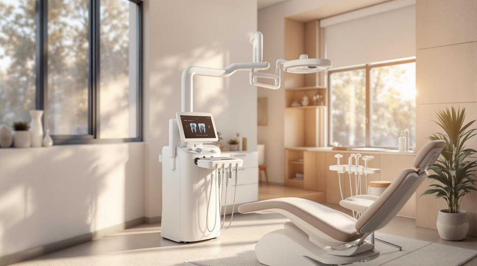
Digital Dental X-Rays: How They Work
Digital dental X-rays are a safer, faster, and more accurate way to diagnose dental issues compared to older film-based methods. Here’s what you need to know:
- Instant Results: Images are available immediately, with no need for chemical processing.
- Reduced Radiation: Up to 90% less radiation exposure than traditional X-rays.
- Enhanced Clarity: Displays over 200 shades of grey, improving diagnostic precision.
- Eco-Friendly: Eliminates chemical waste and uses reusable components.
- Advanced Sensors: Uses CCD, CMOS, or PSP sensors for high-quality imaging.
- Seamless Integration: Works with dental software for easy sharing, treatment planning, and AI-assisted diagnostics.
- Safety Standards: Adheres to Australia’s strict radiation safety guidelines, including the ALARA principle.
Digital X-rays are essential for detecting cavities, gum disease, infections, and more, while also guiding treatments like orthodontics, implants, and root canals. They’re faster, safer, and better for both patients and the environment.
Safe Digital Dental X-Rays
How Digital Dental X-Rays Work
Digital dental X-rays capture, process, and display images within seconds, making it easier and faster for dentists to diagnose issues. This speed and accuracy are thanks to advanced sensor technology.
Image Sensor Technology
Digital X-rays use three primary types of sensors to capture images: Charge-Coupled Device (CCD), Complementary Metal-Oxide-Semiconductor (CMOS), and Photostimulable Phosphor Plate (PSP) [3][4][7]. Each type has its own strengths:
- CCD Sensors: These convert X-ray energy directly into electronic signals, offering high image quality with minimal noise [5][6][7].
- CMOS Sensors: Known for being more affordable, these sensors provide fast data transfer, consume less power, and have a broad dynamic range [5][7].
- PSP Sensors: These work differently by capturing data in an analogue format. The data is then scanned and converted into digital images. PSP plates are reusable after being exposed to light, which erases any residual energy [3][4].
Modern systems often use Active Pixel Sensor CMOS, which delivers high-resolution images suitable for both intraoral (inside the mouth) and extraoral (outside the mouth) imaging [3][7].
Image Processing and Display
After the sensor captures the X-ray energy, the data is sent to a computer, where specialised software transforms it into a visual image [6][8]. This process happens almost instantly, allowing dentists to review the results right away.
The software also lets practitioners enhance the images by adjusting contrast, zooming, and applying filters. These tools make subtle details more visible compared to traditional film X-rays [8]. High-resolution images can be displayed on a monitor, shared with specialists, or used for patient consultations. Features like digital subtraction radiography enable comparisons between past and current images, helping to spot even minor changes over time [10].
Integration with Dental Software
Digital X-ray systems integrate seamlessly with dental practice management software, improving efficiency and patient care [11]. These systems allow practices to work with various sensor types while maintaining a smooth workflow. Cloud-based platforms make storing and accessing X-rays easier, enabling team collaboration and real-time updates [11].
By linking digital X-rays to electronic health records (EHRs), practices can reduce errors and streamline operations [11]. Artificial intelligence further enhances diagnostics by providing real-time analysis and insights [11]. These integrated systems also simplify file management, treatment planning, and image sharing, all while maintaining data security [11][12].
When selecting analysis software, practices should weigh their specific needs, budget, and goals [13]. These integrations not only improve diagnostic accuracy but also make patient care more efficient and organised, showcasing the many advantages of digital radiography.
Radiation Safety and Patient Care
Digital dental X-rays have transformed patient safety by significantly reducing radiation exposure while still delivering high-quality diagnostic images. In Australia, dental practices adhere to strict guidelines to ensure the safety of patients during all radiographic procedures.
Reducing Radiation Exposure
One of the major benefits of digital X-ray technology is its ability to dramatically lower radiation doses compared to traditional film-based systems. In fact, digital dental X-rays use 80% to 90% less radiation than conventional machines [1]. This means patients are exposed to minimal radiation while dentists still receive the detailed images they need for accurate diagnoses.
Australian dental practices follow the ALARA principle, which stands for "As Low As Reasonably Achievable." This principle ensures that X-rays are only used when absolutely necessary and that the benefits to the patient outweigh any potential risks.
"The ALARA principle stands for ‘as low as reasonably achievable’ and places the responsibility on practitioners to ensure they only use ionising radiation on a patient when required, and when the reward to the patient outweighs the risk of the radiation." [16]
Digital sensors play a key role in reducing radiation exposure. These sensors are highly sensitive to X-ray energy, allowing them to capture diagnostic-quality images with much lower doses of radiation. Unlike film, which requires specific exposure levels for proper development, digital systems allow post-capture adjustments like brightness and contrast enhancement. This reduces the need for retakes, which were more common with older film systems [2].
Beyond the technology itself, dental clinics in Australia implement robust safety protocols to further minimise risks for both patients and staff.
Safety Measures in Australian Clinics
While digital X-rays already reduce radiation exposure, Australian clinics go a step further by enforcing rigorous safety measures. Oversight from the Australian Radiation Protection and Nuclear Safety Agency (ARPANSA) ensures that dental radiation doses remain well within safe limits.
"It is still important to have regulatory oversight to ensure that dental doses continue to remain low, and to provide assurance to the community." – Dr Ivan Williams, ARPANSA’s Chief Medical Radiation Scientist [14]
Protective equipment like lead aprons and thyroid collars remains essential, even with advancements in digital systems. These barriers can reduce radiation exposure by up to 95% [18], adding an extra layer of protection during X-ray procedures.
Radiation safety extends to dental staff as well. Occupational exposure limits are strictly regulated, with staff exposure capped at 20 mSv per year, averaged over five years, and no more than 50 mSv in any single year [16]. To provide context, dental staff in the UK report average exposure levels of less than 0.1 mSv per year, while their counterparts in the USA typically receive 0.2 mSv annually [15].
State and territory regulations further strengthen this framework. Practices must secure appropriate licences, undergo regular equipment checks, and ensure all X-ray machines are calibrated to operate within safe limits [20]. Continuous staff training ensures that teams stay updated on the latest safety protocols [18].
Modern clinics also use dosimeters to monitor staff radiation exposure over time. These devices provide an early warning if exposure levels approach regulatory thresholds [18]. For advanced imaging technologies like Cone-beam CT (CBCT), practitioners carefully limit the field of view (FOV) to the smallest area necessary, reducing radiation exposure while still obtaining the required diagnostic information [16].
The foundational principles of radiation safety – time, distance, and shielding – are central to Australian dental practices. By limiting exposure time, maintaining a safe distance when possible, and using appropriate protective barriers, clinics prioritise the safety of both patients and staff during every radiographic procedure [17].
sbb-itb-2be92ed
Uses in Dental Care
Digital dental X-rays have become a cornerstone of modern dentistry, offering critical insights for diagnosing and monitoring conditions that might otherwise go unnoticed during routine exams. They provide a detailed view of what lies beneath the surface of teeth and gums, enabling more accurate and effective treatment.
"X‐rays are the primary method for diagnosing the majority of conditions we deal with in dentistry including cavities, gum disease, and various types of dental infections." – DrManduzzi [23]
These imaging systems go beyond the basics, allowing dentists to examine key structures like the jawbone, nerves, sinuses, and tooth roots [1].
Diagnostic Uses
Digital X-rays play a vital role in uncovering dental issues that aren’t visible to the naked eye:
- Cavity detection: They help identify decay in hard-to-see areas, such as between teeth or under existing fillings. Bitewing X-rays are especially useful for spotting decay in these tricky spots [1].
- Gum disease diagnosis: Periapical X-rays reveal bone loss and the extent of periodontal damage. Catching these issues early can lead to less invasive and more affordable treatments compared to managing advanced gum disease [26].
- Infection identification: X-rays are indispensable for locating infections or abscesses, whether at the tooth root or between the gums and teeth. Periapical X-rays are particularly effective in detecting abnormalities in teeth and surrounding bone [1].
- Detection of cysts and tumours: Beyond routine dental concerns, digital X-rays can uncover cysts, tumours, and other irregularities that may not be apparent during a clinical exam [22].
Routine X-rays, taken every six to 18 months, allow dentists to catch problems early, often preventing them from escalating into more serious and expensive issues [1]. Alongside diagnosis, they are also essential for monitoring the progress of ongoing dental treatments.
Treatment Monitoring
The precision of digital X-rays makes them invaluable for guiding and adjusting treatments across various dental procedures:
- Orthodontic care: X-rays are integral to planning treatments like braces or Invisalign, ensuring teeth and jaws are properly aligned. Cephalometric X-rays assess jaw relationships, while Cone Beam Computed Tomography (CBCT) offers detailed 3D imaging for more complex cases [9][24].
- Dental implants: X-rays guide the placement of implants, ensuring they are positioned correctly within the jawbone. They also monitor the integration of the implant with the surrounding bone during the healing process [24][25].
- Root canal therapy: Throughout the procedure, X-rays confirm that infected tissue has been fully removed and that the tooth structure remains stable, ensuring a successful outcome [24].
- Dental restorations: Whether it’s a crown, filling, or another type of restoration, X-rays help verify proper fit, identify any leakage, and track long-term success [25].
Maintenance and Compliance for Digital X-Ray Systems
Keeping digital X-ray systems in top shape is essential for ensuring safety, accuracy, and reliability in Australian clinics. Proper maintenance and adherence to legal requirements protect patients and staff while maintaining the high imaging quality that sets digital radiography apart from traditional film methods.
"Proper disinfection of and asepsis with technology – including radiologic equipment – is vital to delivering safe dental care." [27]
These systems demand more than just routine cleaning. Regular software updates, hardware calibration, and strict radiation safety protocols are critical. Together, these practices support the longevity and diagnostic accuracy of digital X-ray equipment.
Cleaning and Maintenance
Digital X-ray sensors and equipment require careful cleaning to prevent cross-contamination while protecting their delicate electronic components. Disposable gloves and FDA-approved barriers should be used on sensors and holders for every patient to minimise risks [27] [28].
After removing the barriers, sensors and cords need to be disinfected with an intermediate-level disinfectant using a wipe-discard-wipe or spray-wipe-spray method. Directly applying cleaning solutions to sensors should be avoided to prevent damage [27] [28].
Clinical contact surfaces like control panels, touch screens, and exposure buttons must also be disinfected between patients. Use EPA-registered hospital disinfectants and ensure the solution remains on the surface for the recommended contact time. If blood or bodily fluids are present, clean the area before disinfecting [27] [28] [29].
Regular technical maintenance is equally important. For instance, the phosphor chassis should be cleaned with manufacturer-recommended solutions, and equipment should be stored at the specified temperature outlined in the operation manuals [30].
"Preventive maintenance is critical to help keep your equipment running at peak performance, in compliance of all regulatory and safety standards, and within the operating standards determined by the manufacturer." [30]
Authorised service personnel play a key role in preventive maintenance, addressing issues like guide rail jamming or automatic restarts before they escalate [30] [31]. Additionally, ongoing staff training on proper equipment use is crucial to maintaining the system’s integrity and performance [30].
Meeting Australian Regulations
While advanced sensor technology enhances diagnostic capabilities, compliance with Australian regulations ensures the safety of patients and staff. Each state and territory has its own specific radiation safety guidelines, so understanding local requirements is essential [20].
| State/Territory | Compliance Testing Frequency | Dental Shielding Assessment Required |
|---|---|---|
| NSW | Every 5 years | No (self-assessment possible) |
| Queensland | Every 5 years | Yes |
| Western Australia | Every 5 years | Yes |
| Tasmania | Initial + every 4 years | Yes |
| South Australia | Initial + change of ownership | Yes |
| Victoria | Initial installation only | Yes |
William Green, a well-known Australian dental supply company, highlights the importance of compliance testing:
"It’s critical that all practices are familiar with these requirements, so that compliance checks can be booked in on time. Failure to carry out compliance testing could compromise the health and safety of dentists, staff and patients who come within range of your dental X-ray imaging equipment." [21]
The company integrates compliance testing into its support services, helping clinics meet testing schedules and providing necessary reports. These services are often paired with autoclave validation and calibration [21].
Non-compliance carries heavy penalties, making it essential to engage experienced radiation consultants. These professionals can assist in developing safety management plans, training staff, and ensuring ongoing compliance [20]. The Australian Radiation Protection and Nuclear Safety Agency (ARPANSA) provides standard guidelines that form the basis for state-specific regulations [19].
Regular compliance testing goes beyond legal requirements. It ensures that equipment operates safely and meets manufacturer specifications, protecting the health and safety of everyone in the dental environment.
Conclusion
Digital dental X-rays have revolutionised dental care by combining advanced sensor technology with cutting-edge software. One of the standout advantages is the dramatic reduction in radiation exposure, offering a safer option for patients. Beyond safety, digital systems provide highly detailed images that allow dentists to spot even the smallest issues early, leading to more accurate and timely diagnoses.
By doing away with film, chemical processing, and bulky physical storage, digital X-rays not only cut down on ongoing costs but also align with environmentally conscious practices. Additionally, storing images electronically enables instant sharing, making collaboration between dental professionals faster and more efficient.
The integration of artificial intelligence and advanced imaging tools is further enhancing diagnostic precision. Research even suggests that AI could safely handle the interpretation of up to 7.8% of all radiographs [32], showcasing its potential to streamline workflows.
For patients, the benefits are clear. Digital X-ray technology provides quicker imaging, better quality results, and minimal radiation exposure. The ability to process images instantly eliminates the waiting times associated with traditional film, ensuring a smoother and safer experience. In Australia, digital dental radiography remains a cornerstone of delivering accurate, patient-focused care.
FAQs
What are the safety and environmental benefits of digital dental X-rays compared to traditional film X-rays?
Digital dental X-rays bring notable benefits in terms of patient safety and environmental care when compared to traditional film-based X-rays. One standout feature is their ability to use 80–90% less radiation, offering a safer imaging option – particularly important for children or individuals who need frequent scans.
On the environmental front, digital X-rays do away with the need for harmful chemicals traditionally used to develop film. This means less toxic waste and a reduced impact on the environment. Plus, they cut down on physical waste, making dental care more aligned with sustainable practices.
By prioritising patient safety and reducing environmental harm, digital dental X-rays reflect a forward-thinking step in modern oral healthcare.
What dental issues can digital X-rays detect that might not be visible during a regular dental check-up?
Digital X-rays offer a detailed glimpse into dental health, uncovering issues that might not be visible during a routine check-up. They are incredibly useful for spotting early tooth decay, gum disease, bone infections, tumours, and cysts. Beyond that, they help detect structural concerns like jaw fractures, impacted teeth, and misalignment of teeth or jaws.
With their sharp, high-resolution images, digital X-rays empower dentists to catch problems early and take action before they escalate, leading to improved oral health over time.
How do digital X-rays and dental software improve patient care and treatment planning?
Digital X-rays paired with advanced dental software are transforming patient care by delivering sharp, high-resolution images that dentists can view and analyse immediately. This speedy access to clear visuals enables quicker and more precise detection of dental issues, leading to better-informed treatment decisions. Plus, digital X-rays come with the added benefit of exposing patients to less radiation compared to traditional methods, putting safety at the forefront.
When combined with dental software, digital X-rays offer a seamless way to access a patient’s dental records and current oral health status. This integration makes it easier to track changes over time, refine treatment plans, and provide care tailored to each individual. The ability to compare previous and current images not only helps monitor progress but also allows for early identification of potential problems. Together, these tools are reshaping dentistry by improving outcomes and streamlining the care process.
Related Blog Posts
- Wear-Resistant Materials in Dental Restorations
- Advances in Dental Adhesives: What to Know
- How X-Rays Help in Dental Treatment Planning
- Flapless Implant Surgery: Benefits and Process
Important Notice: Any surgical or invasive procedure carries risks. Before proceeding, you should seek a second opinion from an appropriately qualified health practitioner.
Individual results may vary. The information provided in this article is for educational purposes only and does not constitute medical advice.
Checkout Related Blogs


Get in touch with us
For more information, call us now to start feeling better. Or fill the form below to make appointment
The Latest News from Complete Smiles
How to Clean Clear Plastic Retainers
Checklist for Choosing Wearable Dental Devices
Checklist for Choosing Cloud AI Platforms in Dentistry

Complete Smiles Bella VistaAccepts All Major Health Funds, Including


