


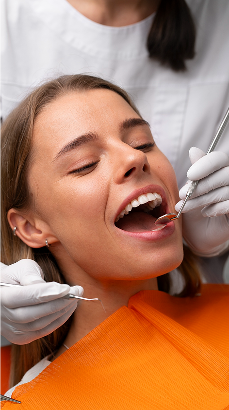
Minimally Invasive Gum Surgery Techniques Explained

Gum recession affects nearly everyone over 35, with causes ranging from brushing too hard to periodontal disease. Traditional treatments often involve tissue grafts, causing discomfort and long recovery times. Newer approaches like the Pinhole Surgical Technique (PST) and LANAP offer gentler options with faster healing, no scalpels, and excellent outcomes. These methods are especially effective for mild-to-moderate cases and focus on preserving existing gum tissue while reducing pain and downtime.
Key Takeaways:
- PST: Uses a tiny pinhole to reposition gum tissue; no sutures or grafts needed.
- LANAP: A laser-based technique for regenerating gum and bone tissue in periodontitis cases.
- PRF (Platelet-Rich Fibrin): Utilises the patient’s blood to promote natural healing and tissue repair.
- Recovery is faster, and these methods avoid the need for tissue harvesting from the palate.
While these techniques are promising, their success depends on factors like gum thickness, oral hygiene, and the severity of the recession. Consulting a qualified periodontist is essential to determine the best option for your needs.
LANAP vs. Pinhole: Why You Might Need BOTH for Healthy Gums!
sbb-itb-2be92ed
Pinhole Surgical Technique (PST)
As dentistry moves towards less invasive and more patient-friendly methods, the Pinhole Surgical Technique (PST) emerges as a promising option for treating gum recession. Known as the "Lunchtime Gum Lift", this method avoids scalpels and sutures, instead focusing on repositioning the patient’s existing gum tissue to cover exposed tooth roots. Instead of making large cuts or grafting tissue from the palate, a small pinhole (just 2–3 mm) is created in the gum near the affected area [5][7][9].
Using this tiny entry point, specialised tools gently loosen the gum tissue and release fibrous attachments. The gum is then carefully moved upwards to protect the exposed roots. To help stabilise the tissue during recovery, dissolvable collagen strips, such as Bio-Gide, are inserted through the pinhole and tucked between the teeth [5][7][9].
"The real genius of this procedure is – collagen strips! … The tissue is supported at the new position, and the strips assist in wound stabilisation during healing to ensure root coverage." – Tina Beck, DDS, MS [7]
One of the key advantages of PST is that it can treat multiple teeth – or even the entire mouth – in one session. Traditional grafting procedures often rely on tissue taken from the palate, which limits the number of teeth that can be treated at once. PST bypasses this limitation by using the gum tissue already present in the treatment area [7][9].
How PST Works
The procedure begins with local anaesthesia to ensure patient comfort. The dentist then creates a small pinhole in the vestibular mucosa (the area where the gum meets the cheek). Using specialised instruments, the gum tissue is gently separated from the bone through a technique called supraperiosteal blunt dissection. This approach minimises trauma and preserves blood supply, allowing the gum to be repositioned towards the tooth crown without creating tension. Collagen strips are inserted through the pinhole into the spaces between the teeth to stabilise the repositioned gum and promote healing as they dissolve [5][7][9].
PST is most effective for Miller Class I and II recession cases, where there’s no major bone loss between the teeth. Patients undergoing this treatment should have healthy gums and follow strict aftercare, including sticking to soft foods and avoiding aggressive brushing near the treated areas [5][6][9].
Research Findings and Success Rates
Studies have highlighted PST’s effectiveness and long-term stability. In Dr. John Chao’s 2012 study, which evaluated 121 recession sites, a 94% reduction in gum recession was observed, with 81% of cases achieving full root coverage [9]. Similarly, a 2017 case series by Dr. Saravanan Sampoornam Pape Reddy treated 18 recession sites in five patients aged 25–40, achieving an average root coverage of 96.7% at six months, with 88.8% of the sites showing complete root coverage [5].
Another study in 2020 by Dr. Manvi Chandra Agarwal combined PST with titanium-prepared platelet-rich fibrin (T-PRF) membranes. This approach treated 20 recession sites in 10 patients, reducing recession depth from 2.65 mm to just 0.35 mm within six months, achieving an average root coverage of 87% [6].
"The high amount success in PST can be attributed to being the least invasive procedure with no incisions/sutures." – Saravanan Sampoornam Pape Reddy, Graded Specialist Periodontology, Army Dental Corps India [5]
Long-term studies further reinforce PST’s reliability, with results remaining stable for up to 19 years [9].
LANAP Protocol for Periodontal Regeneration
LANAP (Laser-Assisted New Attachment Procedure) is a laser-based treatment designed to tackle moderate-to-advanced periodontitis. Unlike traditional flap surgery, LANAP is a less invasive option that uses laser technology to reduce pocket depth and regenerate the periodontal attachment apparatus. When paired with other techniques like PST (Pinhole Surgical Technique) vs traditional grafting, it broadens the scope of minimally invasive treatments available to dental professionals.
The LANAP protocol relies on the PerioLase MVP-7, a pulsed Nd:YAG laser with a 1064 nm wavelength. This laser precisely targets diseased tissue through a process called Selective Photoantisepsis, sparing healthy connective tissue. It is particularly effective against dark-pigmented bacteria like Porphyromonas gingivalis, eliminating harmful pathogens while protecting surrounding tissues [12].
What makes LANAP stand out is its ability to achieve true regeneration. In 2016, the FDA approved LANAP for "true regeneration", which includes the formation of new alveolar bone, periodontal ligament (PDL), and cementum on previously diseased root surfaces. It remains the only laser protocol with this level of clearance, supported by human histologic evidence [12][13].
"Using the Nd:YAG laser with this [LANAP] procedure, periodontal regeneration is achievable on a previously diseased root surface." – Kao et al., Systematic Review from the AAP Regeneration Workshop [13]
LANAP is widely accepted by patients, with 91% choosing it over traditional osseous surgery, which only sees a 33% acceptance rate [17]. Additionally, the PerioLase MVP-7 can reduce oral inflammation by up to 90% in just one session [10].
Procedure Overview
The LANAP protocol follows a structured approach. After applying local anaesthesia, the laser is used to create a trough that selectively removes diseased tissue. Ultrasonic scaling then removes tartar and calculus from the root surface, and the bone is reshaped to encourage healing [11][13].
A second pass with the laser interacts with haemoglobin to form a stable fibrin clot, or ‘red thrombus,’ which seals the periodontal pocket. This clot contains the patient’s own stem cells and growth factors, aiding the healing process. The entire procedure avoids scalpels, incisions, and sutures [10][15].
A critical step in the LANAP protocol is occlusal adjustment, where the dentist modifies the patient’s bite to prevent trauma to the regenerating tissue. Post-procedure care often includes antibiotics like Doxycycline and chlorhexidine rinses to manage bacteria. Patients are also instructed to avoid mechanical plaque removal in the treated area for 2–3 weeks to protect the fibrin clot [11][13][14].
"LANAP is a precise treatment protocol combining laser surgery with the well-established principles of traditional periodontal therapy, all based on biologic and clinical principles." – Raymond A. Yukna, DMD, MS [11]
Treatment Outcomes and Comparisons
LANAP has shown impressive clinical results. In a study by Dr Raymond A. Yukna at the University of Colorado, 22 patients with moderate-to-severe periodontitis underwent LANAP treatment. After 12–18 months, 93.5% of probing depth measurements were reduced to 3 millimetres or less, and 40% of grade 2 furcation involvements were resolved [11][14].
Other studies highlight additional benefits: 54% of treated sites gained at least 2 millimetres of clinical attachment, bleeding on probing was reduced by 92.85%, and visible plaque decreased by 96.87% [14][16]. Bone density at treated sites also increased by an average of 38% [13].
When compared to traditional flap surgery, LANAP offers several advantages. Gingival recession is minimal, averaging just 0.1 millimetres, compared to the tissue shrinkage often seen with flap surgery. Recovery is quicker too, with most patients resuming regular activities within 24 hours, unlike the extended downtime required for conventional surgery [10][15].
| Feature | LANAP Protocol | Conventional Flap Surgery |
|---|---|---|
| Tools Used | PerioLase MVP-7 Nd:YAG Laser | Scalpels and Sutures |
| Tissue Impact | Selective removal of diseased tissue only | Removal of both diseased and healthy tissue |
| Regeneration | FDA-cleared for true regeneration (bone, PDL, cementum) | Focus on repair via long junctional epithelium |
| Recession | Minimal (approx. 0.1 mm) | Often significant |
| Recovery | Immediate (24 hours) | Several days to weeks |
| Pain/Bleeding | Minimal to none | Moderate to significant |
RejuvaGum Lift with Platelet-Rich Fibrin (PRF)
In addition to methods like PST and LANAP, PRF-based techniques bring new possibilities to minimally invasive gum surgery. The RejuvaGum Lift addresses gum recession by leveraging the body’s natural healing processes. At the heart of this method is Platelet-Rich Fibrin (PRF), a material created from the patient’s own blood. Unlike synthetic materials or donor grafts, PRF significantly reduces the risk of allergic reactions or disease transmission, while serving as a natural framework for tissue repair and regeneration [18].
How PRF Works in Gum Surgery
The preparation of PRF is both simple and efficient. A dental professional collects approximately 20 mL of venous blood, which is then centrifuged at 1,500 rpm for 14 minutes. This process produces a fibrin clot enriched with platelets and growth factors, which is then compressed into a membrane [4][22]. The entire process takes less than 20 minutes [18].
PRF’s preparation method distinguishes it from materials like Platelet-Rich Plasma (PRP). Unlike PRP, PRF is created without the use of anticoagulants or additives, forming a stable fibrin matrix that gradually releases growth factors to aid tissue healing [18].
During the RejuvaGum Lift procedure, the PRF membrane is inserted through small incisions near the affected areas. Acting as a scaffold, it supports healing, preserves tissue volume, and reduces inflammation [8]. Additionally, PRF boosts blood flow to the healing site and enhances the integration of grafts [19]. These attributes contribute to the improved clinical outcomes discussed below.
Evidence-Based Benefits
Research supports the clinical advantages of PRF. In a randomised clinical trial conducted by Mansoura University‘s Faculty of Dentistry, 24 patients (46 teeth) with Cairo Class I gingival recession were treated using the VISTA technique combined with A-PRF. After six months, the A-PRF group achieved an average root coverage of 67.26%, compared to 50.09% for those treated with collagen matrix. Improvements were also seen in recession depth (0.88 mm) and width (2.30 mm) [4].
PRF is also effective in promoting periodontal regeneration, offering a clinical attachment level gain of 2.5 mm. It reduces the risk of alveolar osteitis by 70% and decreases vertical bone loss by 1.5 mm in socket preservation procedures [20].
"PRF acts as a natural scaffold, promoting soft-tissue healing, maintaining tissue volume, and potentially reducing gingival inflammation." – Scott Froum, DDS, Editorial Director, Perio-Implant Advisory [8]
These outcomes are driven by PRF’s high concentration of growth factors, such as platelet-derived growth factor (PDGF) and insulin-like growth factor 1 (IGF-1). These proteins play a key role in tissue regeneration by stimulating fibroblast activity, angiogenesis, and collagen production [4]. Patients also report less post-operative pain and faster recovery times compared to traditional grafting techniques [4][21].
Comparing Minimally Invasive Techniques
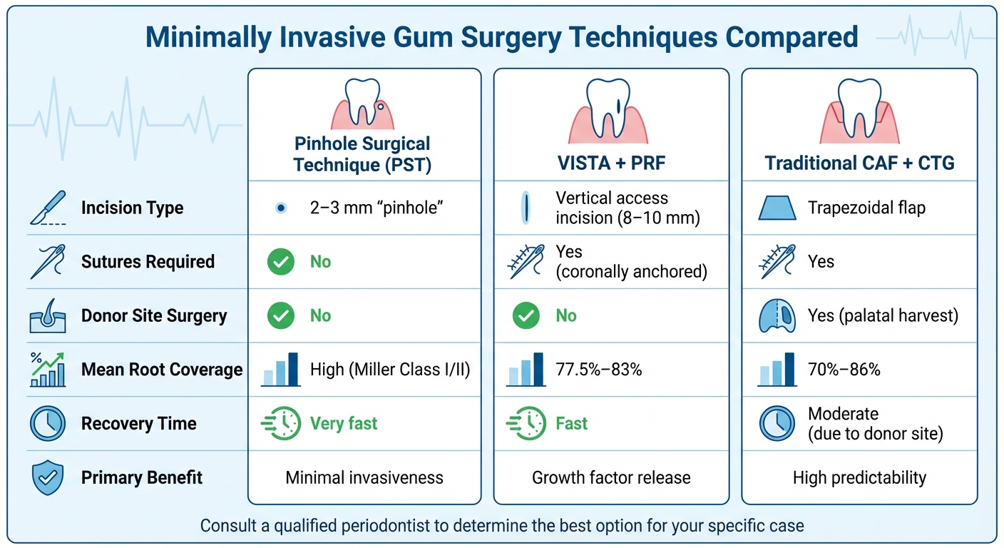
Comparison of Minimally Invasive Gum Surgery Techniques: PST, VISTA, and Traditional Methods
Minimally invasive methods for treating gum recession have gained attention for their tailored approaches, accommodating varying levels of severity and patient preferences. The Pinhole Surgical Technique (PST), for instance, relies on a single, needle-sized entry point (2–3 mm) and eliminates the need for sutures. This technique is particularly effective in the upper jaw, where keratinised tissue is more abundant [2]. On the other hand, the VISTA procedure involves a slightly larger vertical incision (8–10 mm) to create a subperiosteal tunnel. It incorporates platelet-rich fibrin (PRF) membranes to aid healing, preserve the interdental papilla, and maintain blood supply more effectively than traditional flap methods [4].
Recovery processes differ between these techniques. PST patients benefit from the absence of sutures and report minimal post-operative discomfort [8]. VISTA with PRF, however, requires more specific aftercare, including the use of analgesics every 12 hours and avoiding brushing the surgical site for four weeks. Sutures are removed two weeks post-surgery [4]. Both methods spare patients the discomfort of palatal tissue harvesting, a common downside of traditional procedures [4][24].
Technique Comparison Table
Here’s a breakdown of key differences between these approaches:
| Feature | Pinhole Surgical Technique (PST) | VISTA + PRF | Traditional CAF + CTG |
|---|---|---|---|
| Incision Type | 2–3 mm "pinhole" [2] | Vertical access incision [4] | Trapezoidal flap [24] |
| Sutures Required | No [2] | Yes (coronally anchored) [2] | Yes [24] |
| Donor Site Surgery | No [2] | No [4] | Yes (palatal harvest) [24] |
| Mean Root Coverage | High (in Miller Class I/II cases) [2] | 77.5%–83% [4][23] | 70%–86% [23] |
| Recovery Time | Very fast [2] | Fast [4] | Moderate (due to donor site) [24] |
| Primary Benefit | Minimal invasiveness [2] | Growth factor release [4] | High predictability [24] |
Minimally invasive methods like PST and VISTA are proving to be just as effective as traditional approaches. For example, a study conducted in January 2025 at Mansoura University in Egypt treated 24 patients with Cairo Class 1 recession using VISTA combined with advanced PRF. The results showed an impressive mean root coverage of 77.50% after six months [4]. This is comparable to the 70%–86% coverage typically achieved with traditional connective tissue grafts, but without the need for a secondary surgical site.
However, for patients with thin tissue (under 1.1 mm), adding a graft material like PRF is crucial. Flap-only techniques often yield less predictable outcomes in such cases [24]. These findings highlight the growing preference for patient-focused, minimally invasive options that balance effectiveness with comfort.
Research Evidence and Patient Considerations
This section delves into the research evidence and patient-related factors that shape treatment outcomes, building on earlier descriptions of minimally invasive techniques. Systematic reviews have shown these methods can effectively address certain types of gum recession, but careful patient selection remains critical for long-term results. For instance, a 2025 meta-analysis on the VISTA technique revealed an average complete root coverage of 58%, which increased to 69% when combined with Acellular Dermal Matrix (ADM). The study also reported measurable improvements, including a 1.36 mm reduction in recession depth, a 1.32 mm increase in keratinised tissue, and a 2.14 mm improvement in clinical attachment levels [25].
The effectiveness of these treatments largely depends on the classification and severity of the gum recession. Miller Class I/II defects tend to achieve predictable outcomes, while Class III/IV cases are associated with lower success rates [2][23]. Beyond classification, individual anatomical factors also play a crucial role in determining success.
"The selection of the surgical techniques should be dictated by several factors, including the anatomy of the defect site, such as the size of the recession defect, the presence or absence of keratinised tissue adjacent to the defect, the width and height of the interdental soft tissue, and the depth of the vestibule" [1].
Patients with gingival thickness exceeding 1.1 mm generally experience more reliable results [3]. Additionally, upper-jaw procedures like PST are most effective in cases with wider keratinised tissue and favourable papillary shape. On the other hand, techniques such as VISTA can be compromised by limited vestibular depth [2].
While these approaches show promise, certain challenges remain. One major limitation is the scarcity of long-term data beyond 12 months. Although minimally invasive methods reduce postoperative discomfort and eliminate the need for palatal tissue harvesting, large-scale randomised controlled trials are still limited [2][26]. For example, a 2025 study on the NIPSA method reported that 50% to 58.33% of cases achieved residual probing depths of 2 mm after one year. However, some cases still showed 4 mm probing depths, underscoring the importance of managing patient expectations and offering thorough pre-surgical counselling [26].
Patient health and habits also play a substantial role in treatment outcomes. Research often limits participation to systemically healthy non-smokers with excellent oral hygiene [4]. In contrast, heavy smoking and poor oral hygiene significantly reduce the effectiveness of these treatments [26]. To maximise results, patients are encouraged to maintain rigorous oral hygiene and, where applicable, quit smoking at least one year before undergoing treatment [26].
Conclusion
Minimally invasive gum surgery techniques are changing the way gum recession is treated, offering less discomfort, quicker recovery times, and more appealing results compared to older methods. Approaches like the Pinhole Surgical Technique, VISTA, and those using Platelet-Rich Fibrin avoid large incisions and often eliminate the need to take tissue from the palate, which significantly reduces patient discomfort and healing time [2][4].
That said, the success of these procedures isn’t one-size-fits-all. Factors like the type of gum recession, the thickness of the gums, the depth of the vestibule, and the height of the interdental papillae all play a role in determining the outcome.
"The selection of the surgical techniques should be dictated by several factors, including the anatomy of the defect site, such as the size of the recession defect, the presence or absence of keratinised tissue adjacent to the defect, the width and height of the interdental soft tissue, and the depth of the vestibule" [1].
This highlights the need for a customised treatment plan tailored to each patient’s specific oral health needs.
Given the commonality of gum recession [1] and the precision required for these techniques, it’s crucial to consult a qualified periodontist. These specialists, with their advanced training, are equipped to evaluate factors like smoking habits, medical history, oral hygiene, and the root causes of gum recession. Addressing these underlying issues – whether it’s overzealous brushing or ongoing periodontal disease – is key to preventing future problems and ensuring the results last [1].
For anyone dealing with gum recession, a detailed consultation with a dental specialist is the first step in determining if minimally invasive surgery is the right choice. In Australia, where patients often prioritise evidence-based and minimally invasive care, working with a specialist ensures both effective and lasting treatment outcomes.
FAQs
Am I suitable for PST or LANAP?
Your choice between PST (Pinhole Surgical Technique) and LANAP (Laser-Assisted New Attachment Procedure) largely depends on how advanced your gum condition is.
LANAP is often the go-to option for moderate to severe periodontitis. It uses a laser-based approach that’s less invasive than traditional surgery while targeting infected tissue effectively. On the other hand, PST is ideal for addressing mild to moderate gum recession, especially when there’s no significant tissue loss involved.
For the best advice, it’s essential to consult a periodontist. They can evaluate your specific situation and recommend the treatment that suits you best.
How long do results usually last?
Minimally invasive gum procedures, like the Pinhole Surgical Technique, can deliver results that last for several years. Research indicates these benefits often remain for 2–5 years or more, though this depends on factors like individual oral health and consistent care. Maintaining regular dental check-ups and practising good oral hygiene are essential for keeping these results intact.
How can gum recession be prevented from coming back?
Minimally invasive surgical options, like root coverage procedures and connective tissue grafts, can play a crucial role in managing gum recession. When paired with diligent oral hygiene and tackling underlying issues, these techniques can help reduce the chances of the problem returning. Staying proactive with care and addressing concerns early are essential for maintaining healthy gums over time.
Related Blog Posts
- Advances in Gum Grafting Materials and Techniques
- Minimally Invasive vs. Traditional Gum Surgery: Key Differences
- Pinhole Surgery vs Traditional Gum Grafting
- Soft Tissue Grafting: New Techniques Explained
Important Notice: Any surgical or invasive procedure carries risks. Before proceeding, you should seek a second opinion from an appropriately qualified health practitioner.
Individual results may vary. The information provided in this article is for educational purposes only and does not constitute medical advice.
Checkout Related Blogs


Get in touch with us
For more information, call us now to start feeling better. Or fill the form below to make appointment
The Latest News from Complete Smiles
How to Clean Clear Plastic Retainers
Checklist for Choosing Wearable Dental Devices
Checklist for Choosing Cloud AI Platforms in Dentistry
Complete Smiles Bella VistaAccepts All Major Health Funds, Including


