





Soft Tissue Grafting: New Techniques Explained
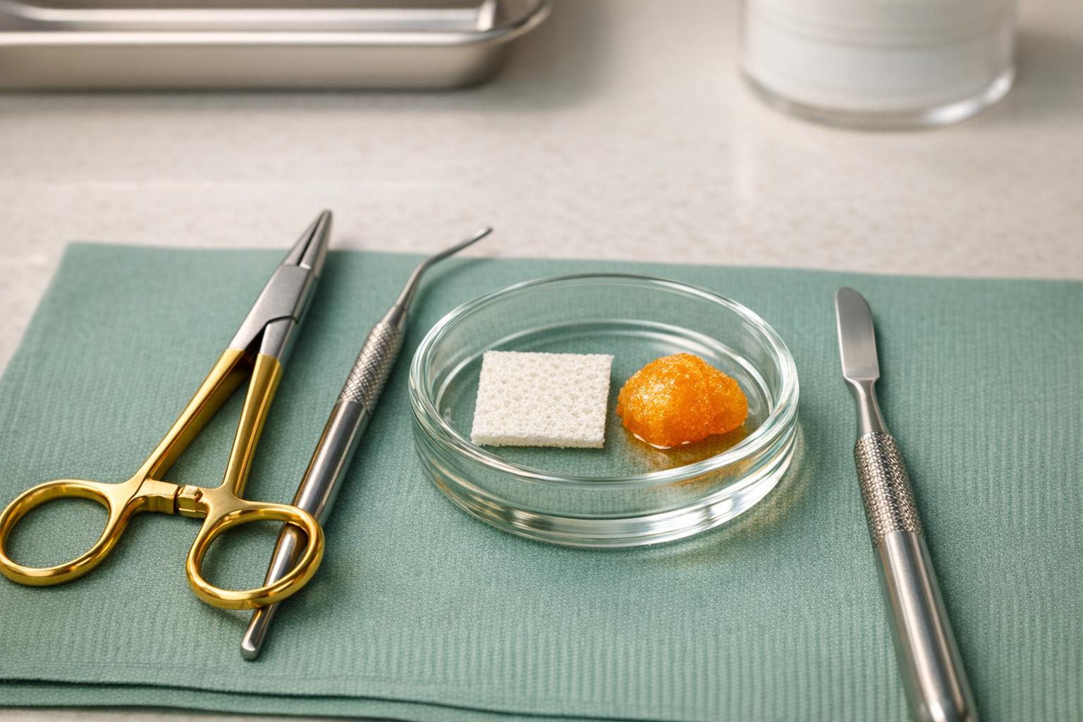
Soft tissue grafting, also known as gum graft surgery, is a procedure to restore receding gums and protect teeth from damage. This involves transferring tissue to areas with gum loss, often using tissue from the patient’s palate or donor materials. The latest methods, like the Pinhole Surgical Technique (PST) and Modified Coronally Advanced Tunnel (MCAT), focus on reducing discomfort and improving recovery. These approaches eliminate the need for traditional tissue harvesting, making the process smoother and less painful. Additionally, new biomaterials and digital tools, such as intraoral scanners and 3D planning, enhance precision and outcomes.
Key Highlights:
- Why it’s done: Prevent sensitivity, decay, and tooth loss caused by gum recession.
- New methods: PST and MCAT avoid cutting and sutures, leading to faster healing.
- Biomaterials: Options like collagen matrices and Platelet-Rich Fibrin (PRF) reduce complications and speed up recovery.
- Digital tools: Scanners and guided surgery improve accuracy and aesthetics.
- Benefits: Less pain, better aesthetics, and long-term durability with success rates over 90%.
If you’re considering soft tissue grafting, consult a qualified periodontist to explore the best options tailored to your needs.
Gum graft – Coronally advanced flap – Receding gums treatment ©
New Techniques in Soft Tissue Grafting
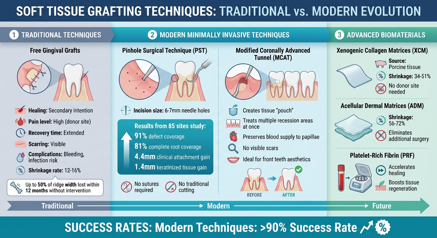
Traditional vs Modern Soft Tissue Grafting Techniques Comparison
Less Invasive Grafting Methods
Advancements in soft tissue grafting have made procedures less invasive, reducing both surgical trauma and patient discomfort. One standout method is the Pinhole Surgical Technique (PST), which involves making tiny needle incisions (6–7 mm above the mucogingival junction). Using specialised tools, clinicians gently elevate and reposition the tissue without the need for traditional cutting or sutures. In a study involving 85 sites with Miller Class I or II recession, PST delivered impressive results: 91% defect coverage, 81% complete root coverage, a 4.4 mm clinical attachment gain, and 1.4 mm of keratinised tissue gain [8].
Another promising approach is the use of tunnelling techniques, such as the Modified Coronally Advanced Tunnel (MCAT) and Subperiosteal Envelope Technique (SET). These methods create a "pouch" beneath the gum tissue, allowing multiple areas of recession to be treated at once. By preserving the blood supply to the interdental papillae and avoiding visible scars, these techniques are particularly effective for front teeth, where aesthetics are crucial [2][8].
"Advances in minimally invasive soft-tissue grafting techniques over the years have improved case acceptance."
- Dr Scott Froum, Editorial Director of Perio-Implant Advisory [8]
These less invasive methods are paving the way for further developments, particularly with the integration of advanced biomaterials and digital technology.
Biomaterials and Growth Factors
Modern grafting techniques are also moving away from traditional donor site harvesting by incorporating advanced materials. Options like Xenogenic Collagen Matrices (XCM), derived from animal sources such as porcine tissue, and Acellular Dermal Matrices (ADM) eliminate the need for additional surgical sites. This shift reduces both pain and complications. However, these materials are not without drawbacks – XCM can shrink by 34% to 51%, while ADM may contract by 56% to 72%, compared to the significantly lower shrinkage rates (12% to 16%) seen in autogenous grafts [8][10][11].
Platelet-Rich Fibrin (PRF) is another innovation gaining traction. By accelerating healing and boosting tissue regeneration, PRF complements grafting procedures effectively [8]. Additionally, nanofibrous scaffolds and hydrogels are being used to deliver growth factors in a controlled manner. These materials aid in cell attachment and seamless tissue integration, enhancing the body’s natural regenerative processes [9]. Given that up to one-third of ridge width can be lost within three months after tooth extraction, using volume-stable materials is critical for maintaining natural-looking outcomes [11].
Digital Planning and Guided Surgery
Technological advancements are revolutionising how grafting procedures are planned and executed. Intraoral scanners now provide detailed digital images, enabling clinicians to track volumetric changes over time. This ensures that reconstruction begins only after the tissue has stabilised, typically around three months post-procedure [11].
"The emergence of 3D bioprinting has further enhanced precision, allowing for patient-specific graft designs that integrate vascular structures for improved survival."
- Dr Maha Assad Munshi, Department of Periodontics [9]
Virtual surgical planning is another game-changer. By combining intraoral scans, cone beam computed tomography (CBCT), and facial scans, clinicians can create a detailed "digital patient" model. This approach is particularly beneficial in aesthetically sensitive areas, ensuring a seamless transition between soft tissue and restorations. Computational modelling tailors treatment strategies to each patient’s unique anatomy and health profile, while real-time monitoring systems provide valuable insights into graft performance and integration [9][11].
Benefits of Modern Techniques
Better Patient Comfort and Recovery
Modern grafting techniques have transformed patient comfort levels. Traditional free gingival grafts often heal by secondary intention, leading to considerable pain at the donor site, bleeding, and extended recovery periods [3]. In contrast, subepithelial connective tissue grafts (SCTG) promote healing by primary intention at both donor and recipient sites. This means the tissue closes immediately, reducing discomfort. As Dr Peter Windisch from Semmelweis University‘s Department of Periodontology states:
"Healing by primary intention can facilitate tissue maturation and decrease postoperative discomfort" [3].
By removing the need for a donor site altogether, techniques using xenografts or allografts (biomaterial substitutes) eliminate risks like donor-site infection, swelling, and necrosis [12][11]. Additionally, advanced tunnelling methods, such as the Subperiosteal Envelope Technique, preserve blood flow to the papillae, leading to minimal post-surgical issues and smoother healing [12].
Better Aesthetic and Functional Results
Beyond recovery, modern grafting techniques also deliver superior aesthetic and functional outcomes. These approaches achieve excellent colour matching and avoid visible scarring, especially in the aesthetic zone where the gums are most noticeable. Subepithelial connective tissue grafts are particularly effective, as the overlying flap retains the same surface characteristics as the surrounding gum tissue, ensuring natural colour blending [3][12]. The Modified Coronally Advanced Tunnel (MCAT) technique further avoids vertical incisions, preventing visible scars and achieving seamless integration [12].
Functionality improves significantly as well. Increasing soft tissue thickness and the width of keratinised mucosa around implants helps maintain crestal bone levels and reduces signs of inflammation [7]. Covering exposed root surfaces not only alleviates hypersensitivity but also strengthens the gums against daily stresses like chewing and brushing [12][7]. This added keratinised tissue acts as a protective shield, reducing the risk of bacterial infections, premature tooth loss, and implant failure.
Long-Term Durability and Success Rates
The combination of subepithelial connective tissue grafts with a coronally advanced flap has proven to deliver reliable, long-term results [12][14]. A Cochrane review of 48 randomised controlled trials involving 1,227 adults found this method to be more effective in reducing gingival recession compared to guided tissue regeneration [13]. Studies tracking soft tissue stability at pontic sites show that volumes remain stable even after 5 to 10 years when SCTG is used [11].
The durability of these techniques lies in their ability to create ideal conditions for graft integration. Bilaminar reconstruction with tension-free closure ensures a strong blood supply and prevents graft retraction, guaranteeing long-term stability [12][3]. Without such interventions, up to 50% of alveolar ridge width can be lost within 12 months after tooth extraction. Early adoption of modern grafting techniques is crucial to preserving natural gum contours [11]. These lasting results highlight the importance of expert, tailored care in soft tissue grafting procedures.
sbb-itb-2be92ed
Risks and Considerations
Common Complications and Management
Like any surgical procedure, soft tissue grafting comes with potential risks that patients should be aware of. These can include infection, pain, swelling, and tissue necrosis (when the graft fails due to loosening or infection) [2]. The specific risks often depend on the type of graft used. For example, free gingival grafts might lead to over-augmentation, where excessive tissue builds up, or poor colour blending, making the graft stand out visually from the surrounding gums [2]. Meanwhile, the Subperiosteal Envelope Technique may occasionally result in epithelial invagination (folding of gum tissue inward) or visible scar lines at the edges of the treatment area [2].
Surgeons take several measures to minimise these risks. For instance, applying pressure to the surgical site for 1–2 minutes helps prevent blood clots from forming between the graft and the recipient site, a common cause of tissue necrosis [2]. Using microsurgical blades and carefully chosen sutures – either resorbable or non-resorbable monofilament – also reduces trauma and ensures the graft remains secure [2]. As highlighted by the Canberra Implant & Periodontal Centre:
"Any surgical or invasive procedure carries risks. Before proceeding, you should seek a second opinion from an appropriately qualified health practitioner" [5].
Understanding these risks highlights the importance of selecting the right patients for the procedure.
Patient Suitability and Pre-Procedure Planning
Soft tissue grafting isn’t suitable for everyone. Dentists assess several factors before recommending it. Physical signs like teeth that appear "too long", exposed roots causing sensitivity to hot or cold, or severe gum recession often indicate a need for grafting [4][5][6]. However, overall health plays a big role. Smoking and uncontrolled diabetes, for example, can significantly hinder recovery and reduce success rates [4]. As the American Association of Oral and Maxillofacial Surgeons explains:
"Patient factors such as diabetes and smoking affect recovery and overall success rates" [4].
Pre-procedure planning involves a detailed evaluation of your oral anatomy. Specialists assess gum recession severity using Miller’s classification (I through IV), examine interproximal bone heights, and measure gingival biotype thickness [2]. Predictability of outcomes depends on these factors. For instance, Miller class I and II cases often achieve complete root coverage, while class III cases may only allow partial coverage [2]. The choice of graft material – whether it’s tissue from your palate, nearby healthy areas, or processed cadaver tissue – depends on your unique situation [4]. Additionally, addressing habits like aggressive brushing or stabilising health conditions before surgery can greatly improve your chances of a successful outcome [4][5].
Thorough assessments ensure that patients are well-prepared for the procedure and equipped to heal effectively.
Post-Procedure Care and Maintenance
Careful pre-procedure planning sets the stage for smoother recovery. The first 24–48 hours are especially crucial. During this time, manage swelling by applying ice packs for 15–20 minutes at a time and keep your head elevated while sleeping [15][16]. Stick to a liquid or soft diet – think yoghurt, smoothies, or mashed potatoes – for the first week. Avoid hard, crunchy, spicy, or sticky foods that could disturb the graft [16][1]. Dr Kent Boon from Kita Dental advises:
"The golden rule is: do not brush or floss directly on the gum graft site until we give you the all-clear. This is essential, as even gentle brushing can disrupt healing" [16].
Other precautions include avoiding strenuous activities like heavy lifting, excessive talking, or using straws for at least a week, as these can lead to bleeding or graft displacement [16][1]. Tobacco use should be avoided for 7–10 days, as it restricts blood flow and significantly increases the risk of graft failure [15][16]. Be vigilant for warning signs such as a fever above 38.3°C, large white patches on the tissue, or separation of the graft from the tooth, as these may indicate infection or compromised blood supply [16][1].
Initial healing generally takes 1–2 weeks, but full integration of the graft into the tissue can take 2–3 months [16][1]. Sutures are typically removed after 14 days [2]. With proper care, soft tissue grafting boasts success rates of over 90% [16][1].
Consulting Professionals for Soft Tissue Grafting
Why Choose a Specialist?
Soft tissue grafting isn’t something to leave to chance – it requires a high level of skill and precision. Specialists like periodontists and oral and maxillofacial surgeons receive an additional three years of focused training after dental school. This advanced education helps them achieve success rates exceeding 90% for gum grafting procedures [1]. Their expertise allows them to carefully navigate around critical structures during tissue harvesting, significantly lowering the risk of complications [3].
These professionals are well-versed in a variety of techniques, ranging from traditional methods to newer, minimally invasive approaches. They tailor each procedure to suit the patient’s unique anatomy, ensuring the best possible results [3]. Beyond the procedure itself, specialists also provide a range of sedation options and highly personalised post-operative care to enhance comfort and recovery [1][4]. When selecting a specialist, it’s wise to confirm their board certification and ask about their experience with modern grafting techniques. Clinics that combine innovative methods with individualised care stand out in delivering exceptional results.
Complete Smiles Bella Vista

One such clinic offering expert care is Complete Smiles Bella Vista. Under the leadership of Dr James Hanna, they provide comprehensive dental services, including advanced soft tissue management and customised treatment plans. For further details, visit completesmilesbv.com.au.
Conclusion and Key Takeaways
Summary of New Techniques
Advancements in grafting techniques have reshaped periodontal care, offering more precise and patient-friendly options. Modern soft tissue grafting incorporates biomaterials such as allografts and xenografts, combined with digital tools like intraoral scanners. These technologies improve accuracy and reduce discomfort for patients [2][11]. Additionally, minimally invasive procedures that eliminate vertical incisions promote faster healing with little to no scarring [2].
The functional benefits are significant. These methods help reduce tooth sensitivity to temperature changes, protect against root decay, and minimise the risk of further bone loss [17]. As Dr Rana Baroudi highlights:
"Soft tissue grafting is not merely a preventive measure; it is a corrective intervention aimed at halting further recession and bone loss" [17].
Beyond improving oral health, these techniques also enhance aesthetics while ensuring a more comfortable recovery experience [17]. The advancements emphasise the value of expert care in achieving the best possible outcomes.
Consult a Professional
If you’re dealing with gum recession or sensitivity, it’s essential to consult a qualified periodontist. These specialists undergo an additional three years of training, equipping them to customise grafting procedures to suit your specific needs [1]. Before committing to any surgical treatment, it’s wise to seek a second opinion from a qualified health professional [5]. A comprehensive consultation should include a digital evaluation of your gum health and a detailed discussion of all available treatment options. From traditional autogenous grafts to biomaterial-based alternatives, the approach should be tailored to your individual oral health requirements.
FAQs
What are the benefits of the Pinhole Surgical Technique compared to traditional gum grafting?
The Pinhole Surgical Technique stands out for its distinct advantages compared to traditional gum grafting. It’s a minimally invasive approach that eliminates the need for incisions, stitches, or grafting tissue from other parts of the mouth. This often means patients experience less pain or discomfort both during and after the procedure.
Another perk? Recovery tends to be quicker, enabling people to get back to their daily routines much sooner. Plus, this method can address multiple areas of gum recession in a single session, offering a more streamlined and convenient solution for many.
How do materials like collagen matrices and Platelet-Rich Fibrin (PRF) help with recovery after soft tissue grafting?
Biomaterials like collagen matrices and Platelet-Rich Fibrin (PRF) play an important role in improving recovery after soft tissue grafting procedures. Collagen matrices serve as scaffolds that support the growth of new cells, helping tissues integrate more smoothly. This can accelerate the healing process and boost the success rate of grafts, especially in cases of gum recession.
On the other hand, PRF, which is rich in growth factors, works by stimulating blood vessel formation, collagen production, and tissue regeneration. This not only helps stabilise the grafted area but also lowers the risk of complications following the procedure. By creating an optimal environment for healing, these materials make soft tissue grafting more efficient and reliable.
How do digital tools improve the success of soft tissue grafting procedures?
Digital tools have transformed the way soft tissue grafting is approached, bringing new levels of precision, planning, and monitoring to the process. With advancements like intraoral scanners, CAD/CAM systems, and digital imaging, clinicians can now design grafts with a higher degree of accuracy, anticipate healing progress, and closely observe tissue changes over time.
These technologies make surgical planning and execution far more precise, which in turn enhances both the aesthetic and functional results of the procedure. They also help minimise risks of complications. By facilitating evidence-based decisions, these tools contribute to more reliable and successful outcomes for patients undergoing soft tissue grafting.
Related Blog Posts
- Advances in Gum Grafting Materials and Techniques
- Soft Tissue Grafting: Indications Explained
- Soft Tissue Grafting: Pre-Surgery Evaluation Explained
- Soft Tissue Grafting: Aesthetic vs. Functional Goals
Important Notice: Any surgical or invasive procedure carries risks. Before proceeding, you should seek a second opinion from an appropriately qualified health practitioner.
Individual results may vary. The information provided in this article is for educational purposes only and does not constitute medical advice.
Checkout Related Blogs




Get in touch with us
For more information, call us now to start feeling better. Or fill the form below to make appointment
The Latest News from Complete Smiles
How to Clean Clear Plastic Retainers
Checklist for Choosing Wearable Dental Devices
Checklist for Choosing Cloud AI Platforms in Dentistry

Complete Smiles Bella VistaAccepts All Major Health Funds, Including





