





Minimally Invasive Bone Regeneration Techniques
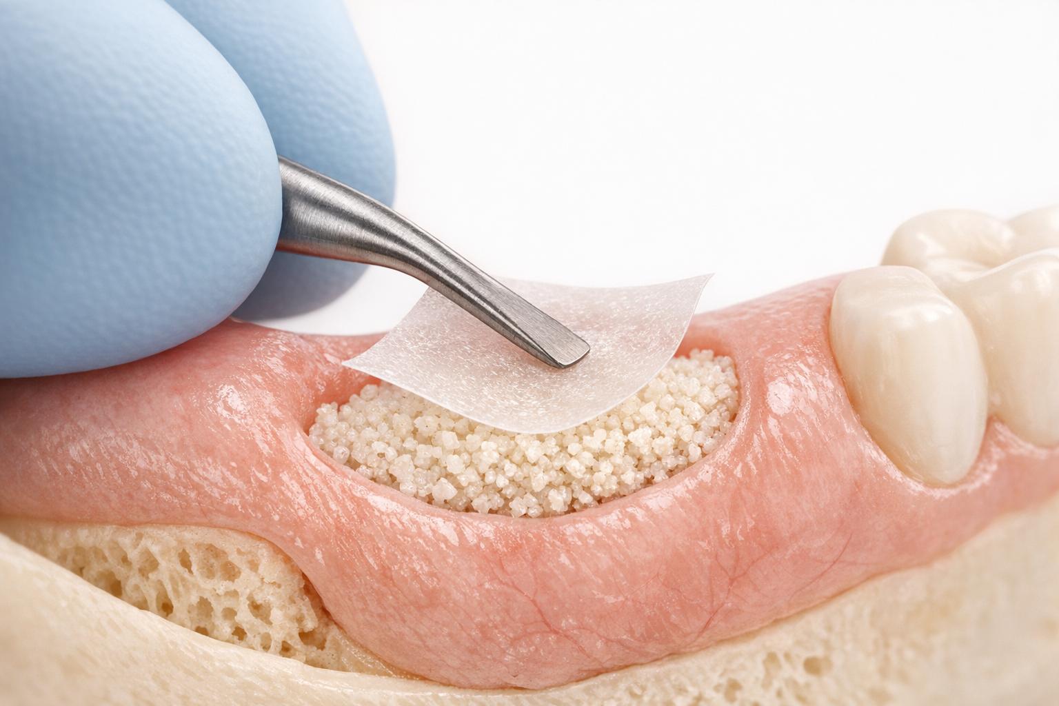
Minimally invasive bone regeneration techniques are transforming dental care by reducing surgical trauma, preserving soft tissues, and improving recovery. These methods use tools like magnification, mini-instruments, and minimal flap reflections to restore lost bone and tissue while maintaining blood supply. Benefits include faster healing, less discomfort, and better aesthetic outcomes compared to traditional methods.
Key highlights:
- Techniques like EPPT, NIPSA, and MINST: Preserve blood supply and soft tissue, ensuring reduced gingival recession and faster recovery.
- S.M.A.R.T. Bone Grafting and VISTA Tunnelling: Avoid flap elevation, using subperiosteal tunnels for grafting, improving stability and reducing complications.
- Growth Factor Integration: Use of rhPDGF-BB enhances bone regeneration without the need for extensive surgical interventions.
- Applications in Periodontal and Implant Dentistry: Effective for managing defects, improving implant success rates, and reducing patient discomfort.
These advancements are reshaping periodontal and implant procedures, offering better outcomes with less surgical impact.
Bone Grafting | Minimally-Invasive S.M.A.R.T. Bone Graft
Advantages of Minimally Invasive Methods
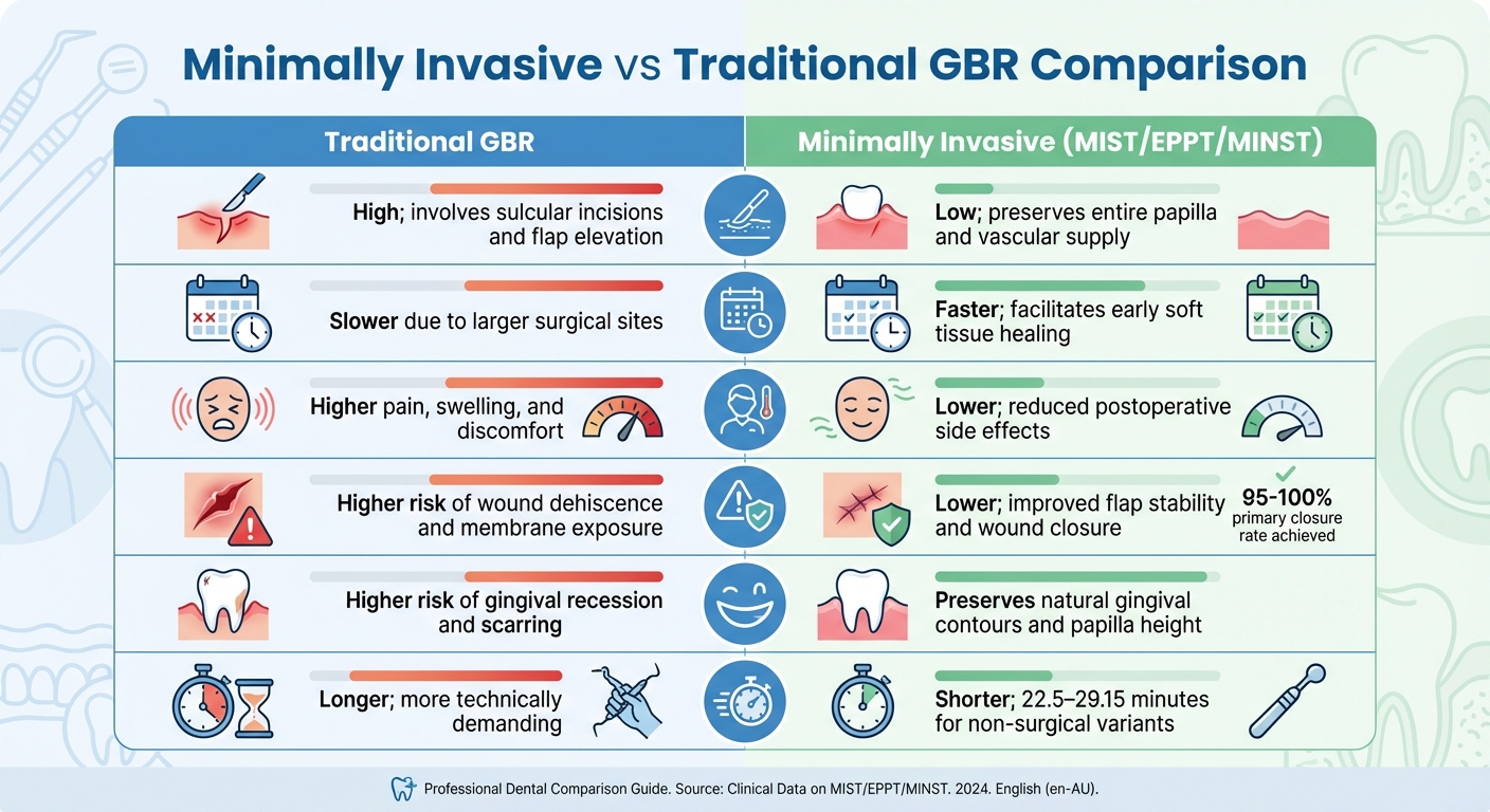
Minimally Invasive vs Traditional GBR: Clinical Comparison Chart
Minimally invasive bone regeneration techniques offer a range of benefits, particularly in reducing trauma to soft tissues and preserving vascular supply. By maintaining blood flow, these methods significantly lower the risks of tissue necrosis and early wound complications like dehiscence [1]. Unlike traditional approaches, these techniques avoid making traumatic incisions at the papilla base, which helps protect the blood supply to interdental tissues. This vascular preservation plays a key role in promoting better healing and reducing complications.
As Sylwia Jakubowska from the Medical University of Warsaw highlights:
Preserving papilla integrity and the soft tissue profile is essential for minimising complications, especially in the esthetic zone [1].
Another major advantage is the high primary closure rates achieved with minimally invasive methods, ranging from 95% to 100%. This is a marked improvement over traditional guided bone regeneration (GBR) techniques, which are more prone to membrane exposure and bacterial contamination [1][3][7].
Patients benefit from less pain, swelling, and discomfort, along with shorter operative times, which further reduces postoperative issues [2][5][6].
Styliani Anoixiadou from Aristotle University adds:
The application of MINST minimises soft and hard tissue trauma achieving better treatment outcomes in comparison with traditional scaling and root planing [5].
These clinical advantages become even more apparent when comparing traditional GBR and minimally invasive methods across key treatment parameters.
Minimally Invasive vs Traditional GBR Comparison
| Feature | Traditional GBR | Minimally Invasive (MIST/EPPT/MINST) |
|---|---|---|
| Tissue Trauma | High; involves sulcular incisions and flap elevation [1] | Low; preserves entire papilla and vascular supply [1] |
| Healing Time | Slower due to larger surgical sites [1] | Faster; facilitates early soft tissue healing [1] |
| Patient Morbidity | Higher pain, swelling, and discomfort [6] | Lower; reduced postoperative side effects [1][2] |
| Complication Rates | Higher risk of wound dehiscence and membrane exposure [1][7] | Lower; improved flap stability and wound closure [1] |
| Aesthetic Outcome | Higher risk of gingival recession and scarring [1] | Preserves natural gingival contours and papilla height [1] |
| Chair Time | Longer; more technically demanding [2][6] | Shorter; 22.5–29.15 minutes for non-surgical variants [2][5] |
One of the standout benefits of minimally invasive techniques is the enhanced flap stability, which shields the blood clot from functional forces during chewing [1]. This stability creates an environment conducive to regeneration. On the other hand, traditional GBR often struggles to achieve stable primary closure, leaving it more vulnerable to complications like wound dehiscence [7][6].
The combination of improved efficiency, faster healing, and fewer complications is driving a shift towards minimally invasive methods in periodontal and implant dentistry. These advancements set the stage for discussing even more refined techniques in upcoming sections.
Primary Minimally Invasive Bone Regeneration Techniques
Modern minimally invasive methods are transforming bone augmentation by reducing surgical trauma and preserving soft tissue structures. Below, we explore three key techniques that exemplify this approach.
S.M.A.R.T. Bone Grafting Technique
The Subperiosteal Minimally Invasive Aesthetic Ridge Augmentation Technique (S.M.A.R.T.) is a flapless approach designed to protect vascular and periosteal integrity. Developed by Dr. Ernesto A. Lee at the University of Pennsylvania, this method is particularly suited for cases where aesthetics are a top priority, avoiding visible gingival changes.
The procedure begins with a remote incision, followed by the creation of a subperiosteal tunnel using specialised laparoscopic instruments. A combination of anorganic bovine bone xenograft and recombinant human platelet-derived growth factor (rhPDGF-BB) is introduced into the tunnel using a tailored carrier system and compacted into place.
As Dr. Lee explains:
The subperiosteal minimally invasive esthetic ridge augmentation technique called SMART – developed as an alternative to traditional guided bone regeneration procedures – has the potential to profoundly impact the future of bone augmentation. [8]
What makes S.M.A.R.T. unique is what it avoids: no flap elevation, no use of cell-occlusive membranes, no tenting screws, and no decortication. By keeping the periosteum intact, this technique preserves a vital source of pluripotent cells, essential for bone regeneration through growth factors [8].
A 2017 case series involving 60 sites across 21 patients demonstrated impressive results. Horizontal augmentation averaged 5.11 mm, with edentulous ridges reaching 6.47 mm, and bone content was measured at 52% over follow-ups ranging from 4 to 30 months. All 25 implants placed in these augmented sites remained stable, with no crestal bone loss [8][9].
VISTA Tunnelling for Hard and Soft Tissue Augmentation
Tunnelling techniques, such as the VISTA approach, extend minimally invasive principles to both hard and soft tissue. By avoiding flap mobilisation, these methods maintain blood supply and periosteal integrity. A subperiosteal pouch is created to securely house bone graft material, eliminating the need for membranes or tenting screws. This approach is especially beneficial when hard tissue augmentation is paired with immediate implant placement [8][9].
In addition to the mechanical precision of these techniques, biological enhancements further improve outcomes, making them a reliable option in challenging cases.
Growth Factor-Enhanced Techniques with PDGF
The use of rhPDGF-BB capitalises on the periosteum’s regenerative potential. This growth factor attracts host stem cells, encourages their transformation into osteoblasts, and supports revascularisation. In techniques like S.M.A.R.T., rhPDGF-BB is mixed with anorganic bovine bone xenograft and delivered into a subperiosteal pouch through laparoscopic methods [8].
Dr. Lee highlights the importance of this approach:
The role of the periosteum as a source of pluripotential cells in growth factor-mediated bone regeneration must be considered, especially when decortication is not performed. [8]
Growth factors also stabilise the blood clot, a crucial element for regeneration in the absence of protective membranes. While these methods require precision, they are highly effective in aesthetic-sensitive cases where traditional surgeries could result in visible tissue changes.
sbb-itb-2be92ed
New Approaches in Bone Regeneration
Recent advancements in bone regeneration focus on refining less invasive techniques that preserve soft tissues while improving outcomes in complex cases. These methods build on established practices but aim to minimise surgical trauma and maintain essential tissue structures.
Intrasocket Guided Bone Regeneration
Intrasocket techniques offer a flapless option for managing extraction sites where maintaining bone is crucial. Unlike traditional guided bone regeneration, which involves extensive flap elevation, these techniques work directly within the socket, reducing surgical impact [11].
This approach uses occlusive membranes to block epithelial and connective tissues from entering the socket. By doing so, it gives bone cells the time needed to repopulate the area. Additionally, sealing the socket with barrier membranes or soft tissue grafts helps stabilise the blood clot, lowering the risk of infection [11][12][13].
Yanko G. Yankov from the Clinic of Maxillofacial Surgery highlights:
Socket preservation is a beneficial technique for maintaining hard and soft tissue volume. It can preserve about 2 mm of both vertical and horizontal ridge dimensions. [11]
Materials like xenografts, especially deproteinised bovine bone mineral, are commonly used due to their durability and ability to maintain space and contour over time [14]. High-density d-PTFE membranes are particularly effective in these cases because they resist bacterial infiltration, ensuring graft stability in the socket [15].
The outcomes are impressive. Socket preservation typically retains around 2 mm of vertical and horizontal ridge dimensions. However, about 10% of cases may still require additional bone grafting [11]. Implant survival rates after guided bone regeneration range from 79% to 100%, with most studies reporting over 90% survival after one year of functional use [11].
Building on these principles, another technique takes a similar minimally invasive approach to address buccal plate reconstruction.
Ice Cream Cone Technique Variations
The Ice Cream Cone Technique is a valuable method for reconstructing the buccal plate, particularly in cases involving socket dehiscence. This technique involves folding a resorbable membrane over the graft material, creating a stable scaffold that aids in buccal augmentation while preserving the papillae [13][12].
This membrane placement provides essential support for the thin or absent buccal wall, while also maintaining the delicate papillae structure, which is critical for achieving aesthetic results.
These variations align with the growing use of computerised tools for creating custom bone blocks, titanium mesh, and zirconia membranes. Such innovations help reduce surgical time and limit trauma [10]. Additionally, high-temperature sintered nonresorbable allografts combine the compatibility of human bone with the stability seen in xenografts [14].
Applications in Periodontal and Implant Dentistry
Minimally invasive bone regeneration is reshaping how dental professionals handle periodontal disease and implant procedures. These approaches tackle complex defects while preserving essential structures critical for long-term success.
Use in Periodontal Defect Repair
Deep intrabony defects pose a significant risk, with untreated cases showing a 68% likelihood of tooth loss over a decade. In contrast, shallower defects have a much lower risk, around 13% [2][5]. Minimally invasive methods enhance regenerative outcomes while reducing trauma during defect repair.
For example, the Non-Incised Papillae Surgical Approach (NIPSA) avoids cutting into the papilla base. This preserves blood supply and reduces tissue damage. A study by Moreno Rodríguez and Raul Caffesse (2019) showed that NIPSA led to a probing depth reduction of 5.53 ± 2.56 mm and a clinical attachment gain of 5.33 ± 2.47 mm after one year, with minimal gingival recession (0.20 ± 0.41 mm) [1].
Similarly, the Entire Papilla Preservation Technique (EPPT) is effective for isolated two- and three-walled defects. When paired with Enamel Matrix Derivative and bovine-derived bone substitutes, EPPT achieves substantial clinical attachment gains with minimal tissue recession [1][16].
"The interdental soft tissues act like a stable ‘roof’, preventing the loss of volume and contributing to blood fill and clot formation and stabilisation, thus preventing the need for additional application of regenerative materials."
– MDPI Dentistry Journal [2]
For patients seeking non-surgical options, the Minimally Invasive Non-Surgical Technique (MINST) offers promising results. Using magnification and specialised mini-curettes, MINST effectively treats deep, narrow defects while cutting down on chair time [2][5].
Integration with Dental Implant Placement
Minimally invasive techniques have also enhanced dental implant procedures, often incorporating bone augmentation to address deficiencies and improve implant positioning. Today, about 50% of implant procedures involve bone grafts [18].
Guided Bone Regeneration (GBR), done alongside implant placement, reduces the need for multiple surgeries. Studies show implants with GBR using bovine-derived bone mineral achieve survival rates above 90% [16]. However, GBR is highly technique-sensitive. For instance, membrane exposure occurs in about 16.8% of lateral augmentation cases, but when avoided, bone gain during early healing can be up to six times greater [17].
The choice between submerged and non-submerged healing protocols also affects outcomes. Submerged healing offers better space maintenance and reduces contamination risks, while non-submerged healing eliminates the need for a follow-up surgery but demands stringent infection control [17]. Additionally, hydrophilic implant surfaces like SLActive can promote bone formation in minor dehiscence areas without additional grafting [17].
In managing peri-implantitis, minimally invasive debridement combined with a 50:50 mix of autogenous bone and allograft has shown success in addressing circumferential defects around implants [16]. Alveolar ridge preservation after extraction also maintains both hard and soft tissue dimensions, typically preserving around 2 mm of ridge height and width for future implants [11].
"Minimally Invasive Procedures minimise postoperative complications and enhance the healing of both soft and hard tissues, leading to superior results."
– Journal of Dentistry, Volume 154 [10]
The use of computerised planning tools for custom bone blocks and titanium mesh further simplifies these procedures. These tools not only reduce surgical time but also make treatments more predictable and comfortable for patients [10].
Conclusion
Minimally invasive bone regeneration techniques are reshaping periodontal and implant dentistry. By preserving soft tissue, reducing surgical trauma, and delivering comparable results to traditional methods, these approaches not only enhance patient comfort but also cut down on chair time.
Research highlights their effectiveness: NIPSA shows just 0.2 mm of recession compared to 0.73 mm with conventional methods, while MINST procedures are completed in only 22.5–29 minutes [1][2]. Globally, around 2.2 million bone grafts are performed each year, contributing to a market valued at over $2.5 billion [14][4].
As Styliani Anoixiadou from the Department of Periodontology at Aristotle University explains:
"The development of periodontal regeneration in the last 30 years has followed two distinctive, though totally different, paths… regenerative materials and products on one side, and on novel surgical approaches on the other side."
– Styliani Anoixiadou [5]
Techniques like EPPT and VISTA, paired with biologic tools such as EMD and growth factors, highlight how advancements in both technology and surgical methods are improving outcomes. In Australia, where 37% of people over 60 experience intrabony defects, these minimally invasive strategies are becoming essential for maintaining oral health while addressing patient and clinical demands [5].
For dental practitioners, successfully adopting these methods requires careful case selection, magnification tools, and a thorough understanding of defect morphology. Looking ahead, innovations like 3D-printed scaffolds and computer-guided surgery promise even greater precision and accessibility, further refining these techniques [19][20]. Together, they represent a step forward in achieving better results with less invasive procedures.
FAQs
Am I a suitable candidate for minimally invasive bone regeneration?
Your eligibility for the procedure depends on several factors, including the condition of your bones and the health of your gums. A dental professional will evaluate these through a thorough clinical assessment. It’s crucial to consult a qualified practitioner who can provide personalised advice based on your specific needs.
How do these techniques affect healing time and pain after surgery?
Minimally invasive bone regeneration techniques are known to cause less pain and swelling following surgery. Recovery times can differ based on the specific procedure and a person’s overall health, but they typically fall within several months to about 6–9 months. These methods aim to make the healing process smoother while promoting successful bone regeneration.
Are growth factors like rhPDGF-BB safe, and when are they used?
Growth factors like rhPDGF-BB are generally regarded as safe when used properly. Clinical studies have shown no reports of serious adverse effects. These factors are mainly utilised to aid in the regeneration of alveolar bone and soft tissue, making them a key component in oral regenerative treatments, especially in periodontal procedures.
Related Blog Posts
- Soft Tissue Grafting: New Techniques Explained
- Advances in Bone Grafting for Implant Stability
- Guided Tissue Regeneration vs Bone Grafting
- Minimally Invasive Gum Surgery Techniques Explained
Important Notice: Any surgical or invasive procedure carries risks. Before proceeding, you should seek a second opinion from an appropriately qualified health practitioner.
Individual results may vary. The information provided in this article is for educational purposes only and does not constitute medical advice.
Checkout Related Blogs




Get in touch with us
For more information, call us now to start feeling better. Or fill the form below to make appointment
The Latest News from Complete Smiles
How to Clean Clear Plastic Retainers
Checklist for Choosing Wearable Dental Devices
Checklist for Choosing Cloud AI Platforms in Dentistry

Complete Smiles Bella VistaAccepts All Major Health Funds, Including





