





3D Bioprinting for Periodontal Regeneration
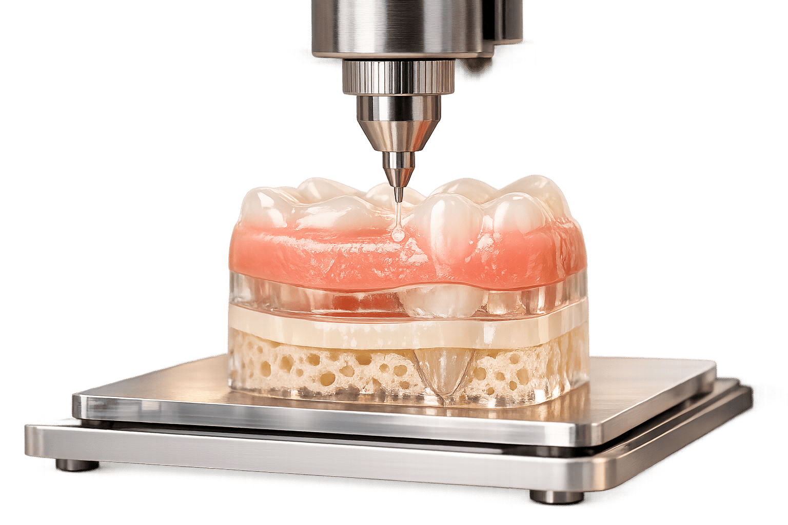
3D bioprinting is reshaping how we treat periodontal disease, which affects nearly half the global population. Unlike conventional methods that focus on repair, this technology uses bio-inks – made from living cells, biocompatible materials, and growth factors – to create patient-specific tissue constructs. This technology is also advancing dental implantology by providing customized structural support. These constructs mimic the natural structure of the periodontium, including gingiva, periodontal ligament, cementum, and alveolar bone.
Here’s why it matters:
- Global Impact: Severe periodontitis is predicted to affect 1.56 billion people by 2050.
- Limitations of Current Treatments: Traditional therapies have a 20–30% failure rate and struggle to regenerate complex tissue connections.
- 3D Bioprinting Advantages: Offers precision, patient-specific designs, and the ability to regenerate multiple tissue types simultaneously.
However, challenges like cost (bioprinters range from $250 to $200,000), mechanical stability, and regulatory hurdles remain. Researchers are working to improve vascularisation, scaffold design, and scalability to bring this technology into clinical practice.
JC 3D BIOPRINTING AND BIOFABRICATION TECHNIQUES IN DENTAL TISSUE ENGINEERING
sbb-itb-2be92ed
How 3D Bioprinting Works in Periodontal Regeneration
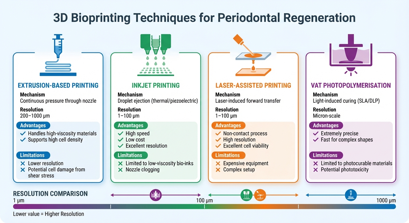
Comparison of 3D Bioprinting Techniques for Periodontal Regeneration
3D bioprinting for periodontal regeneration starts with capturing detailed images of the defect using clinical imaging techniques like CT and CBCT scans or MRI. These images are then transformed into CAD models and converted into STL files, which bioprinters can interpret [3][9].
Bio-Ink Preparation
Bio-inks combine biomaterials, living cells, and bioactive molecules into a printable solution [3]. Hydrogels, such as gelatin methacryloyl (GelMA) and sodium alginate, are commonly used to provide a structural framework. These are mixed with stem cells – typically at a density of 1 × 10⁷ cells per millilitre – along with growth factors like BMP-2 (5 µg/mL) and PDGF (100 ng/mL) [8].
In August 2023, researchers from Guangzhou Medical University developed a specialised bio-ink made of 15% GelMA, 10% sodium alginate, and 5% bioactive glass microspheres. This bio-ink, enhanced with mouse bone marrow mesenchymal stem cells and growth factors, was used to treat 5 mm × 5 mm periodontal defects in Beagle dogs. Within 8 weeks, the treatment successfully reconstructed periodontal structures, including gingiva, periodontal ligament, and alveolar bone [8].
Sodium alginate is often added to improve viscosity and ease of printing. High-viscosity bio-inks (30 to 6 × 10⁷ mPa s) are suitable for extrusion systems, while inkjet systems require lower viscosities (3.5 to 12 mPa s) [3]. The choice of bio-ink depends on the printing method and the desired tissue characteristics.
3D Printing Techniques
Once the bio-ink is prepared, different printing techniques can be employed to create the scaffold. Extrusion-based printing uses continuous pressure to deposit bio-ink layer by layer according to the CAD design. This method is versatile and supports high cell densities and viscous materials but offers lower resolution (200–1000 µm) and may expose cells to shear stress [3][5].
| Technique | Mechanism | Resolution | Advantages | Limitations |
|---|---|---|---|---|
| Extrusion | Continuous pressure through nozzle | 200–1000 µm | Handles high-viscosity materials; supports high cell density [3][5] | Lower resolution; potential cell damage from shear stress [3][5] |
| Inkjet | Droplet ejection (thermal/piezoelectric) | 1–100 µm | High speed; low cost; excellent resolution [3][5] | Limited to low-viscosity bio-inks; nozzle clogging [3] |
| Laser-Assisted | Laser-induced forward transfer | 1–100 µm | Non-contact; high resolution; excellent cell viability [3][5] | Expensive equipment; complex setup [3][5] |
| Vat Photopolymerisation | Light-induced curing (SLA/DLP) | Micron-scale | Extremely precise; fast for complex shapes [3][5] | Limited to photocurable materials; potential phototoxicity [3] |
After printing, the scaffold undergoes crosslinking to maintain its structure. Techniques like calcium chloride treatment for alginate or UV light (365 nm) for GelMA are commonly used [3][8]. Using biocompatible photoinitiators like LAP ensures cell viability remains above 90% [3]. Advanced scaffolds may feature multiple layers of bio-ink, each tailored to mimic specific tissue zones such as cementum, periodontal ligament, and alveolar bone [4].
Integration into Periodontal Defects
Before implantation, the scaffold is matured in vitro to promote cell growth and differentiation. During surgery, its customised design ensures a perfect fit with the patient’s anatomy, supporting adhesion, tissue regeneration, and revascularisation [4][8].
The scaffold’s design plays a vital role in integration. High porosity and interconnected pores promote cell communication and bonding with surrounding tissues [3]. Features like grooves or strand spacing guide the alignment of periodontal ligament fibres, ensuring they attach correctly to both the root and bone. As Arwa Daghrery from the University of Michigan notes:
"Because functional PDL attachment is defined not only by tissue presence but by fibre organisation and insertion, incomplete or misdirected regeneration can result in ankylosis [or] root resorption" [4].
To further enhance healing, some scaffolds are coated with bioactive materials or ion-releasing ceramics (e.g., magnesium, strontium, calcium-phosphates). These stimulate local bone growth and blood vessel formation. The scaffold’s degradation rate is carefully synchronised with the tissue’s healing timeline, providing support during critical phases and gradually transferring load to the regenerating tissue [2][4].
Applications in Periodontal Tissue Regeneration
3D bioprinting is making strides in regenerating the different components of the periodontium – namely, the periodontal ligament, alveolar bone, and gingival tissue. Each approach is specifically designed to address the unique challenges posed by these diverse tissue types.
Periodontal Ligament Regeneration
The periodontal ligament (PDL) is a thin, specialised fibrous tissue that connects the cementum to the alveolar bone. To regenerate this structure, scientists encapsulate human periodontal ligament stem cells (hPDLSCs) or fibroblasts in bio-inks like GelMA, collagen, or decellularised extracellular matrix. Techniques such as Melt Electrowriting (MEW) are used to create scaffolds with directional fibres that mimic the natural alignment of PDL, ensuring proper integration and avoiding complications like ankylosis or root resorption [2].
In a notable study from May 2021, researchers at Seoul National University and T&R Biofab Co. bioprinted hPDLSCs with 5% collagen onto 3D-printed titanium scaffolds. After six weeks in a rat calvarial defect model, the bioprinted group achieved a 91.2% soft tissue-implant interface, compared to just 9.4% in the cell-seeded group. The bioprinted PDL-like tissue also expressed key functional markers such as periostin, vWF, and CEMP1 [11]. For optimal results, bioprinting typically uses a 12.5% GelMA solution with a 0.05% LAP photoinitiator, extruded at 135 kPa through a 25 G needle, maintaining over 90% cell viability [10][3].
Alveolar Bone Regeneration
3D bioprinting facilitates scaffold-guided bone regeneration (SGBR) by creating customised frameworks that combine synthetic polymers like polycaprolactone (PCL) with bioactive ceramics such as hydroxyapatite and β-TCP. These scaffolds provide mechanical support while incorporating growth factors like BMP-2 and living cells, such as osteoblasts or mesenchymal stem cells, to actively promote healing. Achieving an overall porosity of 70% with pore sizes between 150 μm and 500 μm ensures proper vascularisation and nutrient diffusion [13].
In December 2024, a clinical team led by Sašo Ivanovski at the University of Queensland used a 3D-printed PCL scaffold for alveolar ridge augmentation in a 46-year-old male. This resulted in a volumetric bone gain of 364.69 ± 2.53 mm³, equating to 108.4% of the original defect volume. The subsequent dental implant achieved primary stability at 35 Ncm⁻¹, with only a 2.4° deviation from the planned position [12]. Unlike traditional bone grafting, which often involves time-consuming shaping and secondary donor-site surgery, 3D-printed scaffolds are pre-designed for precise anatomical fit, streamlining the surgical process and reducing patient recovery time [12][13].
Gingival Tissue Repair
Bioprinting is also tailored to the specific needs of gingival tissue regeneration. Constructs are created using gingival fibroblasts combined with collagen and extracellular matrix (ECM) proteins. Multi-component hydrogels, such as GelMA and sodium alginate, provide the necessary mechanical stability, while growth factors like VEGF and PDGF encourage vascularisation and soft tissue repair [1][14].
In 2023, researchers led by Guohou Miao and Xuechao Yang developed a composite bio-ink containing GelMA, sodium alginate, and bioactive glass microspheres, loaded with mouse bone marrow mesenchymal stem cells and growth factors (BMP2 and PDGF). When applied to periodontal defects in Beagle dogs, the study achieved complete regeneration of gingival tissue, periodontal ligament, and alveolar bone within 8 weeks [14]. Similarly, in February 2024, another study demonstrated the effectiveness of an extrusion-based bio-ink made from acellular dermal matrix, gelatin, and sodium alginate, seeded with gingival fibroblasts. This approach significantly improved keratinised gingiva in vivo, showing promise for oral soft tissue repair [6].
These advancements highlight the potential of 3D bioprinting to transform periodontal care by offering precise, patient-specific solutions for tissue regeneration. However, ongoing research is essential to refine these techniques and fully assess their long-term impact.
Benefits and Limitations of 3D Bioprinting for Periodontal Regeneration
Benefits
3D bioprinting offers a level of precision and personalisation that far surpasses traditional methods. Using patient-specific CAD models, it creates scaffolds tailored to fit perfectly, removing the need for manual adjustments during surgery [2].
One of its standout features is the ability to regenerate multiple periodontal components simultaneously. By producing multiphasic and anisotropic scaffolds, the technology replicates the intricate cementum-PDL-bone interfaces. This is a challenge for traditional methods like passive barrier membranes used in Guided Tissue Regeneration (GTR) [2]. Instead of merely controlling infection, as seen in conventional scaling and root planing, bioprinting focuses on reconstructing all periodontal components functionally. The controlled microenvironment allows precise adjustments to porosity, interconnectivity, and pore size, fostering better cell communication and integration with surrounding tissues [2].
Another significant advantage is the high cell viability rates. Laser-assisted techniques, for instance, achieve cell viability exceeding 95%, which directly contributes to improved outcomes for periodontal regeneration by combining anatomical precision with biological compatibility [3].
"Compared with conventional methods of tissue engineering, a 3D-printed scaffold holds many advantages, such like high fidelity, customised topographies, and superior production efficiency" [6].
- Fengxiao Zhao, West China Hospital of Stomatology
Limitations
Despite its promise, 3D bioprinting faces several hurdles. One of the most glaring issues is cost. High-end bioprinters can cost up to $200,000, making them inaccessible to many institutions [16]. While more affordable options, like Stanford University’s "Printess" bioprinter priced at $250, exist, these are primarily limited to research settings rather than clinical applications [16].
"The major demerit is the time and cost spent which renders the justified use of this technology in complex cases only" [17].
- Meisha Gul, Aga Khan University Hospital
Technical and biological challenges also pose significant barriers. Achieving reliable vascularisation in the bacteria-rich oral environment is particularly challenging. Mechanical stability over time remains an issue, as seen in a 2025 case where a patient-specific PCL scaffold failed after 14 months due to degradation [2][4]. Natural hydrogels often lack the necessary mechanical strength, while synthetic polymers like PLA and PLGA can release acidic by-products during degradation, causing local inflammation [6][15]. Additionally, cell viability can be compromised by high shear stress during extrusion or phototoxicity from UV light used in some printing methods [3][5].
Another roadblock is the lack of standardised regulatory frameworks for constructs containing living cells, which complicates the transition from lab research to clinical practice.
"3D printing is advancing SGPR toward functional, personalised therapies; however, its translation depends on reliable vascularisation, immune modulation, long-term mechanics, scalable manufacturing, and clear regulatory and safety pathways" [2].
- Arwa Daghrery, Jazan University
For 3D bioprinting to achieve widespread clinical use, these challenges must be addressed, ensuring the technology can meet its full potential.
Future Developments in 3D Bioprinting for Periodontal Care
Researchers are working to tackle one of the biggest hurdles in 3D bioprinting for periodontal care: vascularisation. A key approach being explored is the creation of prevascularised constructs. By co-printing endothelial cells, such as HUVECs, alongside dental stem cells within hydrogel bio-inks, scientists aim to establish functional vascular networks either before or during implantation [4][6]. Advances like electrohydrodynamic (EHD) jetting have made it possible to fabricate microscale structures with resolutions as fine as 1 μm, closely mimicking natural vascular networks [3]. Some scaffold designs even include "lotus-root-mimetic" microchannels – parallel, interconnected channels that allow for faster blood vessel growth and better nutrient transport [4]. These developments are helping to refine biochemical signalling for tissue regeneration.
Another exciting area of progress involves the integration of growth factors. Future scaffolds are expected to use spatiotemporal systems to deposit growth factors layer by layer, replicating the complex structure of the periodontium. For example, tri-layered scaffolds could be functionalised with cementum protein 1 for the cementum layer, fibroblast growth factor 2 (FGF‑2) for the periodontal ligament, and platelet-rich plasma–derived growth factor for the alveolar bone [2]. This precise delivery system ensures that each tissue type receives the right regenerative signals at the right time during the healing process.
A growing trend in the field is the move toward cell-free therapeutics. Instead of using living cells, researchers are loading scaffolds with small extracellular vesicles (sEVs) derived from mesenchymal stem cells. These vesicles provide sustained angiogenic signalling with a lower risk of immune rejection [18]. Additionally, ion-releasing ceramics containing magnesium, strontium, and calcium phosphates are being developed to chemically stimulate both bone and blood vessel growth [4].
Despite these technical breakthroughs, broader clinical adoption faces several barriers. For instance, a PubMed survey conducted in September 2025 identified 1,863 records on 3D printing for periodontal regeneration, demonstrating steady research growth since 2003 [2]. However, challenges remain, including scalable manufacturing, standardised regulatory frameworks for constructs containing cells, and creating workflows that integrate CT imaging with automated biofabrication [2][4]. Emerging innovations like in situ bioprinting – where constructs are printed directly into the periodontal defect during surgery – and 4D bioprinting using smart materials that respond to inflammatory signals, show great promise for further advancements in periodontal care [2][7].
While the field continues to address issues like vascularisation, immune response, and regulatory hurdles, these advancements are driving periodontal bioengineering closer to widespread clinical application. The future of 3D bioprinting in this area is rich with potential, but real-world adoption will depend on systematically solving these challenges.
Conclusion
3D bioprinting is reshaping the way periodontal diseases are treated. Unlike conventional approaches like scaling or passive grafting, this cutting-edge technology focuses on restoring the intricate, layered structure of the periodontium. By using living cells, biocompatible materials, and growth factors, it creates patient-specific constructs that closely replicate the natural interface of the gingiva, periodontal ligament, cementum, and alveolar bone [1][3].
The need for such advancements is pressing. Severe periodontitis is expected to affect around 1.56 billion people globally by 2050 [1]. As Jahnavi R Acharya from Karnavati School of Dentistry explains:
"Integrating 3D bioprinting into periodontal regenerative therapies could revolutionise clinical practices, offering more effective, tailored, and sustainable solutions to address the challenges of periodontal disease" [1].
The potential doesn’t stop here. Beyond current 3D bioprinting techniques, emerging technologies like 4D bioprinting are paving the way for even greater precision. These constructs can adapt their shape or function over time, while in situ bioprinting allows tissue to be printed directly into periodontal defects during surgery [2][4]. Machine learning is also stepping in, improving scaffold design and fine-tuning bioink formulations to meet individual patient needs [2][4].
However, challenges remain. For 3D bioprinting to become a standard in clinical practice, hurdles like reliable vascularisation, long-term mechanical stability, scalable production, and regulatory clarity must be addressed [1][4]. Tackling these issues will be crucial in shifting periodontal care from reactive treatments to proactive tissue regeneration. Encouragingly, research continues to advance, with over 1,863 published studies as of September 2025, bringing the field closer to transforming periodontal treatment [2].
FAQs
Is 3D bioprinting for gum disease available in Australia yet?
3D bioprinting for periodontal regeneration is still in the research and development phase. Although progress in this field looks promising for potential clinical applications down the track, it hasn’t yet become a standard treatment option in Australia.
What risks come with bioprinted periodontal tissues?
One of the key challenges in bioprinting periodontal tissues is ensuring proper vascularisation, especially for larger tissues. Without adequate blood vessel formation, the tissue may struggle to regenerate effectively or integrate seamlessly with the surrounding biological structures.
Another concern lies in the use of UV light during the bioprinting process. While UV exposure is often necessary, improper optimisation can harm cellular health, potentially causing damage to the cells and compromising the overall success of the bioprinted tissue.
How long does a bioprinted scaffold last in the mouth?
The lifespan of a bioprinted scaffold can range from several months to a few years. This depends on various factors, such as the type of material used, how well it integrates with biological tissues, and the conditions within the oral environment. However, research is still ongoing to gather precise data about its durability.
Related Blog Posts
- Stem Cells in Periodontal Regeneration: Current Trials
- Recent Advances in Biomaterials for Periodontal Regeneration
- How 3D Bioprinting Creates Periodontal Scaffolds
- Challenges in Dental 3D Bioprinting
Important Notice: Any surgical or invasive procedure carries risks. Before proceeding, you should seek a second opinion from an appropriately qualified health practitioner.
Individual results may vary. The information provided in this article is for educational purposes only and does not constitute medical advice.
Checkout Related Blogs




Get in touch with us
For more information, call us now to start feeling better. Or fill the form below to make appointment
The Latest News from Complete Smiles
How to Clean Clear Plastic Retainers
Checklist for Choosing Wearable Dental Devices
Checklist for Choosing Cloud AI Platforms in Dentistry

Complete Smiles Bella VistaAccepts All Major Health Funds, Including





