





Improving Fracture Resistance in Dental Nanocomposites
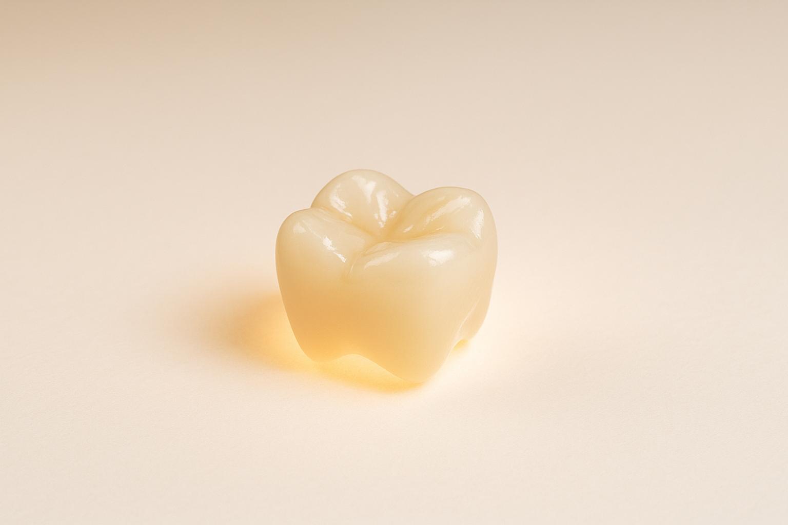
Dental nanocomposites are modern materials used in restorative dentistry to mimic natural teeth. While they offer benefits like better aesthetics and strength, fractures remain a common issue. This article explores how to improve their fracture resistance through:
- Nanoparticle properties: Small, evenly distributed particles enhance strength, but excessive filler can weaken the material.
- Silanisation: Chemical treatment strengthens bonds between nanoparticles and the polymer matrix, reducing crack propagation.
- Short fibre reinforcement: Adding fibres boosts durability, especially for teeth under heavy stress.
- Layered structures: Combining fibre cores with aesthetic surface layers balances strength and appearance.
Improving these materials can extend the lifespan of dental restorations, reduce failure rates, and enhance patient outcomes in Australian clinics.
How Fractures Occur in Nanocomposites
Understanding Fracture Mechanisms
Fractures in nanocomposites often begin within the polymer matrix, where the interaction between nanoparticles can either slow down or speed up the spread of cracks [3]. Two main processes influence how fractures develop: nanofibre pull-out and crack front bowing. In nanofibre pull-out, elongated nanoparticles are pulled from the matrix, which absorbs energy and helps to resist the crack. In crack front bowing, particles with low aspect ratios force the crack to curve, dissipating energy as it propagates [3].
The behaviour of polymer chains also plays a part. As the polymer stretches and deforms around the nanoparticles, it forms an interphase – a layer that buffers the rigid nanoparticles and evenly distributes stress. This highlights the importance of nanoparticle properties in fracture resistance.
External factors, such as chewing and temperature changes, add to the complexity. Chewing applies forces ranging from 30 to 2,900 N at a frequency of 0.4 to 12 Hz [7], which can lead to fatigue damage over time. Additionally, daily thermal cycling – 20 to 50 cycles of expansion and contraction – creates stresses that can initiate cracks [7].
Impact of Nanoparticle Properties
The size, shape, distribution, and surface treatment of nanoparticles directly influence how nanocomposites respond to fractures. Smaller nanoparticles, for instance, provide a larger surface area for interaction with the polymer matrix, which can enhance hardness and compressive strength [4]. Studies show that low filler content (0.1% to 1.5%) can significantly improve fracture toughness – sometimes by hundreds of per cent – but excessive filler can actually weaken the material [3].
Even distribution of nanoparticles is equally important. When nanoparticles clump together, the reduced interfacial area limits stress transfer between the matrix and the fillers, weakening the composite’s resistance to fractures [3].
The shape of the nanoparticles also matters. For example, silicon nitride whiskers offer better strength and toughness compared to silicon carbide whiskers. Meanwhile, silanated carbon nanotubes can boost the flexural strength of composite resins by around 23%, although their dark colour may impact the material’s appearance [6].
| Filler Property | Effect on Fracture Behaviour | Clinical Impact |
|---|---|---|
| Large particle size | Poor wear resistance, increased crack propagation | Early failure, reduced polishability, discolouration |
| Smaller particles | Better wear resistance, increased matrix viscosity | Improved aesthetics, though polymerisation may decrease |
| Whisker shape | Greater strength and toughness | Enhanced ability to bear loads |
| Nanotube geometry | Stronger flexural strength | Better resistance to bending forces |
Surface treatments like silanation further enhance fracture resistance by strengthening the bond between nanoparticles and the polymer matrix. This improves interfacial toughness and increases the energy absorbed during a fracture [3].
Real-world examples back these findings. For instance, adding just 1% by weight of a multiwall carbon nanotube (MWCNT)-MXene hybrid filler to an epoxy matrix increased fracture toughness from 0.97 MPa·m¹ᐟ² to 1.79 MPa·m¹ᐟ² – an 85% improvement due to mechanisms like crack deflection and filler debonding [3]. Similarly, surface-treated carbon nanotubes have shown improvements of 85% to 145% in Mode II interlaminar fracture toughness in epoxy/carbon fabric laminates [3].
To maximise durability in dental restorations, achieving an even dispersion of nanoparticles is essential. When nanoparticles are uniformly distributed and firmly bonded, they create a network that resists crack propagation, significantly improving the composite’s overall strength and longevity.
Fracture toughness and Impact Strength | Dental Materials
Methods to Improve Fracture Resistance
Improving the fracture resistance of dental nanocomposites involves strengthening filler-resin bonding and fine-tuning the integration of nanoparticles and fibres. This allows the material to better handle chewing forces and temperature changes.
Interface Engineering and Silanisation
Silanisation plays a crucial role in creating strong chemical bonds between nanoparticles and the polymer matrix, which significantly boosts fracture resistance.
"Theoretical results indicated that silanisation and nanoparticle loadings improved the fracture toughness of dental nanocomposites by a factor of 2 to 3 through a combination of enhanced interface toughness by silanisation, crack deflection, as well as crack bridging." [8]
Different silane coupling agents produce varying levels of effectiveness. For instance, a study in Dental Materials (March 2023) explored the impact of silane treatment on S-2 Glass fibres in dental composites. It found that composites treated with 8MOTS at an optimal concentration showed a 51.9% and 15.9% improvement in fracture toughness compared to those treated with 3GPS and 3MPS, respectively [9]. Untreated fibres, by contrast, performed poorly, with fracture toughness values 81.6%, 38.6%, and 110.5% lower than those treated with 3MPS, 3GPS, and 8MOTS, respectively [9].
Proper silanisation not only enhances filler dispersion but also strengthens the composite’s mechanical properties. This prevents crack propagation by forming robust interfacial bonds that resist stress concentration.
Optimising Nanoparticle Loading
The right amount of nanoparticles can make all the difference. Overloading leads to clumping, while optimal levels improve mechanical performance. For example, a 1% silica nanoparticle loading increases indentation force by 35%, along with a 21.15% boost in flexural modulus and a 19.20% increase in strength compared to reference samples [10].
"By incorporating nanofillers and nanostructures, composites offer a wide array of benefits, including enhanced mechanical properties, improved aesthetics, and superior antibacterial capabilities." [5]
Achieving uniform nanoparticle dispersion is essential. Clusters of agglomerated particles create weak points that can lead to fractures. Once this balance is achieved, the foundation is set for reinforcing composites with short fibres.
Adding Short Fibres
Short fibre reinforced composites (SFRCs) address the issue of low fracture toughness found in traditional particulate filler composites. These fibres act as barriers, absorbing energy and redirecting cracks away from critical areas.
A study by Garoushi et al. demonstrated the effectiveness of SFRCs. Restorations made from conventional particulate filler composites had a load-bearing capacity of 1,081 N, while those using SFRCs handled 1,733 N [12]. Even with a surface layer of particulate filler composite over the SFRC core, the restorations achieved a load-bearing capacity of 1,405 N, significantly outperforming the control group [12].
SFRCs are particularly valuable for teeth with structural damage. For instance, losing just one marginal ridge reduces tooth rigidity by 46% in premolars, while a mesio-occluso-distal (MOD) cavity can cause a 63% reduction [11].
"Within the limitations of this review, in vitro evidence suggests that short-fibre-reinforced composite materials effectively reinforce structurally compromised teeth." [11]
By deflecting or absorbing crack energy, short fibres slow crack propagation and improve durability.
Layered Structures for Added Strength
Layered structures combine a short fibre core with a particulate filler surface, striking a balance between strength and aesthetics.
"The function of bulk short fibre composite substructure is based on supporting the surface particulate filler composite layer and working as crack stopper layer." [12]
This approach leverages the strength and toughness of the fibre-reinforced core while maintaining a polished, natural appearance with the surface layer. The outer layer not only enhances aesthetics but also ensures better polishability and colour matching.
This design is especially effective for large restorations, where both structural support and a natural look are essential. The fibre-reinforced core provides the durability to handle chewing forces, while the surface layer ensures the restoration blends seamlessly with natural teeth.
sbb-itb-2be92ed
Comparing Different Nanocomposite Systems
Advancing earlier methods to boost fracture resistance, this section takes a closer look at how various nanocomposite systems perform. Gaining insight into these systems helps dental professionals choose the best materials for specific clinical needs. Each system comes with its own strengths and limitations, impacting both immediate results and long-term reliability.
Nanohybrid vs Fibre-Reinforced Composites
When it comes to fracture resistance, there’s a noticeable difference between nanohybrid and fibre-reinforced composites. Research from Saraswati Dental College in Lucknow revealed that EverX Posterior, a fibre-reinforced composite, achieved a mean fracture resistance of 697.62 ± 20.40. In comparison, the nanofiller composite Tetric N-Flow reached 439.16 ± 33.53 – a 58.8% improvement [2].
E-glass fibres in fibre-reinforced composites play a crucial role by absorbing and evenly distributing forces. This creates a monoblock effect with dentine, reducing stress [13]. On the other hand, while nanohybrid composites are strong, they tend to show lower fracture resistance under high-stress conditions [13].
Clinical studies further highlight these differences. A 24-month randomised controlled trial found that nanofilled composites had an average wear depth of 19.4 μm, compared to 49.6 μm for conventional composites [17]. However, short glass fibre-reinforced composites can present challenges with colour matching compared to nanohybrid options [18].
EverX Posterior, a premixed fibre-reinforced composite used alongside conventional enamel composites, has shown superior fracture resistance compared to systems that require manual fibre placement [13] [14]. Additionally, clinical trials have demonstrated that direct fibre-reinforced resin composite Class II MOD restorations can perform as well as nanohybrid resin composites over an 18-month period [18].
Pros and Cons of Reinforcement Methods
Here’s a quick breakdown of the advantages and challenges of different reinforcement methods:
| Reinforcement Method | Advantages | Challenges | Ideal Use Cases |
|---|---|---|---|
| Nanohybrid Composites | Great aesthetics and colour matching; easy to apply; excellent surface finish | Lower fracture resistance; less effective in high-stress areas; may need more frequent replacements | Anterior restorations; small to medium cavities; areas where appearance matters most |
| Short Fibre-Reinforced | 58.8% higher fracture resistance; excellent force distribution; isotropic reinforcement | Colour matching issues; more technique-sensitive; requires a covering layer for aesthetics | Posterior restorations; large MOD cavities; high-stress areas |
| Layered Fibre Systems | Combines strength and aesthetics; prevents crack propagation; supports surface layers well | Complex application; higher material costs; requires precise technique | Extensive restorations; weakened teeth; load-bearing restorations |
Longevity is another critical factor in selecting a composite system. Composite restorations typically last between 5 and 10 years [15], but up to half of them may fail within a decade due to issues like secondary caries or fractures [16]. This highlights the importance of the enhanced fracture resistance offered by fibre-reinforced systems, particularly for long-term outcomes.
Ease of application also matters. Nanohybrid composites are generally straightforward to use, while fibre-reinforced systems – especially those requiring manual fibre placement – can demand more time and skill [18]. Premixed options like EverX Posterior have made this process significantly simpler.
Maintaining marginal integrity and retention is essential to prevent microleakage, recurrent caries, and restoration failure. Choosing the right composite system involves balancing aesthetic appeal with functional durability. Fibre-reinforced systems are especially suitable for posterior restorations that need high fracture resistance, while nanohybrid composites are often the first choice for anterior restorations where appearance is the priority. Selecting the right material, along with advanced treatment techniques, plays a key role in extending the lifespan of restorations.
Clinical Applications and Local Insights
Bridging the gap between laboratory research and clinical practice demands a close look at the specific challenges faced by Australian dental clinics. With dental service spending in Australia reaching around $8.7 billion in 2012–13 [19], the choice of durable, fracture-resistant materials becomes a crucial decision for both patients and practitioners.
Personalised Treatment Planning
The successful use of fracture-resistant nanocomposites starts with a detailed evaluation of each patient’s unique needs. Key considerations include factors like age, cavity size, the amount of remaining tooth structure, and the location of the cavity [19]. According to Australian survey data, caries risk is the primary factor influencing treatment choices [20], with secondary caries rates for composite resin restorations reported between 50% and 60% due to micro-leakage [19]. This highlights the need for nanocomposites with improved marginal seal properties, especially for patients at higher risk.
For high caries-risk patients, antimicrobial nanocomposites that prevent biofilm formation may be the best option [1]. On the other hand, patients with lower risk might prioritise aesthetics, making nanohybrid systems more suitable. The high surface area to volume ratio of nanomaterials [19] allows dental professionals to customise restorative materials by tweaking nanoparticle composition and the polymeric matrix [1]. This means practitioners can focus on strength for posterior teeth or aesthetics for anterior restorations, depending on the patient’s needs.
These individualised assessments enable modern clinics to effectively integrate nanocomposite materials into their treatment strategies.
Role of Advanced Dental Clinics
Modern Australian dental clinics are increasingly adopting advanced diagnostic tools and tailored treatment protocols to optimise the performance of restorations. These practices emphasise comprehensive evaluations and long-term monitoring to ensure the best outcomes for patients.
Take, for instance, Complete Smiles Bella Vista, led by Dr. James Hanna. This clinic combines personalised treatment planning with thoughtful material selection, applying nanocomposite systems in procedures like porcelain crowns, dental implants, and broader restorative care. Their approach reflects how evidence-based material selection can be seamlessly integrated into everyday practice.
The shift towards minimally invasive dentistry in Australia is also shaping how nanocomposites are used [20]. Many clinics now prioritise preserving tooth structure, leveraging the superior bonding and fracture-resistant properties of modern materials. However, professional development remains essential. Survey data show that 55% of dentists believe repaired composite restorations have a worse prognosis compared to replaced ones [20], highlighting the importance of ongoing training.
Australia’s diverse population also brings unique challenges. Communication about nanocomposite options must account for varying health literacy levels and cultural attitudes. Cost and access to advanced equipment can be obstacles for some practices [20]. Yet, the long-term advantages of using fracture-resistant nanocomposites – such as fewer replacements and higher patient satisfaction – often outweigh the initial costs, making them a worthwhile investment for many clinics and patients.
Conclusion: Strengthening the Future of Dental Restorations
Fracture-resistant dental nanocomposites represent a remarkable intersection of materials science and clinical progress. By leveraging techniques like interface engineering and silanisation, researchers have achieved breakthroughs in fracture toughness, improving it by two to three times compared to earlier materials [21]. Additionally, incorporating amorphous calcium and phosphate ions into nanocomposites has doubled the flexural strength and elastic modulus when compared to traditional composites [5]. These advancements set the stage for further progress in the field.
Recent studies highlight the potential of these innovations. For example, Wu et al. (2015) demonstrated that dental composites with antimicrobial, remineralisation, and self-healing properties could recover 65%–81% of their original fracture toughness while retaining their mechanical integrity [5]. Similarly, Mitwalli et al. (2022) introduced rechargeable nCaF₂ composites, which provide sustained fluoride and calcium ion release – key factors for supporting remineralisation [5].
In Australia, these advancements translate to tangible benefits for patients. Approximately 60% of composite resin restorations in the country last over a decade, reflecting the durability of modern materials [1]. Collaboration between material scientists and dental professionals remains essential to ensure that research findings lead to better material choices and optimised treatment outcomes [23].
These innovations also align with Australia’s growing emphasis on bio-based and sustainable healthcare solutions. Such an approach balances environmental responsibility with patient safety and treatment longevity [1]. However, it is equally important to address challenges such as potential cytotoxicity, long-term stability, and economic feasibility [1]. With tooth decay affecting about 3.5 billion people globally [22], the need for continued advancements in dental materials is undeniable.
The future of dental restorations lies in systems that are not only stronger but also smarter, capable of adapting to the complex challenges of the oral environment. Through ongoing research, clinical trials, and the integration of advanced diagnostic technologies, fracture-resistant nanocomposites are poised to redefine dental care in Australia and beyond.
FAQs
What role do nanoparticles play in improving the fracture resistance of dental nanocomposites?
Nanoparticles significantly improve the fracture resistance of dental nanocomposites by boosting their mechanical strength, toughness, and ability to resist cracks. The durability of these materials hinges on key factors like nanoparticle size, surface treatment, and composition.
Take silanisation, for instance. This treatment, along with carefully optimised nanoparticle loadings, strengthens the bond between the nanoparticles and the resin matrix. The result? A tougher, more durable material that can handle the wear and tear of daily dental use, making it a dependable option for long-lasting dental restorations.
How does silanisation improve the strength and longevity of dental restorations?
Silanisation is a key step in boosting the strength and longevity of dental restorations. By forming a chemical bond between resin cements and ceramic or composite surfaces, it enhances adhesion and lowers the likelihood of fractures or debonding over time.
Additionally, silanisation shields restorations from the wear and tear caused by daily use and exposure to oral conditions. This protection ensures they stay functional and dependable for extended periods. By strengthening the bond at a molecular level, silanisation plays a vital role in creating more durable and resilient dental restorations.
How do short fibre reinforcements and layered structures improve the durability of dental nanocomposites in high-stress areas?
Short fibre reinforcements play a key role in improving the durability of dental nanocomposites. They work by deflecting cracks and distributing stress more evenly throughout the material. This makes fractures less likely, even in parts of the mouth that endure intense chewing pressure.
Layered structures add another layer of protection by increasing mechanical strength and enhancing long-term reliability. These layers act as shields, offering extra resistance to wear and damage, which makes them particularly suitable for high-stress dental applications like molars or bridges.
Related Blog Posts
- Wear-Resistant Materials in Dental Restorations
- Wear Resistance of Polymer-Based Restoratives
- Advances in Dental Adhesives: What to Know
- Nanomaterials in Dentistry: Wear Resistance Explained
Important Notice: Any surgical or invasive procedure carries risks. Before proceeding, you should seek a second opinion from an appropriately qualified health practitioner.
Individual results may vary. The information provided in this article is for educational purposes only and does not constitute medical advice.
Checkout Related Blogs




Get in touch with us
For more information, call us now to start feeling better. Or fill the form below to make appointment
The Latest News from Complete Smiles
How to Clean Clear Plastic Retainers
Checklist for Choosing Wearable Dental Devices
Checklist for Choosing Cloud AI Platforms in Dentistry

Complete Smiles Bella VistaAccepts All Major Health Funds, Including





