





Electrochemical Properties of Implant Coatings Explained

When implants are placed in the body, they face a harsh environment that can lead to corrosion, weakening their structure and releasing harmful metal ions. This can cause inflammation, bone loss, or implant failure. Implant coatings, such as titanium dioxide or hydroxyapatite, act as protective barriers to prevent corrosion and support bone integration.
Key points:
- Corrosion Resistance: Coatings form passive layers (e.g., titanium dioxide) to block corrosion caused by chloride ions and pH fluctuations.
- Biocompatibility: Prevents metal ion release, reducing inflammation and supporting tissue growth.
- Material Choices: Titanium, hydroxyapatite coatings, and noble metals like gold or palladium offer varying benefits.
- Testing Methods: Techniques like Electrochemical Impedance Spectroscopy (EIS) and ion release testing ensure coatings perform well in Australia’s healthcare environment.
This article explores how these coatings work, their materials, and testing methods that ensure implants meet long-term performance expectations in demanding conditions.
Materials Science #036 – Corrosion in Medical Implants and the Electrochemical Theory, Demystified!
sbb-itb-2be92ed
How Electrochemical Behaviour Affects Implant Coatings
When an implant is placed in the body, it becomes part of an electrochemical system. Bodily fluids like saliva, blood plasma, and tissue fluids act as electrolytes, setting off surface reactions on the implant. These reactions are critical to understand because they determine whether the implant remains stable or starts to degrade over time.
The oral environment poses unique challenges. Saliva contains chloride ions, and its pH levels fluctuate frequently. Add to this the mechanical forces from chewing, and even the most corrosion-resistant materials face significant stress. Because of this, the electrochemical behaviour of implant coatings plays a key role in preventing the release of metal ions into surrounding tissues.
Corrosion and Passive Layer Formation
One of the most important electrochemical processes is the formation of a passive layer on metallic implants. This thin oxide film forms spontaneously on metals like titanium, cobalt-chromium, and stainless steel, providing both physical and chemical protection against corrosion. Even though it’s only a few nanometres thick, this layer is essential for shielding the implant from bodily fluids [4].
For titanium alloys, this passive layer consists mainly of titanium dioxide (TiO₂). In contrast, stainless steel and cobalt-based alloys develop films rich in chromium oxide (Cr₂O₃) [4]. These oxide layers help block chloride ions from initiating reactions with the metal underneath. However, the oral environment can still cause localised breakdowns in the passive layer, leading to issues like pitting and crevice corrosion, especially in areas where fluids don’t flow freely [4].
"The corrosion resistance of an implant material affects its functionality and durability and is a prime factor governing biocompatibility." – Materials (Journal) [4]
The durability of this passive layer directly impacts the lifespan of an implant. For example, surgical stainless steel is more prone to pitting and crevice corrosion in chloride-rich conditions compared to titanium-based or cobalt-chromium alloys [4]. During coating processes like calcium phosphate deposition, trapped dihydrogen bubbles can create porosities on the surface. These weaken the coating’s mechanical strength and reduce its ability to shield the metal underneath [3]. Using pulsed current during deposition can help release these bubbles, resulting in smoother, less porous, and more corrosion-resistant coatings [3].
Biocompatibility and Electrochemical Reactions
Electrochemical reactions at the implant surface influence not only its structural integrity but also its compatibility with surrounding tissues. When corrosion is excessive, released metal ions can disrupt the local tissue environment, potentially causing inflammation, bone loss, or even implant failure [4].
"The fundamental paradigm of metallic biomaterials, except biodegradable metals, has been ‘the more corrosion resistant, the more biocompatible.’" – Materials (Journal) [4]
The pH at the interface between the implant and tissue also plays a key role in electrochemical stability. For instance, during coating synthesis, a rise in local pH can promote the formation of bioactive phases like hydroxyapatite, which supports bone integration [3].
In dental implants, the combination of electrochemical corrosion and mechanical wear from chewing gives rise to biotribocorrosion. This process damages the passive layer, exposing fresh metal and speeding up ion release [4]. To ensure implants last in such a demanding environment, they need coatings that offer both strong electrochemical and mechanical protection.
Common Materials for Implant Coatings
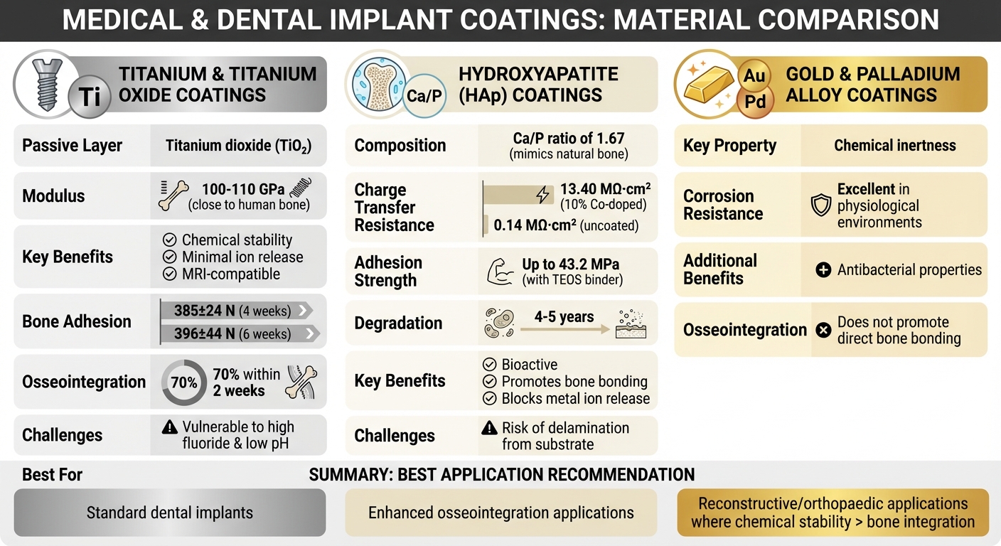
Comparison of Implant Coating Materials: Titanium, Hydroxyapatite, and Noble Metals
When it comes to implant coatings, three key categories stand out: titanium-based materials, ceramic coatings like hydroxyapatite, and noble metal alloys. Each offers distinct advantages in terms of electrochemical properties and clinical performance.
Titanium and Titanium Oxide Coatings
Titanium has become the benchmark for dental implants, thanks to its ability to form a stable titanium dioxide (TiO₂) layer. This passive film ensures chemical stability while minimising the release of metallic ions, which helps reduce toxicity risks [6][1].
"Titanium and its alloys have emerged as a material of choice for dental implants fulfilling all requirements biologically, chemically and mechanically." – Journal of Oral Biosciences [7]
With a modulus of around 100–110 GPa – closer to that of human bone – titanium provides excellent compatibility. Its non-ferromagnetic properties also make it ideal for MRI and radiographic imaging [5][7].
Researchers continue to refine titanium’s performance. For instance, a study in May 2021 highlighted a thermochemical treatment that created a sodium titanate gel layer on titanium implants. This modification, led by Javier Gil from the Bioengineering Institute of Technology, achieved impressive results: bone-implant adhesion forces of 385 ± 24 N at four weeks and 396 ± 44 N at six weeks in porcine models, with 70% osseointegration reached within two weeks [6].
However, the durability of the titanium oxide layer can be challenged by high fluoride concentrations (like those in some toothpastes) and low pH environments, potentially causing localised corrosion [7][8]. While the Ti-6Al-4V alloy offers enhanced mechanical strength, it may release vanadium and aluminium ions over time, which raises biocompatibility concerns. For this reason, commercially pure titanium (CpTi) is often preferred [5][1].
Next, ceramic coatings such as hydroxyapatite bring additional bioactive features to the table.
Hydroxyapatite Coatings
Hydroxyapatite (HAp) is well-known for its ability to mimic natural bone, with an ideal calcium-to-phosphorus molar ratio of 1.67 [9][10]. This bioactive property fosters a strong connection between the implant and bone.
"The ionic exchange between the biomaterial and the organism is the key factor for a material to be bioactive and not biostable." – Javier Gil, Bioengineering Institute of Technology [6]
HAp serves as both a physical and electrochemical barrier, preventing the release of harmful metallic ions like nickel, chromium, aluminium, and vanadium [9][10]. In one study, a 10% cobalt-doped HAp coating showed a charge transfer resistance of 13.40 MΩ·cm², compared to just 0.14 MΩ·cm² for an uncoated Ti-6Al-4V substrate [11].
The porous nature of HAp supports cell migration, vascularisation, and mechanical interlocking with bone. Depending on its crystallinity and porosity, the coating gradually degrades over four to five years through natural cellular processes [9]. While plasma spraying is popular for its efficiency, electrochemical deposition offers greater control over coating thickness and operates at lower temperatures [10].
One challenge with HAp coatings is their tendency to delaminate from the metallic substrate. Solutions include using binders like tetraethyl orthosilicate (TEOS), which can increase adhesion strength to 43.2 MPa [11], or adding intermediate layers like TiO₂ nanoporous coatings to improve bonding [10].
For scenarios where chemical stability outweighs the need for osseointegration, noble metal coatings provide an alternative.
Gold and Palladium Alloy Coatings
Gold and palladium coatings enhance implant surfaces, particularly in reconstructive and orthopaedic applications [12]. Their standout feature is their chemical inertness, which offers excellent corrosion resistance in the body’s harsh physiological environment [12].
These coatings are often applied to traditional implant materials – such as titanium, stainless steel, or ceramics – to address issues like surface degradation and limited bioactivity [12][3]. Beyond their electrochemical stability, noble metal coatings and nanoparticles also exhibit antibacterial properties, reducing the risk of infection at the implant site [12].
While gold and palladium alloys excel in resisting corrosion, they do not encourage direct bone bonding like hydroxyapatite does. As a result, they are better suited for applications where chemical inertness is more critical than osseointegration [3][12]. This chemical stability plays a key role in extending the lifespan of implants.
Testing Methods for Coating Electrochemical Properties
Implant coatings go through extensive lab testing to assess their corrosion resistance, ion release, and stability of the protective layer. These evaluations are critical for ensuring the implants can endure Australia’s unique oral environment and maintain their durability and wear resistance over time.
Potentiodynamic polarisation (PDP) is a widely-used method that applies a gradually increasing electrical potential to provoke redox reactions. The resulting current forms a polarisation curve, which helps determine corrosion rates through Tafel slope extrapolation [13]. While effective, this approach can be destructive, as the high external voltages applied may damage the oxide film protecting the implant [13]. To complement PDP, researchers often turn to non-destructive methods like electrochemical impedance spectroscopy (EIS) for a more detailed view of ion resistance.
When it comes to non-destructive testing, EIS offers valuable insights into a coating’s ability to resist ion movement. In October 2021, researchers Duduzile Nkomo and Nomsombuluko Masia from Mintek examined Grade 4 titanium dental implants with both machined and roughened surfaces. The PDP results showed corrosion rates below 0.02 mm/year for both types, but EIS highlighted that machined titanium had a more stable passive layer. This was evident from its phase angles near –90° and impedance values around 10⁶ Ω·cm² [13][14].
"EIS is the most suitable method for monitoring corrosion rate values due to its reproducibility, it is non-destructive and has reliable determination of small corrosion rates, much lower than those measured by other techniques." – Duduzile Nkomo, Researcher, Mintek [13]
Ion release testing with ICP-MS (Inductively Coupled Plasma Mass Spectrometry) is another critical method, measuring how many metallic ions leach from implants into surrounding fluids. This technique relies on argon plasma heated to 8000–9000°C, allowing for the detection of ions at extremely low concentrations – down to parts per billion [15]. Testing often reveals that ion release increases significantly in acidic environments, which mimic the low pH caused by bacterial plaque. For instance, cobalt release from CoCr alloys can rise to 3600 ppb in such conditions, raising concerns since ions like cobalt and chromium may contribute to oxidative stress and inflammation [15][16]. This method is key to verifying the stability of coatings under simulated physiological conditions.
How Surface Coatings Extend Implant Lifespan
Surface coatings play a crucial role in improving implant longevity by addressing corrosion resistance, promoting tissue compatibility, and enhancing bone integration. Essentially, these coatings form a protective barrier between the metal implant and the corrosive oral environment, helping to prevent the release of harmful ions. The release of such ions has been linked to inflammation and bone loss, both of which can compromise implant success [2][1][17][18].
Advanced materials like titanium nitride (TiN) significantly enhance surface hardness – up to seven times more than uncoated titanium. Similarly, tantalum pentoxide (Ta₂O₅) coatings have been shown to reduce corrosion current density by approximately 65%. When combined with a biphasic calcium phosphate (BCP) layer, this reduction can reach around 80% [19]. These improvements in mechanical strength and corrosion resistance directly support better cellular attachment and integration with bone.
The electrochemical stability of these coatings also plays a key role in how well bone cells attach and grow on the implant surface. Biomimetic bioactive coatings, such as hydroxyapatite (HA) and calcium phosphate, go beyond forming a physical bond with bone tissue – they create a chemical connection that enhances integration [3][18]. To ensure durability, the US-FDA mandates that bioactive coatings on metallic implants must have an adhesion strength of at least 50 MPa [19].
These advancements in surface coatings are particularly important for Australian patients, given the rising rates of primary total hip arthroplasty and peri-implantitis. Projections indicate that primary total hip arthroplasty rates in Australia will increase by 66% between 2013 and 2046. Additionally, about 20% of patients may develop peri-implantitis within five years of implant placement, with one-quarter facing this condition after 11 years [17]. Coatings that combine corrosion resistance with antibacterial properties could help reduce the need for revision surgeries.
Another factor to consider is the implant’s Young’s modulus, which should closely match that of human bone (approximately <30 GPa). A mismatch can lead to stress shielding, a condition that causes bone resorption and implant loosening [18]. To address this, there is a growing preference for β-titanium alloys containing elements like niobium, tantalum, and zirconium. These alloys not only form more stable passive oxide layers but also better mimic the mechanical properties of natural bone compared to traditional Ti-6Al-4V alloys [18].
Some Australian dental and orthopaedic practices, such as Complete Smiles Bella Vista (Complete Smiles Bella Vista), are already incorporating these advancements to improve implant performance and extend their lifespan.
FAQs
How do implant coatings stop corrosion in saliva and blood?
Implant coatings play a crucial role in preventing corrosion caused by exposure to saliva and blood. They achieve this by forming protective passive layers on the implant’s surface. These layers minimise the release of metal ions and inhibit electrochemical reactions, which enhances both the implant’s resistance to corrosion and its compatibility with the body. This ultimately contributes to the durability and safety of implants over time.
What coating is best for bone bonding versus corrosion resistance?
Hydroxyapatite-based coatings are well-suited for bone bonding because they promote bioactivity and support osteointegration. For implants requiring enhanced corrosion resistance, ceramic coatings like Al₂O₃–YSZ work exceptionally well. These materials play a key role in improving the durability and functionality of implants.
What do EIS and ICP-MS tests tell you about a coating?
Electrochemical Impedance Spectroscopy (EIS) is used to analyse how well a coating can protect against corrosion. It does this by examining the coating’s barrier properties and its resistance. On the other hand, Inductively Coupled Plasma Mass Spectrometry (ICP-MS) measures the metal ions that are released from the coating or the substrate. This provides detailed information about corrosion or degradation processes. When combined, these methods offer a comprehensive way to evaluate the performance and durability of coatings.
Related Blog Posts
- Electrochemical Deposition for Titanium Implants
- Electrochemical Deposition for Implant Coatings
- How Hybrid Coatings Improve Osseointegration
- How Surface Coatings Prevent Implant Corrosion
Important Notice: Any surgical or invasive procedure carries risks. Before proceeding, you should seek a second opinion from an appropriately qualified health practitioner.
Individual results may vary. The information provided in this article is for educational purposes only and does not constitute medical advice.
Checkout Related Blogs




Get in touch with us
For more information, call us now to start feeling better. Or fill the form below to make appointment
The Latest News from Complete Smiles
How to Clean Clear Plastic Retainers
Checklist for Choosing Wearable Dental Devices
Checklist for Choosing Cloud AI Platforms in Dentistry

Complete Smiles Bella VistaAccepts All Major Health Funds, Including





