





Hydroxyapatite Coatings via Electrochemical Deposition
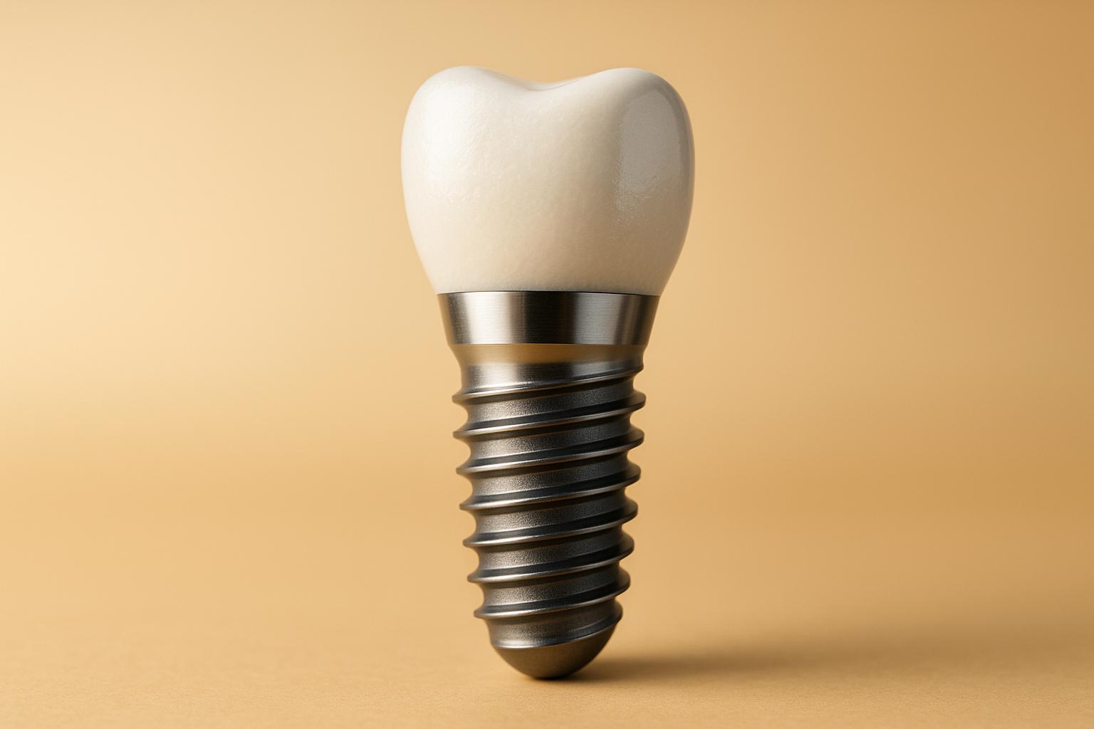
Hydroxyapatite (HA) coatings, created through electrochemical deposition, are transforming dental and orthopaedic implants by improving integration with bone. HA mimics natural bone structure, enhancing stability and healing. This process involves depositing HA onto titanium implants using an electric current, offering precise control over coating properties like thickness, roughness, and crystal structure. Key benefits include:
- Low-temperature processing (60°C), reducing risks of material damage.
- Enhanced hydrophilicity, improving cell attachment and osseointegration.
- Customisable properties, such as surface roughness (208–228 nm) and crystallinity (20%).
In Australia, HA-coated implants are widely used in dentistry and orthopaedics, meeting stringent safety standards. Challenges like coating consistency and long-term durability remain, but advancements in composite materials and antibacterial agents are addressing these issues. Clinics such as Complete Smiles Bella Vista are adopting these coatings to improve patient outcomes.
Electrochemical Deposition Methods for Hydroxyapatite Coatings
Main Steps in the Deposition Process
Electrochemical deposition involves a structured three-stage process to create consistent hydroxyapatite coatings on implant surfaces.
The first step is substrate preparation, which ensures a solid base for the coating. Titanium substrates are carefully machined, polished, and then ultrasonically cleaned using acetone and ultra-pure water. This meticulous cleaning process is critical for achieving strong adhesion [1].
Next comes the electrolyte formulation. A carefully prepared aqueous solution provides calcium and phosphate ions. For this, calcium nitrate (Ca(NO₃)₂·4H₂O) is dissolved to a concentration of 2.5 mM, while ammonium dihydrogen phosphate (NH₄H₂PO₄) is added at 1.5 mM. The pH of the solution is adjusted to 6 with sodium hydroxide to create an environment conducive to hydroxyapatite formation [1].
Finally, the coating is deposited using a three-electrode setup. The titanium substrate acts as the working electrode, a saturated calomel electrode serves as the reference, and a platinum counter electrode completes the circuit. A potentiostat/galvanostat manages the electrical parameters with precision. The deposition process employs a pulsed galvanostatic protocol: applying −3 mA/cm² for 1 second, followed by 5 seconds of zero current, repeated over 300 cycles at 60 °C [1]. This step-by-step approach allows for precise control over the coating process, directly influencing the final quality.
How Deposition Parameters Affect Results
Fine-tuning the deposition parameters is essential, as even minor changes can significantly impact the coating’s properties and performance.
Temperature regulation at 60 °C is critical. This moderate temperature promotes uniform crystal growth while preventing thermal damage to the titanium substrate. Higher temperatures could cause undesirable phase changes, while lower temperatures might lead to incomplete crystallisation [1].
Maintaining the pH at around 6 is equally important. This slightly acidic environment supports the development of plate-like hydroxyapatite crystals that closely resemble natural bone mineral. Deviating from this pH can alter the crystal structure or result in the formation of other calcium phosphate phases [1].
These optimised parameters yield surface roughness values between 208 and 228 nm, ideal for bone cell attachment. Moreover, the process significantly improves the surface’s hydrophilicity, reducing the contact angle of titanium from 72.36° to as low as 16.77°. This increased hydrophilicity enhances cell attachment and integration [1].
Benefits of Electrochemical Deposition
Electrochemical deposition offers a range of advantages for implant coatings, making it particularly appealing for dental and orthopaedic applications in Australia. Its benefits extend beyond surface quality to overall implant performance.
One of the standout features of this method is its low operating temperature. Unlike plasma spraying, which requires temperatures exceeding 1,000 °C, electrochemical deposition operates at just 60 °C. This gentler approach minimises the risk of thermal damage or microcrack formation, issues commonly associated with high-temperature methods [1].
Another benefit is the ability to achieve precise control over coating morphology. Plasma spraying often results in smoother surfaces, whereas electrochemical deposition can create rough, textured coatings that significantly improve bone integration. Coatings produced through this method achieve a crystallinity of about 20%, far exceeding the 12% typically seen in plasma-sprayed coatings [4].
Additionally, this technique excels in coating complex implant geometries. Whether it’s 3D-printed scaffolds or intricate dental implant designs, electrochemical deposition ensures uniform coverage, even on porous or internal surfaces [2] [4]. This capability has led to its growing adoption in Australian dental clinics. For instance, practices like Complete Smiles Bella Vista utilise this technology to improve implant osseointegration and long-term stability.
| Coating Method | Operating Temperature | Crystallinity | Surface Coverage on Complex Geometries | Risk of Microcracks |
|---|---|---|---|---|
| Electrochemical Deposition | 60 °C | 20% | Excellent | Low |
| Plasma Spraying | >1,000 °C | 12% | Moderate | Higher |
Properties of Hydroxyapatite Coatings and What They Mean
Physical and Chemical Properties
The physical and chemical traits of hydroxyapatite (HA) coatings play a crucial role in ensuring successful implant integration, which is a major focus in Australian dental practices.
Crystallinity is a standout feature. Electrochemically deposited HA coatings typically achieve around 20% crystallinity. This level contributes to lower solubility and greater long-term stability, which is critical once the implant is in place [4].
Surface roughness, measured at 208–228 nm Ra, enhances the attachment of bone cells. The textured surface offers more points for cells to latch onto, speeding up the integration process [1].
The thickness of the coating is another controllable factor during deposition, usually measured in micrometres. This precision enables dental professionals to customise the coating to meet specific patient and implant needs, ensuring its bioactivity aligns with clinical requirements [1][3].
Hydrophilicity is significantly improved with a contact angle as low as 16.77°, compared to 72.36° for uncoated titanium. This property aids in protein adsorption and cell adhesion, both of which are vital for successful integration [1].
An optimal calcium-to-phosphate (Ca/P) ratio, ideally close to 1.67, ensures stoichiometric HA formation and maximises bioactivity. The electrochemical deposition process allows for fine adjustments to this ratio by tweaking the electrolyte composition and pH levels. These adjustments directly impact the coating’s dissolution rate and bioactivity [2][3].
These chemical properties create the foundation for the bone-like structural features explored below.
Structure and Bone-Like Features
The crystal structure of electrochemically deposited HA coatings is engineered to replicate natural bone tissue, making them especially effective for dental applications.
Plate-like crystal structures are highly desirable because they closely resemble the microstructure of natural bone apatite. These structures are particularly effective in promoting bone integration and bioactivity [1]. By fine-tuning deposition parameters, the orientation of these crystals can be adjusted to better mimic the hierarchical structure of bone.
Rod-like structures may also form during deposition, but plate-like formations are generally more effective for mimicking bone. The specific crystal morphology depends on factors like pH, temperature, and current density during the deposition process [2].
The resulting surface texture from these crystal formations provides a favourable environment for bone cell attachment. Unlike smooth surfaces, which offer limited contact points, the textured surface created by plate-like crystals offers numerous anchorage sites for cells [1][4].
This bone-like architecture encourages the body to respond to the implant as if it were natural bone, potentially leading to stronger integration and faster healing.
Testing Methods for Coating Quality
To ensure these coatings meet the stringent standards required for dental implants in Australia, a range of analytical techniques is employed.
Scanning Electron Microscopy (SEM) is commonly used to examine surface morphology and measure coating thickness. It allows researchers to observe the crystal structure, surface texture, and any defects, confirming whether the desired plate-like or rod-like morphologies have been achieved [1][4].
X-ray Diffraction (XRD) is used to evaluate crystallinity and identify the coating’s phase. This technique ensures the coating contains the correct HA phase, rather than other calcium phosphate compounds, and measures the degree of crystallinity, which affects the coating’s stability and dissolution rate [4].
X-ray Photoelectron Spectroscopy (XPS) provides detailed information about the surface chemical composition, including the critical calcium-to-phosphate ratio. This method can detect even slight deviations from the ideal stoichiometric composition, ensuring the coating performs as expected [3].
Australian laboratories adhere to ISO 13779-2 standards for HA coatings, which outline requirements for phase purity, thickness, and adhesion [3].
Simulated body fluid (SBF) immersion testing at 37°C is used to replicate physiological conditions and evaluate the bioactivity of the coating. This test provides valuable insights into how the coating will behave once implanted [3].
| Property | Electrochemical Deposition | Plasma Spray | Testing Method |
|---|---|---|---|
| Crystallinity | 20% | 12% | XRD |
| Surface Roughness | 208–228 nm Ra | Lower | SEM/Profilometry |
| Contact Angle | 16.77° | Higher | Contact Angle Measurement |
| Coating Continuity | Complete coverage | Some microcracks | SEM |
| Bioactivity | Enhanced | Good | SBF Testing |
Impact on Implant Performance: Bioactivity and Bone Integration
Bioactivity and Bone Cell Response
Using the refined properties mentioned earlier, hydroxyapatite (HA) coatings created through the electrochemical method can significantly boost implant performance by enhancing bioactivity and integration. For instance, these coatings reduce the contact angle from 72.36° to 16.77°, which makes it easier for proteins to adhere to the surface. This, in turn, improves the attachment, growth, and mineralisation of osteoblasts – key cells involved in bone formation [1].
The textured surface of these coatings offers numerous anchorage points for bone cells, simulating the natural bone structure [1][3]. When the pH is optimised, the mineralisation process is further improved, which may speed up early bone healing and integration [3]. These bioactive features lay the groundwork for strong and lasting bone integration.
Bone Integration and Implant Stability
The plate-like crystal structure and 20% crystallinity of these coatings closely resemble natural bone apatite, encouraging rapid bone growth on the implant and enhancing stability [1][4]. Additionally, the reduced solubility of these coatings ensures they remain intact throughout the implant’s lifespan.
This added stability is especially beneficial for advanced dental procedures in Australia. Clinics like Complete Smiles Bella Vista, which focus on complex implant treatments, could see improved outcomes with these coatings. They might lead to more predictable implant longevity and quicker healing times, making them a standout choice in modern dentistry.
Comparison with Other Coating Methods
To understand the advantages of electrochemical deposition, it’s helpful to compare it with other coating techniques. The table below highlights key differences:
| Coating Method | Cost | Bioactivity & Integration Performance | Durability | Key Advantages | Limitations |
|---|---|---|---|---|---|
| Electrochemical Deposition | Low–Medium | High | Moderate | Precise control for complex geometries | Requires careful parameter optimisation |
| Plasma Spray | High | Moderate–High | High | Thicker coatings; well-established method | Limited surface precision; risk of microcracks |
| Sol-Gel | Low | Moderate | Low–Moderate | Uniform thin films; chemical flexibility | Lower mechanical strength; inconsistent results |
Electrochemical deposition stands out for its cost-effectiveness and its ability to deliver high bioactivity while coating complex shapes uniformly. It also offers precise control over important surface properties like roughness and crystallinity, setting it apart from plasma spray and sol-gel methods. However, achieving consistent clinical results requires fine-tuning the process parameters for optimal performance.
sbb-itb-2be92ed
Challenges and Future Developments in Hydroxyapatite Coatings
Current Problems and Limitations
While electrochemical deposition of hydroxyapatite (HA) coatings shows potential, it’s not without its hurdles. Issues like delamination, cracking under stress, and inconsistent adhesion remain significant challenges [1][3][4]. Even small changes in pH, temperature, or current density can drastically affect the coating’s structure, thickness, and calcium-to-phosphorus (Ca/P) ratio [1][2][3][5].
Pure HA also struggles with limited mechanical strength, making it prone to wear and gradual dissolution, which complicates its long-term stability [2][3][5]. Additionally, the sensitivity of coating features – such as thickness, crystal orientation, and morphology – to processing conditions makes achieving consistent results tricky. Testing methods like scanning electron microscopy (SEM), X-ray diffraction (XRD), and adhesion strength assessments provide valuable data, but they fall short when it comes to replicating the complex conditions inside the human body. This leaves some uncertainty about how these coatings will perform in the long run [1][3][4]. These limitations have driven researchers to explore new ways to improve HA coatings.
New Research and Improvements
To address these challenges, researchers are focusing on material enhancements and advanced deposition techniques. One promising approach involves functionalising HA coatings. For instance, doping HA with antibacterial agents like silver or zinc has shown potential in reducing infection risks – an essential factor for avoiding implant-related complications [2][6].
Another promising development is the use of composite coatings. By combining HA with materials like chitosan or collagen, these composites gain added flexibility and toughness. This reduces the likelihood of cracking or delamination while also improving cell attachment and proliferation, which are critical for successful implants [2][6].
Refinements in deposition techniques are also making strides. Methods such as pulsed galvanostatic deposition, paired with precise pH control, are producing more uniform coatings with predictable properties. These improved protocols have achieved crystallinity levels of approximately 20%, enhancing long-term stability and reducing solubility compared to plasma-sprayed coatings [4]. Additionally, composite coatings help address thermal expansion mismatches between titanium implants and HA, minimising the risk of microcracks [2][6][4].
Applications in Australian Dental Practice
The next step is translating these advancements into practical applications for Australian dental clinics. All implant coatings must meet the rigorous safety and quality standards set by the Therapeutic Goods Administration (TGA). Australian dental practices are increasingly adopting evidence-based approaches, and advanced HA coatings are gaining attention for their potential to accelerate osseointegration, lower infection risks, and extend the lifespan of implants.
For clinics like Complete Smiles Bella Vista (https://completesmilesbv.com.au), which focus on complex implant procedures, these advancements could lead to more predictable outcomes. However, challenges remain. Scaling up production while maintaining quality, gathering robust clinical evidence, and ensuring compatibility with existing implant systems are crucial hurdles that need to be overcome before these coatings see widespread use.
Collaborations between Australian universities and dental clinics are playing a key role in gathering local data on the safety, effectiveness, and patient outcomes of these advanced coatings. Looking ahead, multifunctional coatings that incorporate antibacterial agents and growth factors may take implant technology to the next level, improving stability and overall patient care even further.
ESR 8 – John Michael Ahmed Escobar Hernandez
Conclusion: Improving Implant Performance with Hydroxyapatite Coatings
Electrochemically deposited hydroxyapatite (HA) coatings have brought notable improvements to implant technology, particularly in enhancing surface properties and overall performance. Studies reveal that these coatings significantly increase hydrophilicity, with contact angles dropping from 72.36° on uncoated titanium to just 16.77°, creating an ideal surface for better cell attachment and bone integration [1].
With a crystallinity of 20% – higher than the 12% seen in plasma-sprayed coatings – HA coatings offer reduced solubility and greater stability over time. Additionally, a surface roughness of around 228 nm provides an optimal environment for bone cells to adhere and grow [4][1].
One of the standout features of the electrochemical deposition method is its precision. This technique allows for meticulous control over coating thickness, composition, and crystal orientation, even on implants with intricate geometries [1][2].
However, challenges remain. Fine-tuning deposition conditions, ensuring strong adhesion, and maintaining consistent quality across different implant designs are areas that require further investigation and rigorous quality control measures [2][3].
For dental practices in Australia, these advancements hold great promise for improving patient outcomes. By incorporating these innovations into clinical protocols, practices can enhance implant stability and accelerate healing. Clinics like Complete Smiles Bella Vista are well-positioned to adopt these advancements, supporting evidence-based care and better results for patients.
FAQs
What are the benefits of using electrochemical deposition for applying hydroxyapatite coatings to implants?
Electrochemical deposition stands out as an effective approach for applying hydroxyapatite coatings to implants. It provides precise control over the coating’s thickness, composition, and uniformity – key factors that boost the implant’s bioactivity and help it integrate seamlessly with surrounding bone tissue.
This method comes with additional perks. It’s more budget-friendly compared to other techniques, operates at lower temperatures, and reduces thermal stress on the implant material. These advantages make it a reliable choice for improving the durability and stability of implants in medical settings.
What are the challenges in applying hydroxyapatite coatings through electrochemical deposition, and how are researchers addressing them?
Hydroxyapatite coatings created through electrochemical deposition come with their own set of hurdles. Ensuring a uniform coating thickness, achieving strong adhesion to implant surfaces, and preserving the material’s bioactivity are some of the key challenges that need to be tackled. These aspects are essential for extending the lifespan and improving the overall performance of implants.
To address these challenges, researchers are fine-tuning deposition parameters like voltage, temperature, and the composition of electrolytes. This helps achieve better coating consistency and adhesion. Additionally, advanced surface treatments and hybrid coating methods are being investigated to boost both durability and functionality. These innovations aim to make implants more dependable and effective for patients.
How do factors like temperature and pH influence the properties of hydroxyapatite coatings applied through electrochemical deposition?
The characteristics of hydroxyapatite coatings – such as their thickness, crystallinity, and how well they adhere to implants – are heavily shaped by factors like temperature and pH during the electrochemical deposition process. For instance, higher temperatures tend to encourage crystal growth, leading to more uniform coatings. Meanwhile, pH levels play a critical role in determining the chemical composition and stability of the layer being deposited.
Fine-tuning these variables is key to producing coatings that are both long-lasting and compatible with the human body, which is especially important for implants used in dental and orthopaedic treatments. Ongoing research is focused on improving these methods to deliver better outcomes for patients.
Related Blog Posts
- Electrochemical Deposition for Titanium Implants
- Electrochemical Deposition for Implant Coatings
- How Hybrid Coatings Improve Osseointegration
- Electrochemical Deposition: Key Benefits for Implants
Important Notice: Any surgical or invasive procedure carries risks. Before proceeding, you should seek a second opinion from an appropriately qualified health practitioner.
Individual results may vary. The information provided in this article is for educational purposes only and does not constitute medical advice.
Checkout Related Blogs




Get in touch with us
For more information, call us now to start feeling better. Or fill the form below to make appointment
The Latest News from Complete Smiles
How to Clean Clear Plastic Retainers
Checklist for Choosing Wearable Dental Devices
Checklist for Choosing Cloud AI Platforms in Dentistry

Complete Smiles Bella VistaAccepts All Major Health Funds, Including





