


Advances in Biomimetic Enamel Repair

Fixing damaged enamel is no longer just about fillings or fluoride. Scientists are now using cutting-edge materials to rebuild enamel naturally.
Enamel, the hardest tissue in your body, doesn’t regenerate once damaged. Traditional solutions like fillings often remove healthy tooth structure, and fluoride has limits in repairing deep lesions. But biomimetic approaches are changing the game by regrowing enamel at the molecular level. Here’s what you need to know:
- New methods mimic natural enamel growth: Protein-based matrices and calcium phosphate clusters can repair enamel defects up to 500 µm deep.
- Nanotechnology speeds up repair: Techniques like laser-assisted biomineralisation can regrow enamel layers in minutes.
- Stronger and more natural results: These methods replicate enamel’s structure, improving durability and reducing risks like microleakage.
- Challenges remain: High costs, scalability, and long-term validation are barriers to widespread clinical use.
Biomimetic enamel repair holds promise for non-invasive, natural tooth restoration. Researchers in Australia and globally are working to bring these solutions to dental clinics soon.
China’s Miraculous Biomimetic Enamel Regeneration Liquid
While these breakthroughs offer a glimpse into the future of non-invasive care, maintaining your current oral health through general dental treatments remains the most effective way to protect your natural enamel.
sbb-itb-2be92ed
The Science Behind Biomimetic Enamel Repair
Enamel Structure and Biomimetic Repair Technologies Comparison
Enamel Structure and Composition
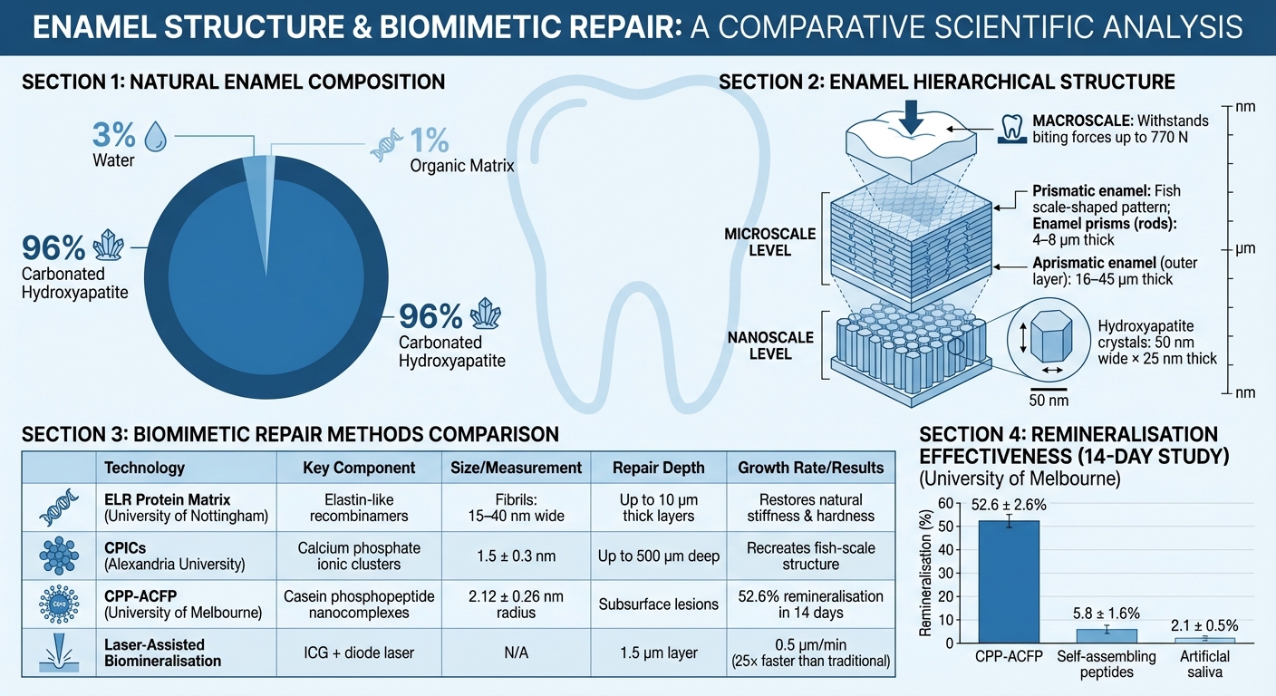
Enamel is made up of about 96% carbonated hydroxyapatite, 1% organic matrix, and 3% water [5][2]. This mineral-heavy composition makes it the hardest tissue in the human body, capable of withstanding biting forces of up to 770 N [5]. Its strength comes from a unique hierarchical structure, ranging from nanoscale crystals to microscale prisms.
At the nanoscale, hydroxyapatite crystals are approximately 50 nm wide and 25 nm thick. These crystals bundle together into enamel prisms, also known as rods, which are 4–8 μm thick [5][2]. The outermost layer, called aprismatic enamel, is 16–45 μm thick and features nanocrystals aligned perpendicular to the tooth surface, creating a dense and protective barrier [5]. Beneath this layer lies prismatic enamel, where crystals are interwoven into a "fish scale-shaped" pattern. This intricate design prevents cracks from spreading and gives enamel its impressive fracture resistance [5][4].
Understanding this complex structure is key to developing biomimetic techniques that replicate natural enamel formation.
How Biomimetic Repair Works
Biomimetic repair methods aim to replicate the natural enamel formation process, known as amelogenesis, at a molecular level. During amelogenesis, the protein amelogenin transitions from a disordered state into structured β-rich fibrils. These fibrils guide the growth and organisation of hydroxyapatite crystals [5].
In November 2025, researchers at the University of Nottingham, led by Alvaro Mata and Abshar Hasan, developed a protein-based matrix using elastin-like recombinamers (ELRs). These ELRs form fibrils that are 15–40 nm wide, mimicking the role of amelogenin [5]. When combined with calcium ions (1.5 mM Ca²⁺) and applied to eroded human teeth, the matrix promoted epitaxial growth. This process allowed new hydroxyapatite crystals to grow from the existing enamel structure while maintaining its alignment. The team successfully regrew mineralised layers up to 10 μm thick, restoring the enamel’s stiffness and hardness to match natural levels [5].
"Formation of dental enamel, the hardest and most mineralised tissue of vertebrates, relies on the 3D assembly and organisation of the protein amelogenin." – Abshar Hasan, School of Pharmacy, University of Nottingham [5]
An alternative approach involves calcium phosphate ionic clusters (CPICs), which are tiny aggregates measuring just 1.5 ± 0.3 nm [4]. These clusters, stabilised by capping agents like triethylamine, allow controlled nanostructure formation [3][4]. Researchers at Alexandria University used CPICs to repair enamel defects as deep as 500 μm, recreating the "fish scale" hierarchical structure through epitaxial growth [4][3]. The capping agents prevent uncontrolled aggregation, ensuring precise nanostructure alignment that integrates seamlessly with natural enamel. Unlike traditional restorative materials, these methods replicate enamel’s natural organisation, providing a seamless and functional repair.
New Technologies for Enamel Repair
Calcium Phosphate-Based Methods
Calcium phosphate-based techniques focus on using mineral ions to rebuild enamel by encouraging crystal alignment directly within the damaged area. A notable method involves casein phosphopeptide-amorphous calcium fluoride phosphate (CPP-ACFP) nanocomplexes. These nanocomplexes, with a hydrodynamic radius of about 2.12 ± 0.26 nm, have a neutral charge, allowing them to penetrate deeply into subsurface lesions. They stabilise calcium and phosphate ions, preventing premature clumping while ensuring a steady supply for controlled enamel mineralisation.
In a January 2023 study at the University of Melbourne, researchers tested four enamel repair technologies over 14 days. Results showed that CPP-ACFP achieved 52.6 ± 2.6% remineralisation, far outpacing self-assembling peptide methods (5.8 ± 1.6%) and artificial saliva (2.1 ± 0.5%) [1].
For repairing deeper enamel defects, researchers at Alexandria University explored a combination of calcium phosphate ionic clusters (CPICs) and bone-derived hydroxyapatite (BHA) nanoparticles. This premixed paste can address defects up to 500 µm deep. The bone-derived material includes trace elements like magnesium and carbonate, closely resembling the natural composition of enamel [3]. While these mineral-based strategies are promising, integrating nanotechnology has further improved the speed and precision of enamel repair.
Nanotechnology Applications
Nanotechnology is revolutionising enamel repair by offering faster and more precise solutions. One standout approach is laser-assisted biomineralisation (LAB). This method uses indocyanine green (ICG) as a light-absorbing agent combined with a dental diode laser to rapidly crystallise hydroxyapatite. A research team led by Kazuo Onuma and Ayako Oyane successfully regenerated a 1.5-µm-thick hydroxyapatite layer on human enamel in just 3 minutes. This corresponds to a growth rate of approximately 0.5 µm per minute – 25 times faster than traditional protein-mediated methods [6].
"The proposed method [LAB] has the potential to replace conventional resin-based treatments for the repair of early caries lesions." – Materials Today Communications [6]
Nanotechnology also enables multifunctional repair. For instance, incorporating silver nanoparticles (smaller than 50 nm) into regenerated hydroxyapatite layers not only aids enamel reconstruction but also provides antimicrobial protection against Streptococcus mutans [6].
Peptide and Organic Matrix Methods
Peptide-based methods aim to mimic the body’s natural enamel formation by replicating amelogenin, the protein responsible for guiding crystal growth during tooth development. One example is the elastin-like recombinamer (ELR) technology developed at the University of Nottingham, which is being advanced for clinical applications by Mintech-Bio Ltd [7].
However, peptide-based solutions face challenges. Replicating the complex post-translational modifications and dynamic behaviours of ameloblasts (cells involved in enamel formation) remains a significant hurdle [2]. This explains why peptide-based products like Curodont Repair (P11-4) have shown lower remineralisation rates compared to calcium phosphate systems in comparative studies [1]. These limitations highlight the need for hybrid approaches that combine the strengths of peptides, calcium phosphate, and nanotechnology for more effective enamel repair solutions.
Clinical Translation and Challenges
Current Clinical Applications
In Australia, several biomimetic enamel repair products have moved from the lab into clinical settings. One standout is Tooth Mousse Plus (CPP-ACFP), which has shown impressive results, achieving over 50% remineralisation in subsurface lesions. It has also been found to outperform alternatives like BioMin F and Curodont Repair in clinical efficacy [1].
That said, cell-based enamel regeneration remains an area with no clinical trial validation to date [2][8]. As Ahmed A. Holiel from Alexandria University’s Conservative Dentistry Department points out:
"Enamel repair relies primarily on acellular remineralisation of superficial demineralised defects… no prospective clinical trial has yet explored viable cell-based enamel tissue engineering." [8]
With enamel subsurface lesions affecting up to 96% of orthodontic patients [1], there’s a clear need for more advanced and reliable solutions. While early results are promising, significant challenges remain.
Research Gaps and Limitations
Despite the progress, several obstacles hinder the broader clinical adoption of biomimetic approaches. While these methods show potential, issues like cost, scalability, and technical limitations continue to pose significant barriers [2][8]. Manufacturing techniques that perform well in controlled research environments often struggle to scale efficiently for everyday clinical use. Researchers at Melbourne Dental School have noted:
"BioMin F and Curodont Repair are new oral care products and there is insufficient independent evidence to assess their true clinical potential." [1]
From a technical standpoint, the methods currently available, such as those using calcium phosphate ionic clusters, can only regenerate thin enamel layers – typically repairing defects up to around 500 µm in depth [3]. Reproducing enamel’s intricate structure and its mechanical strength remains a significant challenge. As Eman M. Sedek from Alexandria University’s Dental Biomaterials Department explains:
"Despite advancements, replicating the hierarchical structure and mechanical properties of natural enamel remains challenging." [2]
On top of these hurdles, regulatory frameworks add another layer of complexity. In Australia, fluoride-based remineralisation remains the cornerstone of clinical guidelines for managing caries [1]. This reliance on fluoride can slow the adoption of newer biomimetic therapies. Moreover, the lack of long-term clinical validation data for these emerging technologies creates hesitation among practitioners, making it difficult to transition from research breakthroughs to routine dental care.
The Future of Biomimetic Enamel Repair
The future of biomimetic enamel repair is shaping up to revolutionise non-invasive dental treatments. In Australia, where minimally invasive dentistry is gaining momentum, these advanced methods are set to play a key role. Unlike the traditional "drill and fill" techniques, biomimetic approaches focus on remineralising early enamel lesions without removing healthy tooth structure. Technologies such as CPP-ACP and protein-based gels are already helping address enamel subsurface lesions, which are common in orthodontic patients, before they progress to cavities [1].
At the forefront of this innovation, researchers at the University of Nottingham, led by Professor Alvaro Mata and Dr. Abshar Hasan, have developed a fluoride-free, protein-based gel that mimics the natural proteins involved in enamel growth. Their work has led to the creation of a start-up, Mintech-Bio, which aims to introduce its first clinical product by 2026 [9]. Professor Mata highlights the practicality of their design:
"We are excited because the technology was designed with both clinicians and patients in mind. It is safe, can be easily and rapidly applied, and it is scalable" [9].
What makes this approach stand out is its ability to integrate seamlessly into routine dental care. Future biomimetic products are expected to be as simple to use as fluoride treatments, making them ideal for regular dental check-ups. Unlike traditional restorative materials like amalgam or composite, biomimetic materials promote epitaxial growth – a process where new mineral naturally bonds with the existing enamel, matching its properties. This not only restores the tooth but also addresses dental hypersensitivity by forming an enamel-like layer over exposed dentine, effectively sealing dentinal tubules [9].
Another major benefit is the extended lifespan of restorative work. Biomimetic layers improve the bonding of restorations and minimise microleakage, which is a common cause of restoration failure. With nearly 50% of the global population affected by enamel degradation [9], the potential to improve oral health outcomes is immense. Australia’s contribution to this field is noteworthy, with the University of Melbourne playing a key role in validating CPP-ACP technologies for clinical use. This highlights Australia’s position as a leader in bringing these advancements into everyday dental practice [1].
Conclusion
Biomimetic enamel repair is reshaping the way we think about dental care. Instead of relying on the conventional "drill and fill" techniques that use synthetic materials, these cutting-edge methods aim to regenerate enamel with substances that closely mimic the natural composition and structure of teeth. For example, calcium phosphate ion clusters recreate the intricate "fish-scale" structure of enamel [4], while protein-based gels guide the organised growth of minerals [9]. These advancements are gradually transitioning from laboratory research to practical clinical use, signalling a shift towards biologically integrated dental treatments.
Research teams, such as those at the University of Nottingham, have shown that biomimetic materials can restore not just the look but also the strength and durability of natural enamel [8]. Considering that nearly half of the global population suffers from enamel degradation [9], the potential benefits for oral health are immense.
However, challenges remain. While some technologies are already being used in clinics and others, like Mintech-Bio’s protein-based gel, are gearing up for commercial release by 2026 [9], many promising approaches still face obstacles. Issues like scalability, affordability, and long-term clinical validation need to be addressed before these solutions can reach their full potential.
To overcome these hurdles, ongoing collaboration between dental professionals, material scientists, and bioengineers is essential. Exploring cost-effective sources, such as hydroxyapatite derived from biological waste like bone [3], could make these treatments more accessible. Independent clinical trials are also critical to ensure these innovations can withstand real-world conditions, including the stresses of chewing, exposure to acidic foods, and daily brushing [9]. Only through rigorous testing and development can these technologies fulfil their promise of transforming dental care into a system that preserves natural tooth structure while restoring both function and aesthetics.
FAQs
Who is biomimetic enamel repair suitable for?
Biomimetic enamel repair works well for people dealing with enamel loss due to cavities, erosion, or physical damage. It’s especially helpful for early-stage issues, where remineralisation can rebuild the enamel’s structure and resilience. This method aims to replicate the body’s natural enamel repair processes, offering a way to support better dental health.
How long do biomimetic enamel repairs last?
Biomimetic enamel repairs are designed to last for several years, typically ranging from 2–3 years or even longer. How long they hold up largely depends on the type of repair technology used and an individual’s oral care routine. Maintaining good oral hygiene – like brushing, flossing, and regular dental check-ups – can play a big role in extending the life of these repairs.
When will these treatments be available in Australian clinics?
Biomimetic enamel repair treatments, including nanotechnology-based remineralisation and bioinspired gels, are currently in advanced stages of research. These innovative approaches are expected to be introduced in Australian dental clinics by 2026–2027, as ongoing studies work to validate their effectiveness in clinical settings.
Related Blog Posts
- Wear Resistance of Polymer-Based Restoratives
- Antimicrobial Nanocomposites for Tooth Regeneration
- How Nano-Hydroxyapatite Repairs Enamel
- Future Trends in Nanodentistry for Tooth Decay
Important Notice: Any surgical or invasive procedure carries risks. Before proceeding, you should seek a second opinion from an appropriately qualified health practitioner.
Individual results may vary. The information provided in this article is for educational purposes only and does not constitute medical advice.
Checkout Related Blogs


Get in touch with us
For more information, call us now to start feeling better. Or fill the form below to make appointment
The Latest News from Complete Smiles
How to Clean Clear Plastic Retainers
Checklist for Choosing Wearable Dental Devices
Checklist for Choosing Cloud AI Platforms in Dentistry
Complete Smiles Bella VistaAccepts All Major Health Funds, Including


