





CBCT in Detecting Root Fractures
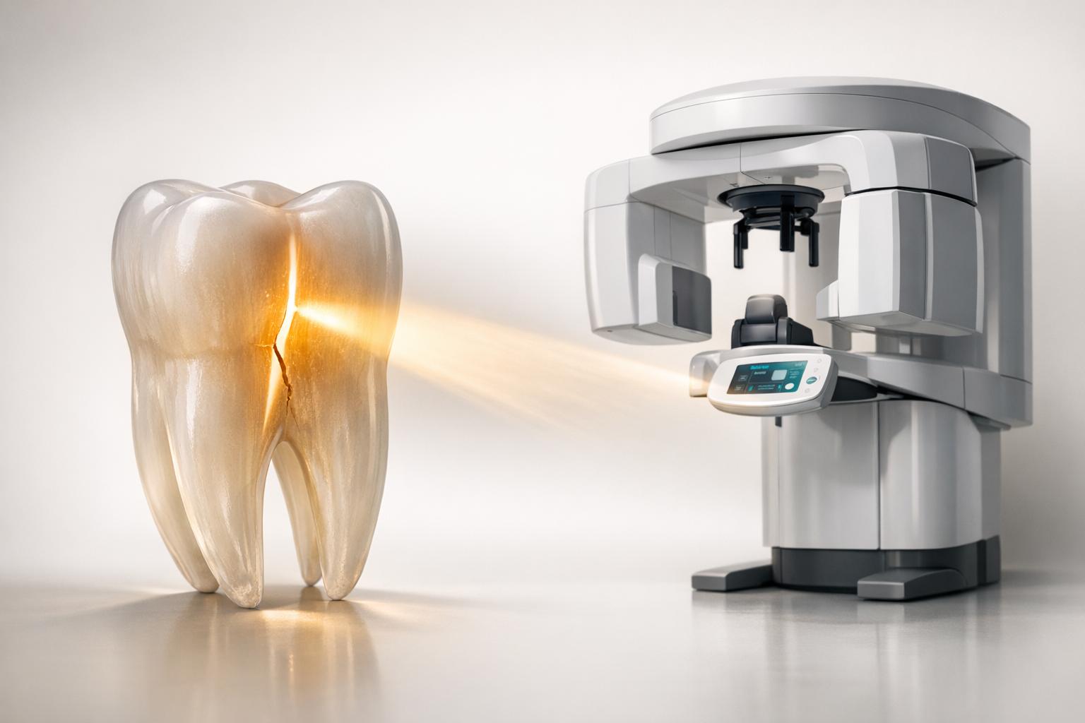
CBCT (Cone Beam Computed Tomography) is a 3D imaging tool that offers better accuracy than 2D X-rays for identifying root fractures. While conventional X-rays often miss fractures due to alignment issues or interference from dental materials, CBCT captures detailed images from multiple angles, improving detection rates significantly.
Key Points:
- Detection Rates: CBCT sensitivity reaches ~89.5%, compared to ~26.3% for traditional X-rays.
- Challenges with X-rays: Limited by beam alignment, overlapping structures, and artefacts from materials like gutta-percha or metal posts.
- CBCT Advantages: Offers multiplanar views (axial, sagittal, coronal), reducing missed fractures and overcoming interference.
- Research Findings: Studies report CBCT’s pooled sensitivity at 92% and specificity at 85%, with better performance when metallic artefacts are absent.
- Device Performance: Systems like the 3D Accuitomo 170 outperform others due to smaller voxel sizes, enhancing fracture detection accuracy.
CBCT is particularly useful in cases where X-rays fail but clinical symptoms suggest fractures. It aids in treatment planning by pinpointing fracture locations and severity, helping clinicians decide between preservation, extraction, or implants. However, artefacts from dental materials and imaging settings like voxel size can affect accuracy, so optimising protocols is crucial for reliable results.
Research Studies on CBCT Diagnostic Performance
Study 1: Detecting Different Fracture Types
Studies consistently highlight CBCT’s ability to detect various root fracture types with impressive accuracy. A meta-analysis of 12 studies reported a pooled sensitivity of 0.92 (95% CI = 0.89–0.94) and a pooled specificity of 0.85 (95% CI = 0.75–0.92) across different fracture types [8]. The summary receiver operating characteristic (SROC) was 0.94, showcasing excellent overall diagnostic performance [8].
Interestingly, CBCT’s accuracy improves when metallic interference is absent. Sensitivity and specificity were recorded at 0.82 and 0.88, respectively, under these conditions. However, these values dropped to 0.73 and 0.72 in the presence of materials like gutta-percha or posts [5]. For horizontal root fractures specifically, sensitivity reached 0.93 without metallic artifacts [5].
Further analysis of CBCT devices highlights variations in diagnostic performance.
Study 2: Comparing CBCT Devices
Comparisons between CBCT devices provide deeper insight into their diagnostic capabilities. Research from Ondokuz Mayıs University revealed significant differences in performance. The 3D Accuitomo 170 outperformed the NewTom 3G in detecting induced vertical root fractures [10]. With a 4×4-cm field of view, the 3D Accuitomo 170 achieved a diagnostic accuracy of 93%, compared to the NewTom system’s 68% [3].
Dr Kaan Gunduz from the Department of Dentomaxillofacial Radiology stated, "The 3D Accuitomo 170 was significantly superior to the NewTom 3G, VistaScan PSP, CCD sensor, and conventional film images in the detection of artificially induced vertical root fractures" [10].
This performance gap is largely due to voxel size. The 80-micrometre voxel size of the 3D Accuitomo 170 allowed it to detect narrow fracture lines that other systems missed [3].
Clinical studies further validate these findings by examining sensitivity and specificity in real-world applications.
Study 3: Sensitivity and Specificity Review
Clinical research using the i-CAT and 3D Accuitomo 80 systems confirms CBCT’s reliability in practice. These devices demonstrated a sensitivity of 88% and specificity of 75% for detecting root fractures in actual patient cases [3]. Additionally, a study from Tufts University School of Dental Medicine found that when periapical radiographs failed to detect fractures but clinical suspicion remained high, the pooled prevalence of confirmed fractures was 91% [8].
Dr David M Leader highlighted the clinical relevance: "CBCT has a high diagnostic accuracy for tooth fractures and could be used in clinical settings. We can be very confident with positive test results but should be very cautious with negative test results" [8].
For endodontically treated teeth, the negative predictive value was 0.28, compared to 0.77 for non-treated teeth [8]. However, the positive predictive value remained excellent, at 0.98 for treated teeth and 0.99 for non-treated teeth [8].
Cracked Teeth & Root Fractures: Advanced CBCT Analysis | Case Bites
CBCT vs Traditional Radiography
CBCT vs Traditional X-Ray Performance in Detecting Root Fractures
Diagnostic Performance Comparison
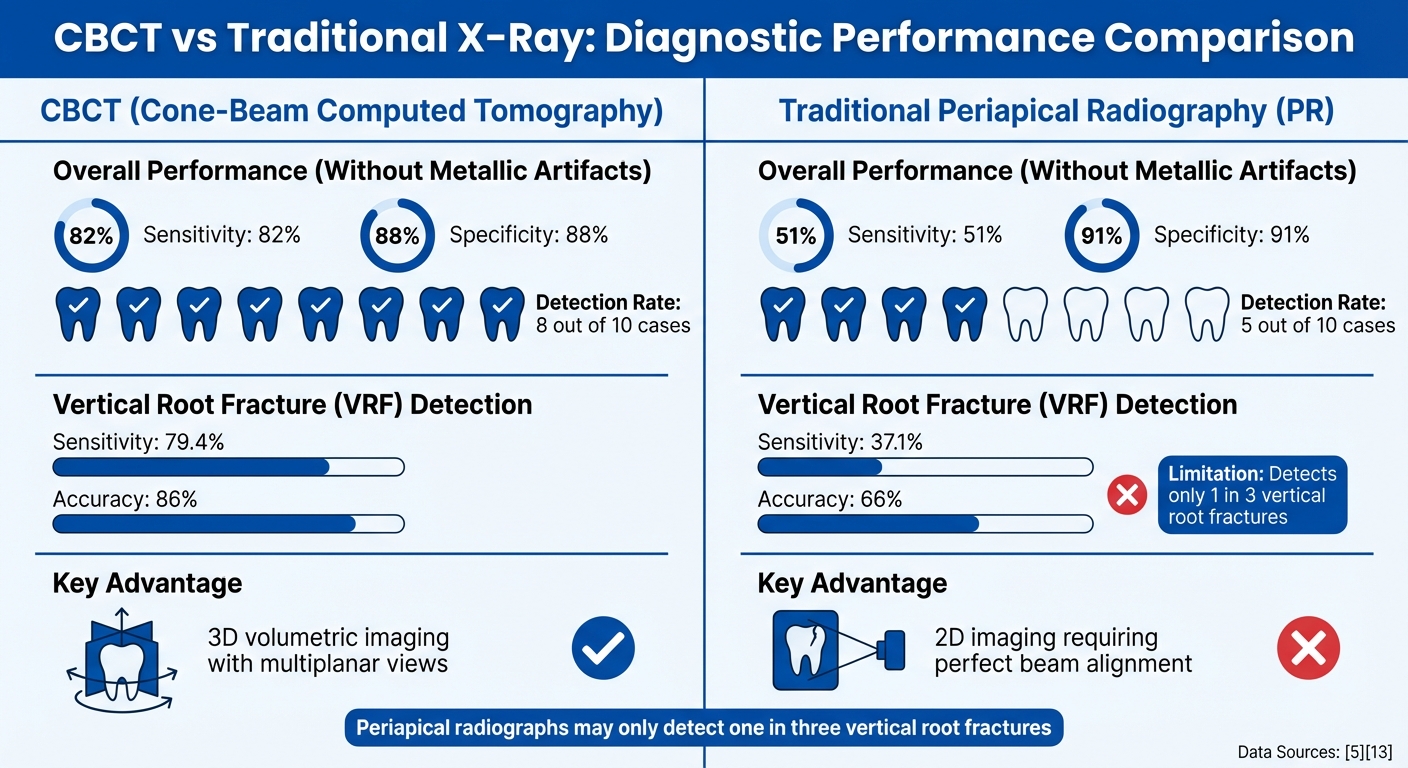
When it comes to diagnostic accuracy, CBCT (Cone Beam Computed Tomography) clearly outshines traditional periapical radiography. Without metallic artifacts interfering, CBCT achieves a sensitivity of 0.82 and a specificity of 0.88. In contrast, periapical radiographs fall short, with a sensitivity of just 0.51 but a slightly higher specificity of 0.91[5]. This means CBCT successfully identifies fractures in about 8 out of 10 cases, while traditional radiographs manage only 5 out of 10.
The difference becomes even more apparent in detecting vertical root fractures (VRFs). CBCT delivers a sensitivity of 79.4% for these fractures, whereas periapical radiographs reach only 37.1%[12]. Additionally, CBCT achieves an accuracy rate of 0.86 for VRF detection, compared to 0.66 for traditional radiography[12]. Geometric constraints inherent to traditional radiographs mean they often miss fractures, detecting just one in three cases[1].
| Imaging Method | Sensitivity (No Artifacts) | Specificity (No Artifacts) | VRF Detection Accuracy |
|---|---|---|---|
| CBCT | 0.82 [5] | 0.88 [5] | 0.86 [12] |
| Traditional PR | 0.51 [5] | 0.91 [5] | 0.66 [12] |
These numbers highlight CBCT’s diagnostic edge, largely thanks to its advanced imaging capabilities.
Why CBCT Performs Better
The secret to CBCT’s superior performance lies in its 3D volumetric imaging, which overcomes the inherent limitations of 2D radiography. CBCT provides multiplanar reconstructions – axial, coronal, and sagittal views – allowing clinicians to assess fractures from various angles[2][11].
"Periapical radiographs may only detect one in three vertical root fractures" [1].
This limitation stems from the fact that, for a fracture to appear on a periapical radiograph, the X-ray beam must align almost perfectly parallel to the fracture plane[1][5]. CBCT, on the other hand, captures multiplanar data, eliminating the risk of missing fractures due to beam misalignment.
As Zahra Dalili Kajan from Guilan University of Medical Sciences explained, reconstructed axial views from CBCT were especially effective in confirming specific diagnoses compared to other views[2].
sbb-itb-2be92ed
Factors That Affect CBCT Accuracy
Getting the most accurate results from CBCT imaging depends on managing artefact-causing materials and fine-tuning imaging protocols.
Effects of Root Canal Materials
Root canal materials can make CBCT imaging tricky. For example, gutta-percha and metal posts create beam hardening artefacts, which can either mimic or hide fracture lines [3][13]. Specifically, gutta-percha reduces sensitivity for detecting vertical root fractures from 72% to just 32–40%, and specificity drops from 96% to 68% [13]. Metal posts are even more problematic, causing white streak artefacts and blurring due to detector saturation [3]. On the other hand, fibreglass posts cause minimal artefacts, allowing for more accurate diagnoses – even with lower radiation doses [14].
"The intracanal filling materials such as gutta-percha reduce the diagnostic ability of the vertical root fractures. Hence, it is recommended to remove those materials from root canals before imaging to improve the diagnostic potential of CBCT."
– Ehsan Hekmatian et al. [13]
If removing filling materials isn’t an option, tweaking the imaging settings can help. For instance, increasing the tube current to 8 mA for gutta-percha or 10 mA for metal posts can improve image clarity [14].
But artefacts aren’t the only challenge – imaging settings also play a huge role in CBCT accuracy.
Imaging Protocol Considerations
Voxel size is a key factor in detecting fractures. Smaller voxels (between 0.08 mm and 0.1 mm) significantly improve accuracy, achieving a sensitivity of 0.84 and a specificity of 0.79. In contrast, larger voxels (0.125 mm to 0.2 mm) drop these figures to 0.70 and 0.55, respectively [16]. For the highest accuracy – up to 93% – an 80-micrometre voxel setting is ideal [3]. This precision matters because vertical root fractures are typically only 20 to 200 micrometres wide [3].
The field of view (FOV) also makes a difference. A smaller FOV (about 4 cm) focused on the area of interest can reduce artefacts, lower radiation exposure, and improve image quality [3]. Additionally, axial slices reconstructed perpendicular to the tooth’s long axis provide the most reliable diagnostic insights [2][3]. However, metal artefact reduction software often falls short, sometimes even blurring critical details [9][15].
Another factor is the fracture width. CBCT achieves around 90% accuracy when the fracture space is 400 micrometres wide or more. But as the fracture narrows, accuracy declines [3]. For the best results, CBCT findings should be combined with clinical observations, such as vertical bone resorption patterns that are typical of root fractures [9].
Clinical Applications and Treatment Planning
CBCT takes diagnostic precision to another level, offering 3D imaging that plays a pivotal role in managing root fractures. This advanced imaging allows clinicians to assess the fracture’s location and severity, which is crucial when weighing treatment options. For example, it helps determine whether to preserve the tooth through procedures like root-end resection or root amputation, or to proceed with extraction and implant placement instead [4].
CBCT becomes particularly valuable in cases where clinical symptoms strongly suggest a fracture, but conventional periapical radiographs fail to confirm it. Studies show that when traditional X-rays fall short, CBCT successfully identifies fractures in the majority of cases [8]. Early detection is key here – it can prevent further alveolar bone loss, preserving bone for future treatments [7]. These capabilities make CBCT an essential tool for guiding treatment decisions, as explored further below.
Using CBCT for Treatment Decisions
The 3D imaging capabilities of CBCT allow for tailored surgical approaches, minimising unnecessary tissue damage. For multi-rooted teeth, CBCT pinpoints the specific root affected, helping to avoid excessive tissue removal. Its diagnostic accuracy for detecting root fractures stands at 91.9%, a stark contrast to the 48.1% accuracy of conventional dental radiography [4].
"CBCT may be helpful in the assessment of the pattern of a fracture line and can help dentists and endodontists to make extraction and/or implant decisions that affect their patients by having reliable documentation and strong enough evidence."
– Z Dalili Kajan, Professor, Guilan University of Medical Sciences [2]
That said, imaging alone isn’t enough. For teeth that have undergone endodontic treatment, CBCT results must be considered alongside clinical signs like J-shaped radiolucencies or vertical bone resorption patterns. If CBCT results are negative but symptoms persist, close monitoring is advised rather than immediate invasive action, as fractures may initially be too narrow to detect [9][2][6]. This approach ensures that CBCT findings integrate seamlessly with clinical evaluations, providing a comprehensive framework for diagnosis and treatment planning.
CBCT’s precision not only aids in surgical decision-making but also ensures its use is targeted to cases where it can make the biggest difference.
Cases Where CBCT Proves Most Useful
Certain clinical scenarios highlight CBCT’s strengths. Premolars, for instance, are particularly prone to vertical root fractures due to their thin, oval-shaped roots and small mesio-distal diameter. In fact, bucco-lingual fractures account for 76.2% of fracture cases in premolars [7]. Endodontically treated teeth also benefit from CBCT imaging, though root canal filling materials can reduce its sensitivity – from 97% in untreated teeth to 71.4% [4].
CBCT is especially helpful in diagnosing vertical root fractures in patients with symptoms like pain during chewing, recurring fistulas, or isolated deep periodontal pockets – symptoms that are often inconclusive on traditional radiographs [2][11]. By enabling early and accurate detection in such cases, CBCT significantly improves treatment outcomes and overall prognosis.
Conclusion
CBCT has transformed the way root fractures are diagnosed by addressing the limitations of two-dimensional radiography. A meta-analysis highlighted CBCT’s impressive performance, showing 92% sensitivity, 85% specificity, and an SROC of 0.94 – a clear improvement over traditional methods, which can only detect one in three vertical root fractures [1][8]. With its ability to provide 3D axial, sagittal, and coronal views, CBCT allows clinicians to pinpoint fractures with greater precision [8].
This enhanced accuracy has a direct impact on clinical decisions. It enables tailored treatment plans, whether that involves preserving a tooth, opting for extraction, or planning an implant. Early and accurate detection also helps avoid unnecessary procedures and prevents progressive bone loss.
As David M Leader from Tufts University School of Dental Medicine explains, "We can be very confident with positive test results but should be very cautious with negative test results" [8].
For patients experiencing persistent dental pain or unexplained symptoms, CBCT combined with clinical evaluation ensures a more reliable diagnosis and a well-informed treatment approach. This technology represents a significant step forward in dental care.
FAQs
When should a dentist order a CBCT for a suspected root fracture?
When traditional X-rays fall short of offering enough clarity to confirm a suspected root fracture, a dentist might turn to a CBCT scan. This advanced imaging method becomes especially useful if clinical signs such as bifurcation radiolucency or swelling are observed. However, it’s essential to balance the advantage of CBCT’s detailed imaging with the consideration of its radiation exposure.
Can CBCT miss a root fracture even if I have symptoms?
Yes, it can. While CBCT (Cone Beam Computed Tomography) is a powerful diagnostic tool, it isn’t foolproof. Studies show that CBCT has limitations, especially when it comes to identifying vertical root fractures. Even when symptoms suggest a fracture, CBCT might not always detect it. This is why relying solely on CBCT isn’t enough – clinical evaluation and other diagnostic methods are often needed to confirm the issue.
Do root canal fillings or metal posts affect CBCT results?
When root canal fillings and metal posts are present, they can create artefacts in CBCT images. These artefacts may interfere with the clarity of the images, potentially affecting diagnostic precision. However, employing metal artefact reduction algorithms can improve the ability to identify vertical root fractures in these situations.
Related Blog Posts
- CT Scans in Bone Graft Planning
- CBCT in Endodontics: Accuracy and Limitations
- Guide to Periapical X-rays for Root Canal Therapy
- CBCT Technology: Mechanisms Behind 3D Imaging
Important Notice: Any surgical or invasive procedure carries risks. Before proceeding, you should seek a second opinion from an appropriately qualified health practitioner.
Individual results may vary. The information provided in this article is for educational purposes only and does not constitute medical advice.
Checkout Related Blogs




Get in touch with us
For more information, call us now to start feeling better. Or fill the form below to make appointment
The Latest News from Complete Smiles
How to Clean Clear Plastic Retainers
Checklist for Choosing Wearable Dental Devices
Checklist for Choosing Cloud AI Platforms in Dentistry

Complete Smiles Bella VistaAccepts All Major Health Funds, Including





