


AI vs Traditional Periodontal Diagnosis
AI is reshaping how dentists detect periodontal disease, offering faster and more consistent results compared to manual methods. While traditional diagnosis relies on probing and X-rays, it can be slow, subjective, and limited by human error. AI tools, on the other hand, use advanced algorithms to analyse radiographs with accuracy rates as high as 94.2%, often outperforming specialists.
Key takeaways:
- Periodontal disease affects millions globally and can lead to severe health issues if undetected.
- Traditional methods like probing and X-rays are effective but rely heavily on clinician expertise, leading to variability and delays.
- AI-powered tools provide precise, real-time analysis, improving early detection and helping less experienced dentists achieve specialist-level accuracy.
- Challenges include ensuring data privacy, avoiding bias in AI systems, and maintaining human oversight.
AI and manual methods complement each other. AI excels in analysing radiographs and speeding up workflows, while clinicians provide the nuanced judgement and patient care AI lacks. For Australian dentists, integrating AI could improve care accessibility, especially in remote regions, while reducing diagnostic delays.
Your Perio Education Sidekick: Detecting and Treating Periodontal Disease with AI
sbb-itb-2be92ed
Conventional Periodontal Diagnosis Methods
In Australia, dental professionals typically rely on a combination of physical examinations and imaging to diagnose periodontal disease. However, these traditional methods come with challenges that can affect both their accuracy and efficiency. By looking at these approaches, we can better understand how they compare to newer, AI-based diagnostic tools.
Manual Probing and Clinical Assessments
The backbone of traditional periodontal diagnosis is manual probing. This involves a dentist using a slim, millimetre-marked periodontal probe to measure probing pocket depth (PPD) at six points around each tooth. During this process, they also check for bleeding on probing (BOP) and record clinical attachment levels (CAL) to assess gum health and disease progression [7].
While this method is widely used, its accuracy depends heavily on the clinician’s skill. Factors like probing pressure, angulation, and precise landmark identification can all influence results [12]. Additionally, the physical markings on the probe only allow measurements to the nearest millimetre. This makes the process not only invasive and technically challenging but also time-consuming. As noted by npj Digital Medicine:
"Diagnosis relies on an invasive, time‐consuming, and technically demanding physical examination to detect periodontal bleeding upon probing and clinical attachment loss."
– npj Digital Medicine [1]
To complement these clinical examinations, imaging is used to assess bone loss. For those due for a check-up, our gap-free new patient pack includes the necessary X-rays and clinical exams to evaluate gum health.
Radiographic Imaging
Radiographic imaging plays a vital role in diagnosing periodontal disease, particularly for evaluating bone loss – a key sign of disease progression. Dentists commonly use periapical radiographs, panoramic radiographs (OPGs), bitewing X-rays, and occasionally Cone Beam Computed Tomography (CBCT) [6].
In these images, radiographic bone loss (RBL) is measured by examining the distance between the cementoenamel junction (CEJ) and the marginal alveolar bone (MAB) [1]. Based on this measurement, bone loss is classified as:
- Mild: Less than 3 millimetres from the CEJ
- Moderate: Between 3 and 5 millimetres
- Severe: Greater than 5 millimetres [11]
Bone loss can also be categorised as horizontal (even reduction) or vertical (angular defects). However, interpreting these two-dimensional images of complex three-dimensional structures can be challenging, leading to variations in accuracy [8].
Limitations of Conventional Methods
Traditional diagnostic methods come with notable limitations. For instance, studies show only moderate agreement between different examiners when identifying radiographic bone loss, with Cohen’s kappa values ranging from 0.454 to 0.482 [1]. Even when the same dentist reviews the same image on separate occasions, the reproducibility (intra-examiner reliability) is only moderately improved, with a kappa value of 0.739 [1].
Several factors contribute to this variability. As highlighted in the Journal of Dentistry:
"Oral and gingival examinations are inherently subjective. Variations in judgement criteria and technical proficiency among different dentists may lead to discrepancies in diagnosis."
– Journal of Dentistry [6]
In addition, human factors like fatigue can affect performance. A dentist reviewing their 30th set of X-rays late in the day may have reduced visual sharpness compared to earlier in their schedule [8]. Moreover, traditional methods are largely retrospective, meaning they often detect periodontal disease only after significant and irreversible damage – such as tissue destruction or bone loss – has already occurred [12][1]. This delay in diagnosis means many Australians are only identified in advanced stages of the disease, leading to more complex and expensive treatment options [1].
AI-Powered Periodontal Diagnosis Tools
Artificial intelligence is reshaping the way dental professionals detect and evaluate periodontal disease. By complementing traditional diagnostic methods, AI introduces a level of precision and consistency that goes beyond human capabilities. These tools utilise advanced algorithms to analyse radiographic images and clinical data, uncovering details often missed by the human eye.
How AI Processes Diagnostic Data
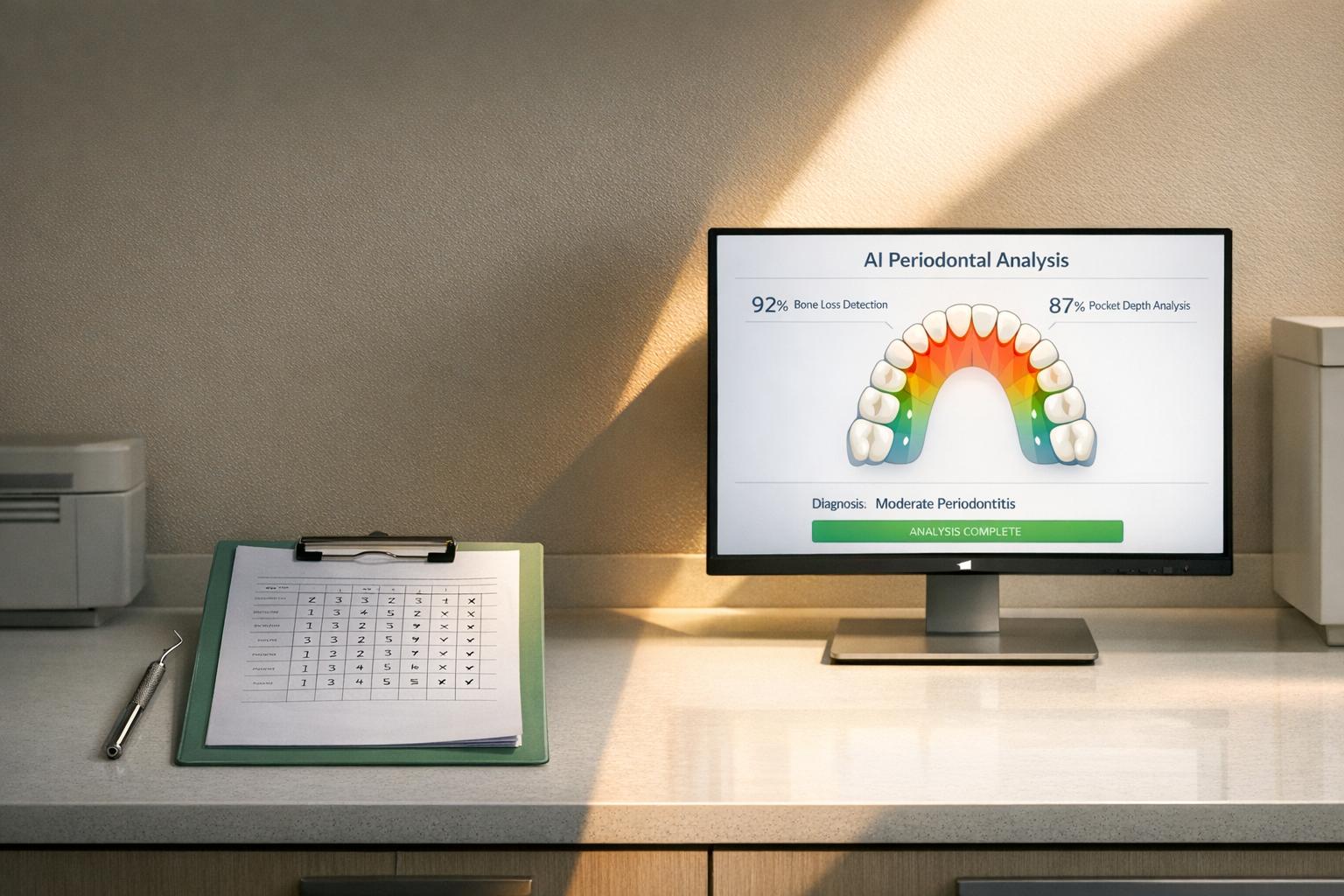
AI systems use technologies like Deep Learning and Convolutional Neural Networks (CNNs) to evaluate dental radiographs. These networks examine millions of pixels, identifying subtle patterns that might escape human detection [3]. The diagnostic workflow involves multiple stages: segmenting individual teeth, identifying anatomical landmarks such as the cemento-enamel junction (CEJ) and marginal alveolar bone, and calculating the radiographic bone loss (RBL) percentage automatically [13][14].
A key feature of advanced AI models is their training process. For instance, HC-Net+, developed at the Prince Philip Dental Hospital in Hong Kong, uses clinical probing data as its "ground truth" instead of relying solely on radiographic readings [1]. This allows the AI to detect minor tissue breakdowns that may not be visible in standard two-dimensional X-rays. The model combines detailed tooth-level analysis with an overall assessment of the patient’s periodontal health, closely imitating the approach of skilled clinicians [1].
These systems also integrate patient-specific risk factors – like age, smoking habits, and diabetes – with radiographic data. This combination supports automated disease staging (severity) and grading (progression rate) in line with the 2017 guidelines from the American Academy of Periodontology (AAP) and the European Federation of Periodontology (EFP) [14][10].
Such capabilities are paving the way for practical applications in dental clinics.
AI Applications in Dental Clinics
Leveraging these analytical methods, AI systems have already been adopted in clinical settings. Between 2018 and 2024, the University of Medicine and Pharmacy at Ho Chi Minh City developed a YOLOv8-based framework. This system demonstrated precision rates between 0.95 and 0.97 in detecting bone levels and the CEJ. By incorporating patient data – such as age, smoking history, and HbA1c levels – it automated periodontitis staging and grading. The system was accessible through a web-based platform, enabling clinicians to upload radiographs and receive instant diagnostic overlays and staging recommendations [14][10].
Similarly, the Dental Department at Fang Hospital in Chiang Mai, Thailand, created a YOLOv8 model between 2015 and 2023. This model achieved an impressive 98% accuracy in segmenting the CEJ and alveolar bone levels, providing tailored prognoses based on Thai Association of Periodontology standards [13].
In a multicentre diagnostic study (NCT05513599) involving 10,881 orthopantomograms from centres in Hong Kong, Shanghai, and Rome, the HC-Net+ model achieved a diagnostic accuracy (AUROC) of 94.2%. This performance far exceeded that of periodontal specialists, who averaged 85.6%. Interestingly, the study also revealed that AI assistance allowed junior dentists to match the diagnostic accuracy of experienced professionals [1].
Benefits of AI in Periodontal Diagnosis
The adoption of AI in periodontal diagnosis brings several practical advantages, addressing key challenges of traditional methods. One major benefit is speed. While manual diagnosis by experts can take 29 to 62 seconds per case, AI systems deliver results almost instantly [1][13]. This efficiency is especially beneficial in busy clinics or areas with limited access to periodontal specialists.
AI also excels in early detection. Conventional methods often identify periodontal disease only after significant damage has occurred. AI models, however, have a pooled diagnostic odds ratio of 29.30, significantly improving the likelihood of accurate early detection compared to manual methods [4]. Additionally, AI tools empower less experienced dentists to reach diagnostic standards comparable to seasoned specialists, broadening access to high-quality care [1].
To address concerns about transparency, modern AI systems include explainable features. For example, heatmaps or Class Activation Maps (CAMs) highlight diagnostic areas, making the process more understandable for clinicians [1][15]. As noted in npj Digital Medicine:
"HC-Net+’s ability to surpass specialist accuracy while making diagnostic expertise more accessible positions it as a transformative tool for precision dentistry." [1]
For dental practices in Australia, these AI tools offer a promising way to enhance diagnostic precision and efficiency, especially in remote areas where access to periodontal specialists can be limited.
AI vs Conventional Methods: A Direct Comparison
AI vs Traditional Periodontal Diagnosis: Performance Metrics Comparison
When you compare AI-powered tools with traditional methods, the differences in performance, reliability, and application become clear. These distinctions highlight the trade-offs and complementary aspects of both approaches.
Accuracy and Performance Metrics
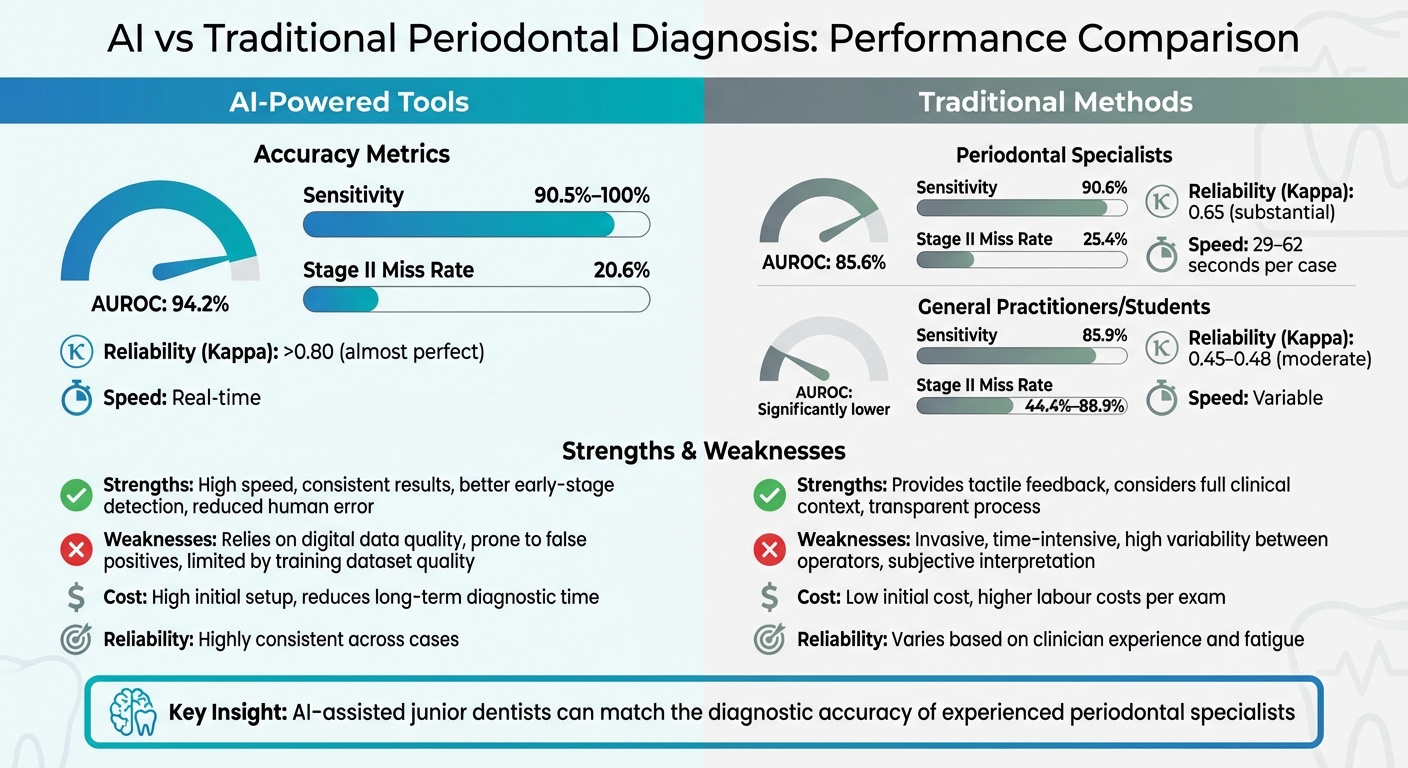
A multicentre study conducted in January 2025 involving 382 OPGs revealed that the HC-Net+ model achieved an AUROC of 94.2%. This surpassed the performance of specialists, who scored 85.6%, and had a Stage II miss rate of just 20.6%. For comparison, specialists had a miss rate of 25.4%, while general practitioners and students ranged from 44.4% to 88.9% [1].
At Fang Hospital in Chiang Mai, Thailand, researchers tested AI against human practitioners using 90 OPG images. The YOLOv8 model achieved 94.4% accuracy and 100% sensitivity, outperforming periodontists, who reached 91.1% accuracy and 90.6% sensitivity. However, the study noted that AI struggled with specificity, making human verification essential to rule out false positives [5].
| Metric | AI-Powered Tools | Periodontal Specialists | General Practitioners/Students |
|---|---|---|---|
| AUROC | 94.2% [1] | 85.6% [1] | Significantly lower [1] |
| Sensitivity | 90.5%–100% [1][5] | 90.6% [5] | 85.9% [5] |
| Stage II Missed Rate | 20.6% [1] | 25.4% [1] | 44.4%–88.9% [1] |
| Reliability (Kappa) | >0.80 (almost perfect) [4] | 0.65 (substantial) [1] | 0.45–0.48 (moderate) [1] |
| Speed | Real-time [5] | 29–62 seconds per case [1] | Variable [1] |
These findings reinforce the idea that AI significantly enhances diagnostic precision, while traditional methods excel in capturing critical clinical nuances.
Strengths and Weaknesses
The performance metrics underscore the unique advantages and challenges of each approach.
AI tools are fast and consistent, delivering results almost instantly. They standardise outcomes regardless of the operator’s experience, allowing junior dentists to perform at levels comparable to seasoned specialists [1]. This makes them particularly appealing for streamlining diagnostics.
On the other hand, conventional methods provide tactile feedback that AI cannot replicate. This feedback reveals essential details like tissue quality and inflammation, which radiographs alone cannot show. Traditional examinations also allow clinicians to apply empathy and moral judgement, considering patient-specific factors. As highlighted in MDPI Diagnostics:
"AI should be seen as a tool that helps doctors make better decisions and not as a way to replace their knowledge and skills." [2]
| Feature | AI-Powered Tools | Conventional Methods |
|---|---|---|
| Strengths | High speed, consistent results, better early-stage detection, reduced human error [1][14][5] | Provides tactile feedback, considers full clinical context, transparent process [1] |
| Weaknesses | Relies on digital data quality, prone to false positives, limited by training dataset quality [2][5] | Invasive, time-intensive, high variability between operators, subjective interpretation [1][14] |
| Cost-Effectiveness | High initial setup but reduces long-term diagnostic time [1][5] | Low initial cost but higher labour costs per exam [1] |
| Reliability | Highly consistent across cases [1] | Varies based on clinician experience and fatigue [1] |
For dental practices in Australia, particularly in rural or regional areas with limited access to specialists, AI tools can enhance diagnostic capabilities while still preserving the human touch essential for patient care. These tools bridge gaps in expertise, ensuring more consistent and accessible dental diagnostics.
Future Integration and Clinical Applications
With AI already showing strong diagnostic performance, the next step is integrating its precision with human clinical expertise. The aim isn’t to replace dentists but to work alongside them, enhancing their capabilities. The Australian Dental Association underscores that patient safety must always come first, with practitioners maintaining full responsibility for clinical decisions [18]. Researchers often describe AI in this context as "complementary intelligence" – a tool that supports, rather than substitutes, human expertise [3].
How AI Can Work Alongside Conventional Methods
In March 2024, researchers from the University of Hong Kong and Shanghai Ninth People’s Hospital tested the HC-Net+ AI system using data from 760 patients across multiple centres. This study, registered under NCT06306677, found that junior dentists assisted by AI could match the diagnostic accuracy of experienced periodontal specialists. The AI system itself achieved over 92.4% accuracy across diverse international sites [1].
This partnership between AI and traditional methods leverages AI’s ability to process data quickly and consistently, while still relying on the nuanced judgement of human practitioners. For example, AI can flag critical radiographs for review, cutting analysis time from 20–25 minutes to just 5–10 minutes [17]. Future workflows may evolve into integrated platforms that combine clinical data, imaging, and biological signals [16].
AI also offers explainable tools like classification activation maps, which visually highlight areas of concern, such as bone loss. These features not only help dentists understand AI reasoning but also improve patient communication and treatment acceptance [1].
Implementation Challenges and Ethical Considerations
Adopting AI in Australian dental practices comes with its own set of challenges. For one, the Therapeutic Goods Administration (TGA) classifies AI diagnostic software as a medical device, meaning it must meet strict standards for accuracy and reliability. Clinics need to confirm that any AI tools they use are TGA-registered [20].
Data privacy is another critical issue. The Privacy Act 1988 (Cth) and its 13 Australian Privacy Principles regulate how patient data is handled, which means consent forms may need to include provisions for AI use and data processing. According to AHPRA:
"Practitioners must apply human judgement to any output of AI." [20]
Bias in AI systems is another concern. Training datasets must represent diverse populations to avoid skewed results based on ethnicity, gender, or socioeconomic factors [19]. Current AI models also face challenges with complex cases, such as vertical bone loss or certain tooth types like molars, which can lead to reduced accuracy or false positives [21]. This makes human oversight essential.
To address these issues, dental education must include AI literacy, covering topics like data flow, model validation, and algorithmic bias. Clinicians will need to verify AI outputs to ensure they align with clinical realities [15][18].
These challenges highlight the importance of using AI as a tool to complement, not replace, existing practices.
Opportunities for Australian Dental Practices
AI has the potential to fill gaps in expertise and elevate care across all clinical settings. For Australian dental clinics, the technology can standardise diagnostic outcomes, ensuring even general practitioners achieve specialist-level accuracy [1].
While initial implementation costs may be high, the long-term benefits include faster diagnostics and streamlined workflows [1]. AI can also flag advanced periodontitis cases (Stages II–IV), which is particularly useful for less experienced clinicians.
Teledentistry is another area where AI is making an impact, raising unique legal and ethical considerations. AI-driven platforms are extending specialist consultations to remote areas, helping to overcome Australia’s geographic challenges while maintaining quality care standards [17]. The Dental Board of Australia has emphasised that, regardless of technological advancements, the responsibility for safe and effective care ultimately lies with practitioners [20].
The successful integration of AI will require careful ethical and educational planning. As researchers from Shahid Beheshti University of Medical Sciences aptly put it:
"AI and periodontology should not stand as opposing forces, but rather as complementary partners where algorithmic precision is shaped, verified, and humanized by clinical judgment." [16]
Conclusion
AI and traditional periodontal diagnosis work hand-in-hand, each filling gaps the other cannot. AI excels in fast, objective radiographic analysis, boasting accuracy rates between 73% and 99.4%[2]. However, it lacks the nuanced clinical judgement and patient-specific insights that manual vs digital probing provides[1]. On the other hand, while manual probing is still considered the gold standard for detecting subtle clinical changes, it comes with challenges like being time-intensive and prone to variability between examiners, with reliability scores as low as 0.454–0.482[1].
Multicentre trials highlight AI’s potential, with systems like HC-Net+ achieving an impressive 94.2% AUROC, surpassing specialists who scored 85.6%[1]. Even more striking, AI-assisted junior dentists matched the diagnostic accuracy of seasoned professionals. These findings underscore the benefits of blending AI with traditional diagnostic methods to enhance overall outcomes.
AI proves particularly effective for tasks such as radiographic screening and measuring bone loss, allowing clinicians to dedicate more time to treatment planning and patient interaction. This demonstrates that AI is a supportive tool designed to complement – not replace – clinical expertise.
Key Takeaways
These findings pave the way for AI integration into Australian dental practices. The future lies in combining AI with traditional methods, enabling general practitioners to achieve up to 86.7% diagnostic accuracy, effectively bringing specialist-level care to more patients[9]. However, for this integration to succeed in Australia, practitioners must navigate requirements like TGA registration, compliance with the Privacy Act 1988, and ensuring clinical teams receive proper AI literacy training.
FAQs
How does AI enhance the diagnosis of periodontal disease compared to traditional methods?
AI is transforming how periodontal disease is diagnosed by using advanced image analysis to pinpoint and classify the condition with a level of accuracy and consistency that often surpasses traditional methods. Research even shows that AI systems can outperform specialists when it comes to detecting moderate to severe periodontitis. This is largely due to their ability to process complex radiographic images with incredible precision.
One major advantage of AI tools is their ability to minimise human error and inconsistencies, especially when analysing panoramic radiographs. This increased reliability allows dentists to make better-informed decisions, which can lead to improved treatment outcomes for patients. The efficiency and impartiality of AI make it a powerful tool in the world of modern dental care.
What challenges do dental practices face when adopting AI for periodontal diagnosis?
Integrating AI into dental practices for diagnosing periodontal conditions comes with its own set of hurdles, particularly around standardisation, workflow integration, and ethical considerations. One major challenge is ensuring that AI systems deliver consistent results across various clinical environments and imaging technologies – a task that’s far from straightforward. On top of that, adapting current workflows and equipping dental professionals with the skills to use these tools effectively can demand significant time and resources.
Ethical issues are another critical aspect. Protecting patient privacy, obtaining proper informed consent, and ensuring that AI algorithms remain transparent, unbiased, and accountable are all pressing concerns. Although AI holds immense potential to enhance diagnostic precision and streamline processes, more research and fine-tuning are needed before it becomes a practical, everyday tool in Australian dental clinics.
Can artificial intelligence replace dentists in diagnosing gum disease?
AI technology has demonstrated remarkable precision in diagnosing periodontal (gum) disease, often matching the performance of skilled clinicians. In fact, when it comes to analysing dental X-rays, AI sometimes even exceeds the capabilities of specialists. Despite this, it cannot replace dentists entirely. The technology lacks the ability to factor in broader clinical contexts, patient history, and the nuances of personalised treatment planning.
Instead, AI acts as a supportive tool, helping dentists improve diagnostic accuracy and streamline their processes. Dentists remain irreplaceable for delivering tailored care and making well-rounded decisions that consider the full scope of a patient’s oral health.
Related Blog Posts
- AI in Dentistry: Predicting Periodontal Disease
- AI vs Radiographs: Caries Detection Accuracy
- How AI Predicts Periodontal Bone Loss
- AI-Powered Image Analysis: What Dentists Need to Know
Important Notice: Any surgical or invasive procedure carries risks. Before proceeding, you should seek a second opinion from an appropriately qualified health practitioner.
Individual results may vary. The information provided in this article is for educational purposes only and does not constitute medical advice.
Checkout Related Blogs


Get in touch with us
For more information, call us now to start feeling better. Or fill the form below to make appointment
The Latest News from Complete Smiles
How to Clean Clear Plastic Retainers
Checklist for Choosing Wearable Dental Devices
Checklist for Choosing Cloud AI Platforms in Dentistry
Complete Smiles Bella VistaAccepts All Major Health Funds, Including


