





3D Imaging in Orthodontics: How It Works
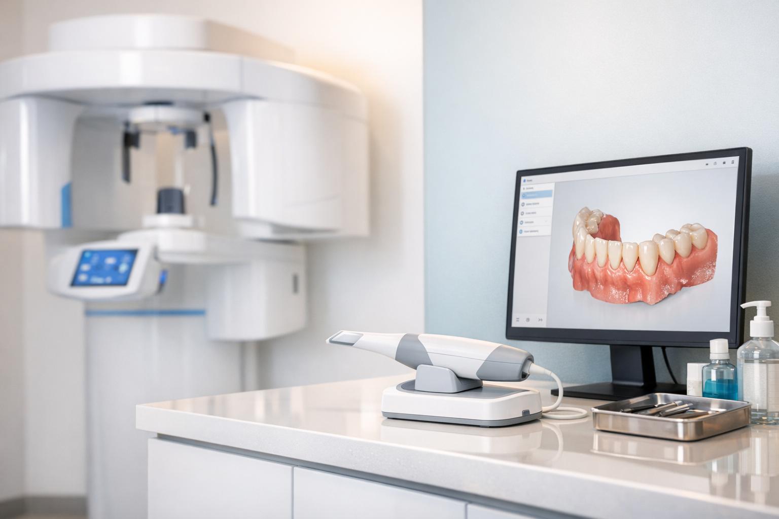
3D imaging has reshaped orthodontics by providing detailed, three-dimensional views of teeth, bones, and soft tissues. Unlike traditional 2D X-rays and physical moulds, it delivers more accurate diagnostics and treatment planning. Key technologies include Cone-Beam Computed Tomography (CBCT) and intraoral scanning, each offering unique benefits:
- CBCT: Captures high-resolution 3D images of internal structures like bone and tooth alignment, with up to 98% less radiation than medical CT scans.
- Intraoral Scanning: Creates precise digital impressions of teeth, replacing uncomfortable moulds with a handheld scanner.
This technology allows orthodontists to simulate treatments, accurately position appliances, and monitor progress. While 3D imaging involves higher costs and radiation exposure than 2D methods, its precision and efficiency make it indispensable for complex cases like impacted teeth or surgical planning.
For patients, it means faster, more comfortable procedures and better outcomes.
Craniofacial Imaging in Orthodontics | From Cephalometry to 3D CBCT Explained
What is 3D Imaging in Orthodontics?
3D imaging is reshaping orthodontic diagnostics by turning flat, 2D data into detailed 3D digital models [3][6]. With this technology, orthodontists can now examine teeth and bone structures from every angle, rather than relying on a flat image. It works using the Cartesian coordinate system, which maps anatomy along three axes: the x-axis (side-to-side), y-axis (up-and-down), and z-axis (front-to-back) [2]. This creates a precise digital replica of a patient’s oral structures, offering a more comprehensive view to guide treatment decisions.
How the Technology Works
The process starts with a Cone-Beam Computed Tomography (CBCT) scanner, which rotates around the patient’s head in just a few seconds, capturing hundreds of 2D images from multiple angles [7]. These images are then processed by specialised software to generate a 3D model. The software first creates a "wireframe" or "polygonal mesh" to outline the structure, then applies texture mapping for surface details [2]. The final step involves converting the data into voxels (three-dimensional pixels) to produce a realistic 3D object [2][1]. This lifelike model can be rotated, zoomed in on, and viewed from any angle on a computer screen, providing orthodontists with an unprecedented level of detail.
"The anatomical data collected from the patient are converted into a lifelike 3D object by the computer, and it can be viewed on the computer screen." – Oya Erten, Department of Orthodontics, Yeditepe University [2]
Main Uses in Orthodontic Practice
3D imaging goes far beyond simply capturing data – it has become a cornerstone of modern orthodontic practice. One of its primary applications is diagnosis, helping to identify issues like impacted or extra teeth and evaluating the interplay between facial soft tissues, the facial skeleton, and dentition [3][6]. It’s also essential for treatment planning, allowing orthodontists to create virtual simulations of patients and design customised appliances [2][4].
In addition, 3D imaging plays a key role in airway analysis, where pharyngeal volumes are measured to assess conditions like sleep apnoea. It’s also invaluable for TMJ assessment, enabling detailed visualisation of the temporomandibular joint to detect issues like erosion or structural irregularities [3][2]. These digital models are often stored as .stl files, making it easy to share them electronically with laboratories worldwide for appliance fabrication [5].
Types of 3D Imaging Technology
Orthodontic practices today utilise two main types of 3D imaging technologies, each tailored for specific roles in diagnosis and treatment planning. Cone-Beam Computed Tomography (CBCT) provides detailed volumetric images of bone and dental structures, while intraoral scanning captures highly accurate digital impressions of tooth surfaces. Together, these tools enhance precision in orthodontic care. Let’s dive into how each technology works and its specific uses.
Cone-Beam Computed Tomography (CBCT)
CBCT is a pivotal tool in orthodontics, offering a detailed look at the internal structures of the mouth and jaw. It works by emitting a cone-shaped X-ray beam through the patient, with a detector capturing the rays. During a single rotation – usually under 60 seconds – the system gathers hundreds of 2D images from different angles [1][9]. These images are then reconstructed into a 3D volumetric dataset using voxels, allowing orthodontists to view the data in multiple planes, such as axial, sagittal, and coronal [1][11].
This technology is especially useful for complex cases where traditional 2D X-rays fall short. For instance, CBCT is commonly used to:
- Locate impacted canines
- Evaluate bone defects in patients with cleft palates
- Plan orthognathic surgeries
- Assess pharyngeal airway volume in sleep apnoea cases
- Determine the best placement for temporary anchorage devices [2]
One major advantage of CBCT is its reduced radiation exposure compared to conventional medical CT scans. Depending on the field of view, radiation doses range from 45 μSv to 650 μSv – up to 98% less than standard CT devices [3][10].
"CBCT imaging provides accurate measurements, improves localisation of impacted teeth, provides visualisation of airway abnormalities, it identifies and quantifies asymmetry, it can be used to assess periodontal structures… and to view condylar positions." – Genevive L Machado, Al Salam Dental Centre [9]
While CBCT excels at internal structural imaging, intraoral scanning focuses on capturing surface-level details with unmatched precision.
Intraoral Scanning
Intraoral scanners offer a modern, patient-friendly alternative to traditional putty-based impressions. Using a handheld device equipped with a camera, orthodontists capture detailed images of the teeth and gums directly inside the mouth. These images are then processed by advanced software to create a high-resolution 3D model. Completing a full-arch scan typically takes about 15 minutes [7].
This digital approach eliminates the discomfort and inaccuracies often associated with traditional impressions, such as gag reflexes and dimensional distortions. The resulting digital models can be instantly shared with dental labs or used in virtual treatment simulations. Orthodontists commonly pair these scans with CAD/CAM technology to create customised appliances like clear aligners, lingual brackets, and retainers [2].
"Intraoral scanning technique has overcome the typical drawbacks of impression techniques like dimensional alterations in the impression materials, storage difficulty, and dental stone errors." – Fahad Abdullah Alshammery, Orthodontic Division, Riyadh Elm University [3]
The 3D Imaging Process: Step by Step
Knowing what to expect during a 3D imaging appointment can make the experience less daunting and more comfortable. The process is efficient, with most scans completed in just a few minutes. Here’s a breakdown of how it all works.
Patient Preparation and Positioning
Before the scan starts, patients need to remove any metal items – like jewellery, glasses, or removable dental appliances – to avoid interference with the image quality. Metal can create artefacts, which may distort the final 3D image. For cone-beam computed tomography (CBCT) scans, patients are positioned either sitting or standing within the machine. Chin supports and ear stabilisers are used to minimise movement, ensuring a clear scan. For intraoral scanning, patients remain seated in a standard dental chair while the clinician uses a handheld wand to capture images. Even minor movements can blur the scan and might require it to be repeated. To ensure safety, protective gear like lead aprons or collars may be provided during the procedure.
Image Capture
Once the patient is prepared and positioned, the scanning process begins. For CBCT scans, the X-ray source and detector rotate around the patient’s head, completing a 180° to 360° revolution in less than a minute [12]. This single revolution produces over 150 images, which are later compiled into a detailed dataset. In contrast, intraoral scanning involves a handheld camera that captures intricate images in just 2 to 5 minutes.
3D Model Creation and Analysis
After the images are captured, specialised software transforms the 2D data into a 3D volumetric dataset made up of voxels. This allows clinicians to examine the model from sagittal, coronal, and axial perspectives. For more intricate cases, like those requiring orthognathic surgery, CBCT skeletal data can be combined with 3D facial surface scans and intraoral dental scans through a process called image fusion. The result is a complete 3D model of the patient. These digital models can also integrate with CAD/CAM technology to design custom tools like clear aligners or to plan the precise placement of orthodontic brackets.
"CBCT imaging in association with computer software allows anatomical structures to be properly represented in all three viewing planes – sagittal, coronal, and transverse." – Genevive L Machado, Al Salam Dental Centre
CBCT scans capture a wide field of view, so orthodontists must thoroughly review the entire dataset for incidental findings – unrelated pathologies that show up in about 25% of scans [9]. These detailed models play a crucial role in accurate treatment planning and execution.
sbb-itb-2be92ed
How 3D Imaging Improves Treatment Planning
When orthodontists create a detailed 3D model of a patient’s dental structures, it opens the door to a level of precision that older methods simply couldn’t achieve. This technology allows clinicians to visualise how teeth, bones, and soft tissues will respond to treatment before any appliances are applied. This means more accurate predictions for tooth movement, better placement of appliances, and the ability to closely monitor treatment progress.
Predicting Tooth Movement
With 3D software, orthodontists can simulate the final outcome of a treatment plan before placing any brackets or aligners [2][3][5]. These virtual models can be rotated and examined to assess how specific tooth movements will affect both hard and soft tissues [3][6]. For more complex cases, combining CBCT scans with facial imaging creates a comprehensive digital model of the patient.
"The triad of teeth, the facial soft and the hard tissues has an extensive role in making plans related to orthodontic treatment" – Fahad Abdullah Alshammery, Orthodontic Division, Riyadh Elm University [3]
These projections also integrate seamlessly with CAD/CAM technology, enabling the design of custom appliances like clear aligners and pre-shaped archwires tailored to each patient [2][4][5].
Accurate Appliance Positioning
3D imaging takes the guesswork out of placing braces, brackets, and aligners. For example, orthodontists use indirect bonding trays, which are 3D-printed guides, to transfer brackets from a digital model onto a patient’s teeth with pinpoint accuracy [2][8]. Systems such as Insignia and SureSmile leverage this data to create personalised brackets and robotically shaped archwires [8]. For clear aligners, intraoral scans provide highly accurate digital impressions.
"The result is a perfectly measured, precisely cut, clear aligner that’s customised to your mouth to ensure it sits correctly over your teeth to help them shift into position" – Orthodontics Australia [4]
Additionally, by overlaying 3D models onto CBCT data, orthodontists can pinpoint the best locations for miniscrews based on bone density. With the help of 3D-printed surgical guides, these screws can be inserted with precision, avoiding sensitive areas like roots and nerves [2][8]. Continuous monitoring ensures that any adjustments remain in line with the original treatment plan.
Tracking Treatment Progress
3D superimposition allows clinicians to compare scans taken at different stages of treatment, making it easier to track tooth movement and evaluate how well appliances are working [2][6]. This is particularly important for ensuring proper root alignment and angulation, which are key to maintaining long-term stability [2]. For patients using clear aligners – often requiring 20 to 30 different aligners – regular scans are critical for ensuring treatment stays on course [4].
"Measurements performed on these images are imported to a computer, and then growth changes and treatment progress are assessed. Thus, stability and post-treatment assessment can be made with the help of 3D superposition" – Kayondo Wahab, Mulago Hospital [6]
If tooth movement doesn’t match initial predictions, orthodontists can make virtual adjustments and create new appliances tailored to the updated plan [2].
3D Imaging Compared to Older Methods
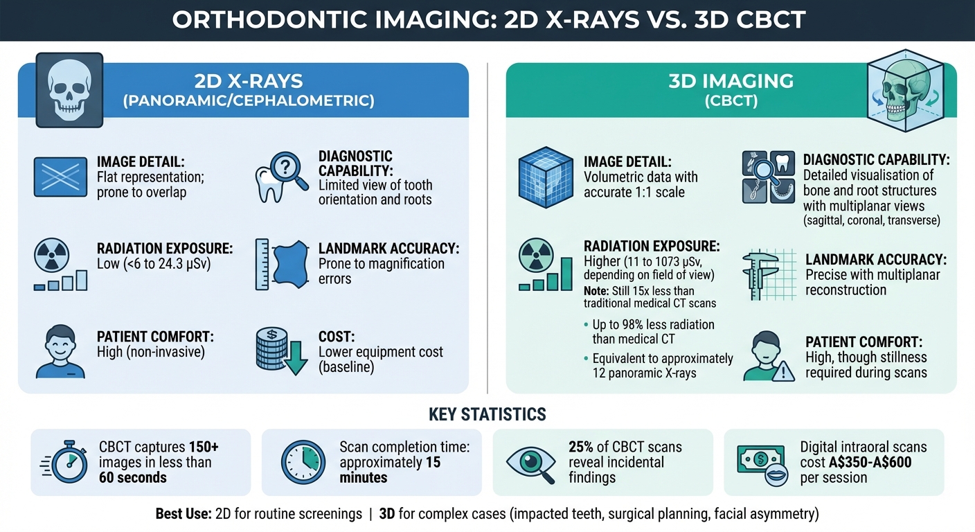
3D Imaging vs 2D X-Rays in Orthodontics: Radiation, Detail & Accuracy Comparison
Traditional methods like 2D X-rays and physical impressions had their shortcomings. With 2D X-rays, three-dimensional structures were flattened into a single plane, leading to overlapping anatomical features and making it tricky to pinpoint specific landmarks. Meanwhile, physical impressions, often taken using putty and trays, could be uncomfortable – especially for those with a strong gag reflex – and were prone to dimensional inaccuracies during the setting process.
Building on the earlier discussion about the precision of 3D imaging, let’s explore how it stacks up against these older techniques. 3D imaging solves many of these issues by capturing volumetric data at a true 1:1 scale with minimal distortion. This allows clinicians to view structures from multiple angles – sagittal, coronal, and transverse – offering unparalleled clarity of root locations and anatomical details. This capability is particularly useful for locating impacted canines and assessing the roots of adjacent teeth.
"3D imaging provided more detailed and realistic diagnostic information about the craniofacial hard as well as soft tissue and allowed to perform easier, faster, and more reliable 3D analyses." – Oya Erten, Department of Orthodontics, Yeditepe University [2]
However, one downside of 3D imaging is the higher radiation exposure. While cephalometric and panoramic X-rays emit between <6 and 24.3 μSv, a maxillofacial CBCT scan can range from 30 to 1073 μSv – roughly equivalent to 12 panoramic X-rays [2] [3]. That said, CBCT still exposes patients to about 15 times less radiation than traditional medical CT scans [6]. For routine screenings, 2D X-rays remain a practical choice, but for complex cases like impacted teeth, facial asymmetry, or surgical planning, the diagnostic advantages of 3D imaging often outweigh the increased exposure.
Digital intraoral scanning has also replaced physical impressions in many cases. These scans are faster, more comfortable, and easier to store or share with labs. Unlike bulky plaster models that can break or degrade, digital scans are durable and efficient. While the cost of a digital scan ranges from A$350 to A$600 per session, CBCT machines are significantly more expensive, costing 10–20 times more than 2D imaging setups [2] [7]. Despite the higher costs, the improved imaging detail and diagnostic capabilities contribute to better treatment planning and outcomes for patients.
Here’s a quick comparison of 2D X-rays and modern 3D imaging:
Comparison Table: 3D Imaging vs. 2D X‐Rays
| Factor | 2D X‐Rays (Panoramic/Cephalometric) | 3D Imaging (CBCT) |
|---|---|---|
| Image Detail | Flat representation; prone to overlap | Volumetric data with accurate 1:1 scale |
| Diagnostic Capability | Limited view of tooth orientation and roots | Detailed visualisation of bone and root structures |
| Radiation Exposure | Low (<6 to 24.3 μSv) | Higher (11 to 1073 μSv, depending on the field) |
| Landmark Accuracy | Prone to magnification errors | Precise with multiplanar reconstruction |
| Patient Comfort | High (non-invasive) | High, though stillness is required during scans |
Conclusion
3D imaging has changed the game in orthodontic diagnosis by overcoming the limitations of 2D methods. It provides clear, volumetric views of dental structures, allowing clinicians to examine tooth roots, bone, and soft tissues from multiple perspectives. This advancement significantly enhances diagnostic accuracy and treatment planning for orthodontic professionals [5].
Beyond diagnostics, the shift to digital workflows has brought a new level of convenience and comfort for patients. Intraoral scanners now replace the often uncomfortable traditional impressions, and CBCT scans can generate detailed diagnostic data in about 15 minutes [7]. This blend of precision, speed, and minimally invasive techniques has made orthodontic care more reliable and less daunting for patients.
"Practices like 3D scanning and printing have helped make treatment faster, more accurate and less invasive." – Orthodontics Australia [4]
For more complex cases, such as impacted canines, craniofacial abnormalities, or surgical planning, the benefits of 3D imaging are undeniable. Clinics like Complete Smiles Bella Vista are leveraging these advanced tools to enhance both treatment outcomes and communication with patients. The investment in such technology is a step towards achieving better results over the long term.
As digital tools continue to advance, the concept of creating "virtual patients" by merging various imaging techniques is on the horizon. This innovation promises to further refine orthodontic care. For now, 3D imaging stands as a cornerstone of precise, evidence-driven treatment in modern orthodontics.
FAQs
How does 3D imaging enhance orthodontic treatments?
3D imaging delivers detailed, three-dimensional visuals of your teeth, jaw, and the surrounding structures. This cutting-edge technology helps orthodontists diagnose issues more precisely, plan treatments with greater accuracy, and create custom-fitted appliances designed specifically for your needs.
With a clearer view of your dental anatomy, 3D imaging enhances treatment outcomes by making procedures more efficient, dependable, and often less invasive. It’s especially beneficial for addressing complex orthodontic cases, providing tailored solutions that lead to a smoother and more effective treatment process.
What’s the difference between CBCT and intraoral scanning in orthodontics?
CBCT (cone-beam computed tomography) and intraoral scanning are two cutting-edge 3D imaging tools widely used in orthodontics. While both are highly advanced, they cater to different needs and offer distinct types of information.
CBCT delivers detailed X-ray images of the teeth, jawbones, and surrounding structures, including bone anatomy, roots, and even the airway. This makes it especially valuable for diagnosing complex conditions, planning surgeries, and understanding skeletal relationships. However, because CBCT involves ionising radiation, it’s typically reserved for cases where its detailed imaging is absolutely necessary.
Intraoral scanning, by contrast, is a radiation-free technology that produces precise digital surface maps of the teeth and gums. It’s commonly used for creating clear aligners, dental restorations, and detailed treatment plans. While it doesn’t reveal internal structures like bone or roots, it’s quick, non-invasive, and extremely precise, making it a go-to option for many orthodontic and prosthetic applications.
In essence, CBCT offers a deeper look at internal structures, while intraoral scanning focuses on surface-level detail, each playing a unique role in modern orthodontic workflows.
Does 3D imaging expose you to more radiation than traditional dental X-rays?
Yes, 3D imaging techniques like cone-beam CT scans do expose patients to higher levels of radiation compared to standard 2D dental X-rays. That said, the radiation levels remain low and are deemed safe when used responsibly and for specific dental purposes.
Dentists take great care in determining whether 3D imaging is necessary, ensuring it’s only employed when the detailed insights it offers are crucial for precise diagnosis and treatment planning. This careful approach prioritises patient safety while leveraging the advantages of advanced imaging technology.
Related Blog Posts
- How 3D Imaging Improves Orthodontic Treatment Outcomes
- Digital Orthodontics: CAD/CAM Workflow Explained
- Recent Advances in Intraoral Scanning Technology
- CBCT Technology: Mechanisms Behind 3D Imaging
Important Notice: Any surgical or invasive procedure carries risks. Before proceeding, you should seek a second opinion from an appropriately qualified health practitioner.
Individual results may vary. The information provided in this article is for educational purposes only and does not constitute medical advice.
Checkout Related Blogs




Get in touch with us
For more information, call us now to start feeling better. Or fill the form below to make appointment
The Latest News from Complete Smiles
How to Clean Clear Plastic Retainers
Checklist for Choosing Wearable Dental Devices
Checklist for Choosing Cloud AI Platforms in Dentistry

Complete Smiles Bella VistaAccepts All Major Health Funds, Including





