


Recent Advances in Intraoral Scanning Technology
Intraoral scanners (IOS) are transforming dental care by replacing outdated impression methods with fast, precise, and patient-friendly digital scans. These devices create high-resolution 3D images of teeth, gums, and bite, improving accuracy and reducing discomfort. Here’s what you need to know:
- Uses: From crowns and bridges to clear aligners and implant planning, IOS supports a wide range of dental procedures with precision.
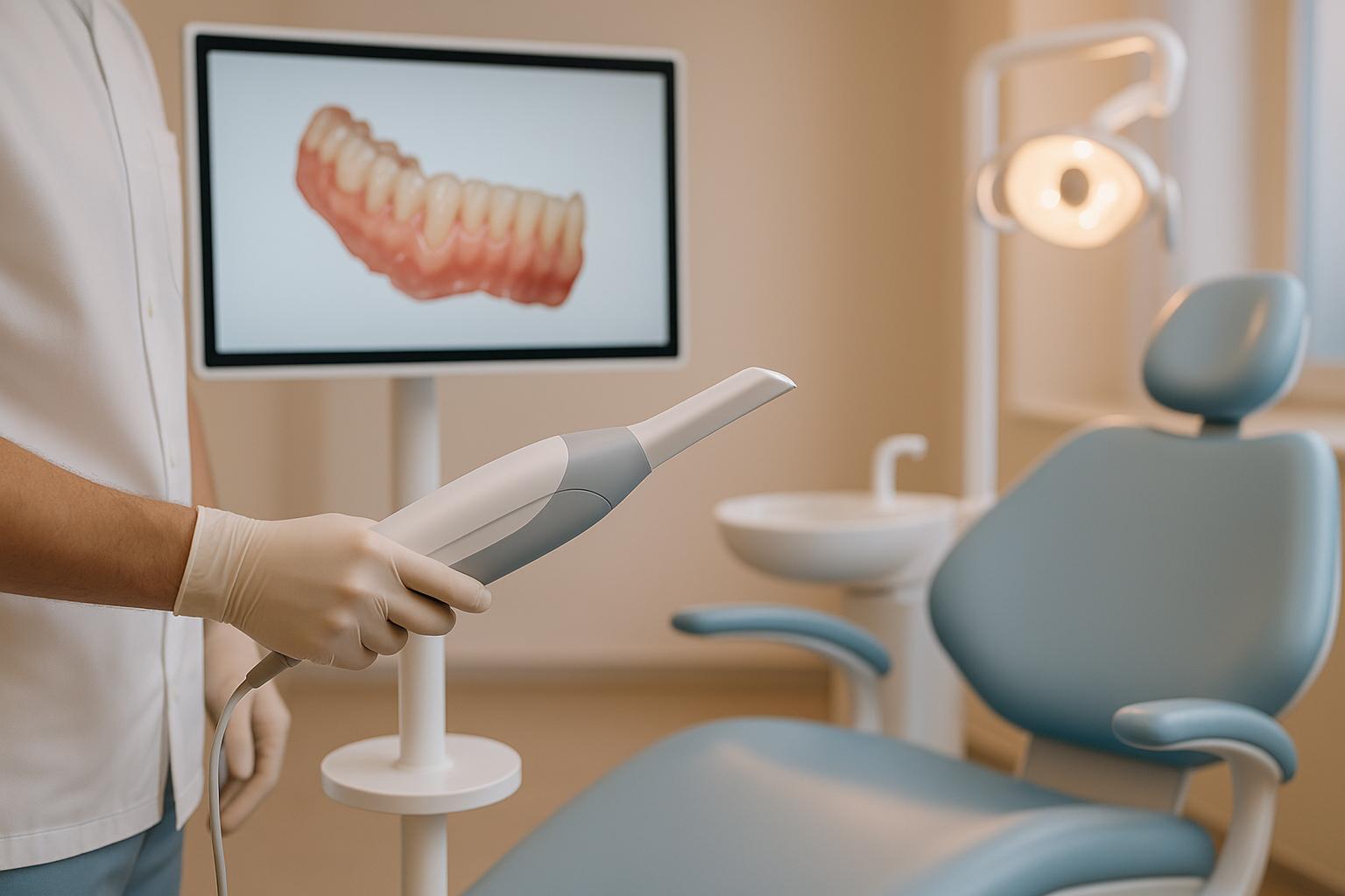
- Benefits for Patients: No more bulky trays or unpleasant materials – scans are quick (often under 30 seconds) and comfortable.
- Australian Context: Many clinics now use IOS as a primary tool, integrating them with CBCT and CAD/CAM systems for efficient workflows.
- Hardware Upgrades: Newer models like the iTero Lumina and Helios 600 feature better sensors, wireless designs, and faster processing, making them easier to use and more accurate.
- AI and Software: Advanced algorithms improve scan quality, detect errors in real-time, and even assist with diagnostics like early cavity detection and tracking tooth wear.
Despite advancements, challenges like full-arch accuracy and operator skill persist. Future developments focus on ergonomic designs, AI-driven analysis, and hybrid imaging systems for even greater precision and efficiency. For Australian clinics, adopting IOS means shorter appointments, improved outcomes, and streamlined workflows.
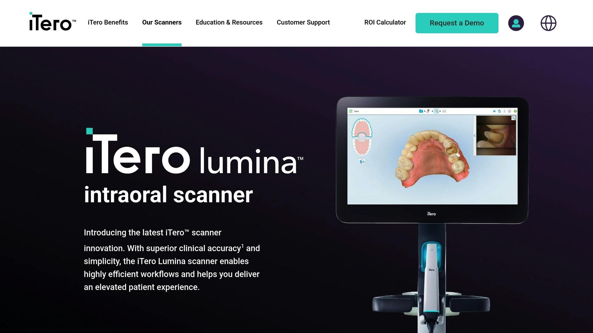
iTero Lumina™ vs. traditional intraoral scanners: A giant leap in dental tech
Hardware Improvements
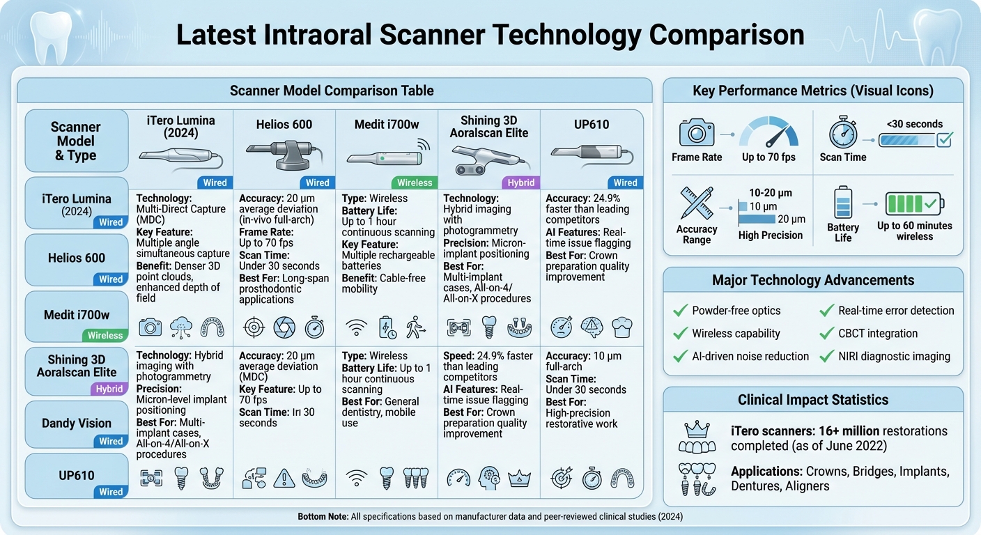
Intraoral Scanner Technology Comparison: Key Models and Performance Metrics
Better Optical and Sensor Technology
The latest intraoral scanners are pushing the boundaries of optical resolution and 3D imaging. With advancements like multi-direct capture (MDC) technology, devices such as the iTero Lumina – introduced in 2024 – are capable of capturing multiple images from various angles simultaneously. This results in denser 3D point clouds and enhances depth of field, making full-arch scans, including complex palatal anatomy and large edentulous spans, more reliable and detailed [1][6]. For example, the Helios 600 achieves an impressive average deviation of 20 μm in in-vivo full-arch scans, showcasing its suitability for precise long-span prosthodontic applications [6].
Incorporating high-resolution CMOS sensors with frame rates up to 70 fps, these scanners enable continuous image capture. This reduces the need for rescans, helping dental practices in Australia stay on schedule. With fewer voids and cleaner margins, lab workflows become smoother, cutting down on adjustment time during crown and bridge fittings [1][5][6].
Ergonomic and Wireless Designs
Beyond imaging advancements, ergonomic innovations are transforming clinical workflows. Newer scanners are smaller, lighter, and feature slimmer tips, making it easier to manoeuvre in hard-to-reach areas such as posterior regions or crowded lower anteriors [6][8]. These refinements reduce wrist strain and operator fatigue, which is especially valuable for clinicians performing multiple full-arch or Invisalign scans in a single session [6][8].
Wireless options like the Medit i700w are also making a splash, offering up to one hour of continuous scanning per charge. With multiple rechargeable batteries, these devices eliminate the hassle of cable management, enhancing mobility in tight-operatory spaces [6]. Additionally, many scanners now feature powder-free optics, removing the need for powder application and further simplifying the process [8][10]. Angled tips, textured grips, and other ergonomic tweaks improve scan stability, even in challenging cases involving children or patients with special needs [8].
These advancements collectively ensure high-quality scans across a broader range of patients while improving comfort for both clinicians and patients.
Hybrid Imaging Methods
Hybrid imaging systems are revolutionising implant dentistry by combining intraoral scanning with photogrammetry. These systems, such as the Shining 3D Aoralscan Elite, precisely measure the spatial position and angulation of multiple implants using specialised scan bodies [2]. This dual capability allows for the capture of implant coordinates with micron-level precision while simultaneously scanning soft tissue and occlusion. By avoiding traditional open-tray impressions or verification jigs, clinicians can create a complete digital record in a fraction of the time [2][1].
This streamlined approach significantly shortens appointments, enhances patient comfort, and minimises costly remakes [2]. It’s particularly beneficial for practices performing All-on-4 or All-on-X procedures, where predictable outcomes are critical. Clinics like Complete Smiles Bella Vista have embraced such workflows to deliver efficient, high-quality implant and restorative treatments [2].
These hardware advancements pave the way for cutting-edge software and AI enhancements, which further refine clinical accuracy and efficiency.
Software and AI Improvements
AI-Driven Noise Reduction and Data Processing
Modern intraoral scanners are equipped with AI-powered algorithms that handle tasks like stitching images together, removing artefacts, and detecting errors in real-time [1][6]. These advanced neural networks can distinguish actual anatomy from artefacts caused by saliva, soft tissue, patient movement, or metal restorations. They automatically clean up scans by deleting irrelevant segments, correcting surface irregularities caused by hand motion, and filling in gaps when the camera temporarily loses its target [1][6]. For busy dental practices in Australia, these advancements mean cleaner digital meshes for CAD designs, fewer rescans, and less time spent on chairside adjustments.
Machine learning also enhances image quality by refining edges, improving soft-tissue clarity, and stabilising colour consistency. This results in more precise margins and occlusal details, rivalling the accuracy of traditional impressions [1][5]. These improvements reduce common issues like voids, double images, and stitching errors, making it easier to achieve a predictable fit for crowns, bridges, and aligners. Beyond image quality, these AI-driven tools are paving the way for more advanced diagnostic applications.
Added Diagnostic Features
Some systems now include diagnostic tools that use fluorescence or near-infrared imaging (NIRI) to detect early interproximal lesions without exposing patients to ionising radiation [5][6]. For example, as of 30 June 2022, iTero digital scanners have been used in over 16 million restorations – including crowns, bridges, implants, and dentures. These scanners employ NIRI technology for real-time imaging, which not only aids in diagnosis but also enhances patient education [6].
Additionally, software modules can track changes over time, such as tooth wear and erosion. By comparing scans taken at different intervals, clinicians can monitor damage from bruxism, acid erosion, or other parafunctional habits. Some platforms go a step further by offering soft-tissue and gingival volume tracking, which helps identify early signs of recession or swelling. These features also provide high-quality visuals for preventive counselling [5][7]. However, while these tools are valuable for preventive care, they should complement rather than replace traditional radiographs. Their findings should be considered alongside a broader clinical and radiographic assessment [5][6]. These diagnostic features integrate seamlessly into digital workflows, as discussed in the next section.
Integration with Digital Workflows
Most modern scanners export files in open formats like STL, PLY, or OBJ, allowing seamless integration with chairside CAD software for designing crowns, onlays, and bridges, or with lab-based CAD systems for more complex restorations [1][7][9]. Cloud-based portals streamline the process by routing scans directly to labs, creating a fully digital workflow that speeds up the production of implant restorations, dentures, and other prostheses [1][7]. For Australian clinics, this digital chain reduces reliance on physical impressions and cuts laboratory costs. Some setups even allow direct integration with in-house 3D printers and milling machines, enabling same-day production of models and surgical guides [7][9].
Advanced systems now offer the ability to merge intraoral scans with CBCT datasets, creating a detailed 3D model that combines high-resolution surface anatomy with volumetric bone and root data [2][3][5]. This integration is especially useful for implant planning, as it aligns soft-tissue and tooth surface data with CBCT bone information, enabling the design of surgical guides and prostheses that balance both functionality and aesthetics [2][5]. For Australian clinics offering treatments like implants, veneers, and clear aligners, this technology supports streamlined, fully digital case management. AI also plays a role in automating tasks like classifying scan types (e.g., crown, bridge, implant, aligner), pre-filling lab prescriptions, and suggesting materials or margin designs. These features save administrative time and reduce the risk of communication errors [1][4][7].
sbb-itb-2be92ed
Clinical Performance and Applications
Restorative and Prosthodontic Uses
Recent studies show that restorations created from digital intraoral scans can match or even outperform those made using conventional impressions when it comes to marginal and internal fit for single crowns and short-span fixed dental prostheses [5]. Modern scanners capture intricate details like tooth preparations and finish lines with high optical resolution, reducing misfits and the need for chairside adjustments [1]. For example, the UP610 and Helios 600 scanners achieve full-arch accuracies of 10 µm and 20 µm respectively, completing scans in under 30 seconds. This not only reduces chair time but also minimises the need for adjustments [6][7].
Advances in optical systems and stitching algorithms have significantly reduced alignment errors, particularly in full-arch and multi-implant cases [1][2]. These scanners can now precisely capture soft-tissue contours and emergence profiles, enabling laboratories to design prostheses with better marginal fit and less soft-tissue irritation [1]. Some hybrid systems, combining photogrammetry with intraoral scanning, can map implant positions with micron-level precision, making them highly effective for multi-implant full-arch restorations [2]. By mid-2022, iTero scanners had been used in over 16 million restorations, including crowns, bridges, implants, and dentures, showcasing their widespread adoption [6].
Speed is another key improvement. Modern scanners are significantly faster than earlier models, directly cutting down chair time [4][7]. For instance, the Dandy Vision scanner is about 24.9% faster than other leading models and includes AI tools that flag issues in real time, helping to improve crown preparation quality [4][7]. This combination of speed and accuracy reduces errors, remakes, and emergency visits [1][5]. For Australian dental practices – where high clinic overheads, lab fees, and patient expectations are common – these advancements translate into shorter appointments and fewer remakes, improving efficiency without compromising quality.
These advancements in restorative procedures provide a strong foundation for improved outcomes in orthodontic treatments.
Orthodontics and Clear Aligners
Digital scans also play a crucial role in clear aligner treatments, where precise segmentation and surface detail are vital for effective tooth movement and aligner fit [5]. Advanced systems now use deep neural-network-based algorithms for accurate tooth segmentation and model creation, ensuring that individual teeth and gingival margins are captured with precision [5]. High-resolution scans eliminate distortions often seen with traditional impressions, leading to better-fitting aligners, fewer pressure points, and consistent force application throughout treatment stages. Clinical studies link accurate digital scanning with fewer mid-course corrections and fewer refinement aligners in complex cases involving tooth rotations or torque movements [5][7]. Additionally, the integration of iTero scanners with Invisalign has been associated with increased aligner case volume and practice growth [6].
Replacing conventional impressions, intraoral scanning enhances patient comfort and avoids common impression-related issues [5][8]. Digital models can be securely stored, eliminating the need for physical cast storage – a significant advantage for space-conscious Australian clinics. These scanners also support long-term monitoring of tooth wear, arch form, and soft-tissue changes, enabling side-by-side comparisons over time for growth assessments or tracking orthodontic relapse [5][7]. Real-time visualisation improves patient and parent understanding, encouraging treatment acceptance and aligning with Australian regulatory standards for informed consent.
Use with Special Populations
These technological advancements are particularly beneficial for patients with special clinical needs, offering safer and more comfortable scanning experiences. Compact, wireless, and powder-free designs make the process easier for children and those with anxiety or sensory sensitivities, as faster capture and a deeper depth of field reduce the need for repeated scans [6][8]. This shortens the time children need to keep their mouths open, improving cooperation in paediatric dentistry for procedures like creating digital records for space maintainers or early assessments [4][7][8].
For patients with intellectual disabilities, sensory processing challenges, or severe dental anxiety, intraoral scanning eliminates the distress often caused by traditional tray-based impressions [5][8]. The ability to pause and resume scans allows clinicians to work in short, manageable intervals, adapting to the patient’s comfort level [4][7]. Compact, wireless wands simplify positioning, particularly when working with patients in wheelchairs or those with limited head and neck mobility [6][8]. Australian clinics can further support these patients by offering desensitisation visits, using visual aids to explain the process, and scheduling longer appointments with breaks.
Intraoral scanning also reduces medical risks compared to traditional impressions, avoiding large trays and viscous materials that could compromise the airway or pose an aspiration risk for patients with neuromuscular or respiratory conditions [5][8]. Faster, less invasive scanning is particularly beneficial for patients who cannot lie flat or who tire quickly, such as those with cardiac or pulmonary conditions [5]. For cases involving cleft lip and palate or other craniofacial anomalies, digital scanning provides a safer alternative to deep palate impressions, avoiding potential airway issues [5]. While clinicians should still monitor vital signs and plan shorter appointments for high-risk patients, intraoral scanning generally offers a safer and more controlled method for data collection in these populations.
Current Challenges and Future Developments
Technical and Clinical Limitations
While digital scanning technology has come a long way, achieving full-arch accuracy remains a hurdle. Issues like cumulative stitching errors over longer spans, artefacts from reflective surfaces (such as metallic restorations, gold, or polished zirconia), and disruptions caused by saliva or soft-tissue movement can compromise precision – especially around subgingival margins [1][5]. Scanning edentulous ridges or mobile soft tissues presents additional difficulties compared to dentate arches, often requiring modified techniques or supplementary imaging for cases like complete dentures or extensive free-end saddles [5][6].
Operator experience plays a big role in scan quality. Inexperienced clinicians may face longer scan times and a higher likelihood of errors [5][6]. Patient-related factors can also complicate the process – limited mouth opening, strong gag reflexes, orthodontic appliances, or difficulty sitting still can all impact scan quality. Interestingly, in many Australian general dental practices, time constraints and diverse case types have led to hybrid approaches. Practitioners often rely on intraoral scans for diagnosis, temporaries, and short-span cases, while sticking to traditional impressions for more complex or high-risk situations [1][5]. These ongoing challenges are shaping the next wave of scanner advancements.
Future Trends and Developments
To tackle these challenges, future developments are focusing on ergonomic design and AI-driven improvements. Hardware innovations include smaller, lighter scan heads to enhance access to posterior areas and reduce operator fatigue. These features are particularly beneficial for paediatric or special-needs patients [6][7][8]. Wireless scanners with longer battery life are also expected, simplifying both positioning and infection control processes [8].
On the software side, AI-powered real-time analysis is being developed to identify issues like voids, insufficient reduction, or missing data during the scan itself. This means clinicians can address problems immediately, reducing the need for remakes or lab adjustments [4][6]. Emerging technologies like augmented reality (AR) and virtual reality (VR) could overlay proposed tooth positions or preparation guidelines directly onto the live scan, streamlining preparation and improving patient communication and consent [6].
Diagnostic capabilities are also evolving. Add-ons like caries detection using near-infrared or fluorescence imaging and tools for monitoring tooth wear and soft-tissue changes are becoming more reliable. These advancements could turn scanners into comprehensive diagnostic hubs [5][6]. For example, the iTero platform’s Near-Infrared Imaging allows for non-ionising detection of interproximal caries, offering a radiation-free alternative to traditional bitewing radiographs in specific cases [6]. Additionally, hybrid imaging systems that combine intraoral scanning with photogrammetry are emerging for multi-implant and full-arch cases. These systems provide micron-level accuracy for implant positioning, reducing the risk of framework misfit [2].
Impact on Multidisciplinary Care
As these technologies evolve, they are transforming collaborative care across dental specialties. Improved scanner accuracy and interoperability make shared digital records a reality, enabling restorative dentists, orthodontists, prosthodontists, and oral surgeons to work from the same 3D datasets [5][7]. This seamless integration supports multidisciplinary planning. For instance, surface scans can be combined with CBCT data for implant planning, orthognathic surgery simulations, or guided soft-tissue and bone procedures, enhancing communication between surgeons and restorative clinicians [2][5]. In orthodontics, precise segmentation and arch-form analysis facilitate coordinated treatment plans, such as creating space for implants or preparing minimally invasive veneers after alignment [5][6].
For paediatric and special-needs patients, digital scanning eliminates the need for traditional impression materials, which can improve cooperation and enable timely interdisciplinary care. This is especially valuable in cases like cleft care or craniofacial anomalies, where digital models can be easily shared among hospital-based and community providers [5]. Australian clinics that adopt structured digital workflows and shared planning platforms are likely to see smoother collaboration across specialties, shorter treatment times, and better outcomes for patients. Standardising scanning protocols – covering aspects like starting points, scan paths, soft-tissue retraction, and moisture control – can also help minimise operator variability and reduce errors, particularly in full-arch and implant cases [1][5].
Conclusion
Intraoral scanning has become a cornerstone of digital dental care in Australia. Today’s scanners offer impressive resolution and built-in diagnostic tools, often matching or even outperforming traditional impression methods [5]. Research shows that restorations created using intraoral scans are just as reliable – if not better – than those made from conventional impressions [5].
These advancements have also enabled seamless integration with CAD/CAM systems, aligner production, and CBCT workflows in Australian dental practices. This connectivity translates to quicker turnaround times, greater consistency, and improved patient comfort. In competitive urban and suburban clinics, where patients expect modern digital care, these benefits are game-changers [7].
When choosing an intraoral scanner, clinicians should rely on systems backed by strong, peer-reviewed evidence. Factors like scanner accuracy, compatibility with local dental labs, and the availability of training resources should guide decisions. Adopting proven scanners and standardised protocols can help clinicians build confidence, especially when managing more complex cases [1]. This shift is essential for improving clinical outcomes and enhancing the patient experience in Australia’s evolving dental landscape.
Looking to the future, emerging technologies such as advanced AI, hybrid photogrammetry, and augmented reality planning tools are set to push the boundaries even further [2]. These innovations promise to make dental workflows more efficient and patient-focused. Ultimately, intraoral scanning represents a practical, research-supported step forward in delivering higher-quality care. Continued research and professional development will be key to unlocking its full potential.
FAQs
How do intraoral scanners enhance dental procedure accuracy?
Intraoral scanners bring a new level of precision to dental procedures by producing detailed digital impressions. Unlike traditional moulds, which can sometimes be prone to inaccuracies, these scanners significantly reduce errors and lead to better-fitting dental restorations like crowns, veneers, and implants.
With their ability to capture accurate 3D images of teeth and oral structures, intraoral scanners also play a crucial role in effective treatment planning. They even assist in spotting dental issues early, contributing to improved results for both patients and dental practitioners.
What are the latest advancements in intraoral scanning technology?
Recent developments in intraoral scanning technology have brought some impressive upgrades. These include sharper imaging accuracy due to high-resolution capabilities, quicker scanning speeds to boost efficiency, and better ergonomic designs that prioritise patient comfort.
The latest models also come equipped with advanced software integration, enabling smooth digital workflows and more efficient data handling. These updates not only aid in delivering precise diagnostics but also simplify treatment planning, making intraoral scanners an essential asset in contemporary dental practices.
How is artificial intelligence improving intraoral scanning technology in dentistry?
Artificial intelligence (AI) is transforming intraoral scanning, bringing a new level of accuracy, efficiency, and precision to the process. Using advanced image processing, real-time data analysis, and automated error detection, AI ensures digital impressions are both highly detailed and dependable.
This technology doesn’t just make life easier for dental professionals – it also improves the patient experience. With fewer manual adjustments and better-quality scans, appointments become quicker and more comfortable. AI is raising the bar for what modern dental care can deliver.
Related Blog Posts
- How Intraoral Scanners Improve Orthodontic Workflows
- Future of Intraoral Scanners in Orthodontics
- 5 Benefits of AI in Laser Dental Treatments
- Training Programs for Intraoral Scanners in Australia
Important Notice: Any surgical or invasive procedure carries risks. Before proceeding, you should seek a second opinion from an appropriately qualified health practitioner.
Individual results may vary. The information provided in this article is for educational purposes only and does not constitute medical advice.
Checkout Related Blogs


Get in touch with us
For more information, call us now to start feeling better. Or fill the form below to make appointment
The Latest News from Complete Smiles
How to Clean Clear Plastic Retainers
Checklist for Choosing Wearable Dental Devices
Checklist for Choosing Cloud AI Platforms in Dentistry

Complete Smiles Bella VistaAccepts All Major Health Funds, Including


