


How 3D Dental Imaging Ensures Radiation Safety

3D dental imaging, specifically Cone Beam Computed Tomography (CBCT), provides detailed views of oral structures but involves higher radiation exposure than traditional X-rays. However, modern advancements and safety protocols significantly reduce risks while maintaining diagnostic accuracy.
Key Takeaways:
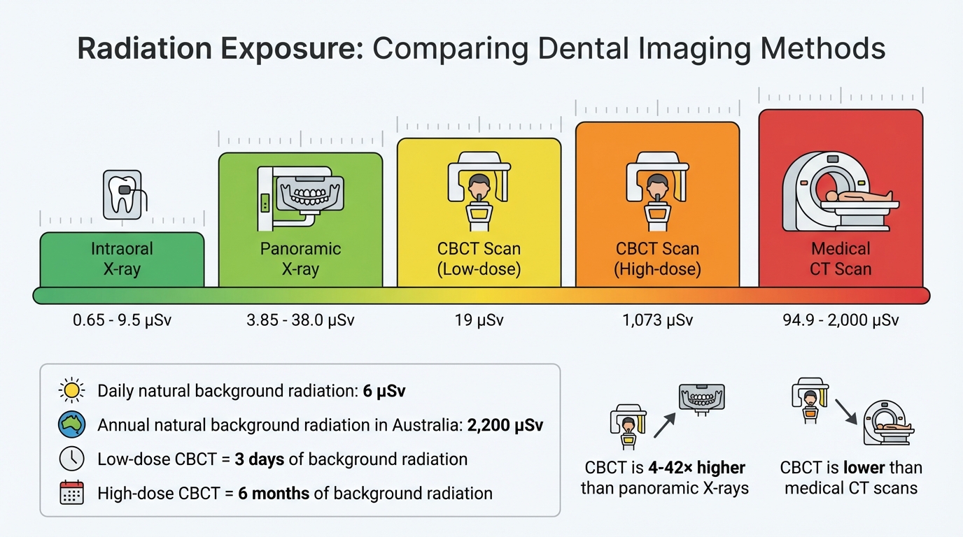
- Radiation Exposure: CBCT doses range from 19 µSv to 1,073 µSv, higher than 2D X-rays but much lower than medical CT scans (up to 2,000 µSv).
- Safety Measures: Following the ALARA/ALADA principle, smaller fields of view, optimised settings, and protective gear like thyroid collars reduce radiation risks.
- Technology Improvements: Features like pulsed radiation and high-sensitivity detectors lower doses while ensuring precise imaging.
- Regulations in Australia: Clinics must comply with strict radiation safety standards, including licensing, equipment registration, and regular calibration.
By combining advanced technology with rigorous safety standards, CBCT offers a safer option for 3D dental imaging while delivering the detail needed for accurate diagnoses.
3D CBCT Dental Imaging and Radiation Safety
Radiation Risks in Dental Imaging
Radiation Exposure Comparison: CBCT vs Other Dental and Medical Imaging
Radiation Dosage in CBCT
The radiation dose from CBCT scans varies depending on the equipment and protocols used, ranging from 19 µSv to 1,073 µSv [3]. This places CBCT between traditional 2D dental X-rays and medical CT scans in terms of radiation exposure. For comparison, a single intraoral X-ray delivers about 0.65 µSv to 9.5 µSv, while a panoramic X-ray typically ranges from 3.85 µSv to 38.0 µSv [3]. Full-field CBCT scans, however, can deliver doses 4 to 42 times higher than a single panoramic X-ray [4], though they still remain below the dose of medical multislice CT scans, which range from 94.9 µSv to 1,066.1 µSv [3].
To put these numbers into perspective, Australians are exposed to approximately 2,200 µSv of natural background radiation annually, equating to about 6 µSv per day [5]. A low-dose CBCT scan (19 µSv) corresponds to about three days of background radiation, while a high-dose scan (1,073 µSv) matches roughly six months of natural exposure [3][5]. This comparison underscores the importance of optimising CBCT protocols to minimise unnecessary radiation exposure while still achieving diagnostic accuracy.
Factors That Affect Radiation Levels
Several technical factors influence the radiation dose in CBCT imaging. Key determinants include the field of view (FOV), voxel size, and exposure settings such as tube voltage, current, and exposure time [2][3]. Larger FOVs increase exposure to radiosensitive tissues, such as the thyroid gland. Expanding the FOV’s height exposes additional sensitive areas, while increasing its width intensifies exposure to tissues already in the beam path [3][4][7]. Smaller voxel sizes, which improve image resolution, require higher radiation doses to maintain quality [3]. To balance safety and diagnostic needs, modern practices follow the ALADA principle (As Low As Diagnostically Acceptable), ensuring radiation doses are optimised for the specific diagnostic task [4].
Patient characteristics also play a significant role in radiation sensitivity. Children and adolescents are particularly vulnerable, with sensitivity levels up to 32 times higher than adults due to their rapidly dividing cells and longer lifespans [6]. Protective measures, such as using a thyroid collar, can significantly reduce exposure. For instance, thyroid collars lower the thyroid gland dose by 48.7% and the oesophageal dose by 41.7% during CBCT scans [7]. These measures highlight the importance of tailoring imaging protocols to protect vulnerable populations while maintaining diagnostic effectiveness.
Safety Protocols in 3D Dental Imaging
The ALARA Principle
Radiation safety in 3D dental imaging relies on the ALARA principle, meaning "As Low As Reasonably Achievable". Over time, this has evolved into ALADA, or "As Low As Diagnostically Acceptable", to strike a balance between minimising radiation exposure and achieving diagnostic quality. The focus is on acquiring images that are adequate for diagnosis without unnecessary detail that could increase radiation exposure [6][7].
The Australian Dental Association underscores this approach, stating:
"Attaining the lowest reasonably achievable radiation exposure and maximum diagnostic outcome is essential to all dental radiology." [8]
In practical terms, applying ALARA/ALADA involves using the smallest field of view (FOV) necessary for the specific diagnostic task. For instance:
- Small detectors (less than 10 cm) are ideal for dento-alveolar imaging.
- Medium detectors (10–15 cm) are used for jaw examinations.
- Large detectors (greater than 15 cm) are reserved for full maxillofacial imaging [7].
Every 3D scan must be justified, meaning it should only be performed when 2D imaging cannot provide the required diagnostic information [1][6]. For paediatric patients, protocols are adjusted with smaller FOVs and reduced exposure settings to account for their heightened sensitivity to radiation [6].
In addition to optimising the dose, physical protective measures are crucial to ensuring patient safety.
Protective Equipment and Measures
Physical shielding plays a key role in reducing radiation exposure during 3D dental imaging. For example:
- A properly fitted thyroid collar can lower radiation doses to the thyroid and oesophagus by 48.7% and 41.7%, respectively [7].
- Lead aprons protect the torso, while collimation ensures the X-ray beam is confined to the intended area [10].
Protecting staff is just as important. Operators should maintain a 2-metre distance from the X-ray source or use lead shielding during exposures [9]. Staff members who may receive an annual radiation dose above 1 mSv are advised to wear personal dosimetry badges to monitor their exposure. For pregnant staff, the exposure limit is set at 0.5 mSv per month [10].
Modern digital image receptors offer an added layer of safety by requiring lower radiation doses compared to traditional film. Regular equipment maintenance is also essential to avoid "dose creep" or malfunctions that could unintentionally increase radiation exposure [9][10].
sbb-itb-2be92ed
Modern CBCT Technology for Radiation Safety
Technology Features for Dose Reduction
Modern CBCT machines are designed with features that help lower radiation exposure without compromising diagnostic quality. One key innovation is pulsed radiation technology, which activates the beam only during specific phases, reducing the total exposure time [11].
Another advancement is the use of high-sensitivity detectors. These detectors efficiently capture X-rays, producing clear 3D images while requiring less radiation. Additionally, the ability to adjust voxel sizes enhances versatility – larger voxels (0.3 mm) are ideal for implant planning, while smaller ones (0.075 mm) provide the detail needed for endodontic analysis [11]. Features like scout views also play a role, helping ensure accurate patient positioning and minimising the need for repeat scans [11].
To put it into perspective, low-dose CBCT scans typically expose patients to 15–100 μSv, significantly lower than the 1,000–2,000 μSv associated with medical CT scans [11]. Some protocols even bring CBCT doses down to the 9–26 μSv range, comparable to panoramic X-rays [11]. These technological advancements, combined with proper equipment calibration, are key to maintaining safety.
Regular Maintenance and Calibration
While advanced imaging features are essential, regular maintenance ensures these benefits are sustained over time. Routine calibration is critical to avoid dose creep, ensuring that kVp, mA settings, and detector sensitivity remain accurate [12].
Daily checks are conducted to monitor CT number accuracy, image noise, and mechanical safety. Monthly tests focus on slice positioning and thickness, while semi-annual evaluations include dose profile width and resolution checks. Additionally, annual dose measurements performed by a medical physicist confirm compliance with safety standards [12].
Keeping a detailed logbook of patient examinations, repairs, and test results is equally important. This documentation not only tracks equipment performance but also demonstrates adherence to Australian radiation safety regulations, reinforcing a commitment to patient safety over time.
Regulatory Compliance and Best Practices in Australia
Australian Radiation Safety Standards
In Australia, radiation safety is managed by state and territory regulations, each with its own licensing, registration, and compliance rules [14]. At the national level, the Radiation Protection Series C-7: Code for Radiation Protection in Dental Exposure, published by the Australian Radiation Protection and Nuclear Safety Agency (ARPANSA), serves as the key guideline for dental imaging safety [13]. This code establishes the core standards to ensure patient safety during dental X-rays.
"The Board and practice accreditation authorities should use the guidelines provided by the Australian Radiation Protection and Nuclear Safety Agency (ARPANSA) as the standard for radiation safety in dentistry." – Australian Dental Association [8]
Before installing a CBCT machine, dental clinics must secure a radiation licence from their respective state or territory regulator. Additionally, all equipment must be registered, and operators are required to hold AHPRA registration and have specific training in 3D volumetric X-ray systems [15]. For instance, Queensland implemented annual compliance testing for CBCT units in September 2021. The cost of maintaining compliance is estimated to be about 0.076% of a three-chair practice’s average annual turnover over a five-year period [14].
This regulatory framework ensures that dental imaging consistently prioritises patient safety, laying the groundwork for robust internal safety protocols.
Clinic Safety Protocols
Before using a CBCT machine, clinics must obtain a shielding report from an accredited consultant. This report assesses the protection required based on factors like machine workload, radiation output, and the surrounding area’s occupancy levels [14]. Even machines advertised as "low-dose" still require this evaluation, as CBCT systems emit significantly more radiation compared to standard OPG (panoramic) units.
In addition, clinics must implement a Radiation Safety Management Plan. This plan should include detailed safety procedures, emergency guidelines, regular compliance testing, certification, and thorough record-keeping to ensure ongoing safety improvements [14]. These measures align with the ALARA/ALADA principles, reinforcing the importance of minimising radiation exposure while leveraging advanced CBCT technologies.
Conclusion
3D dental imaging provides accurate diagnostic capabilities when used alongside stringent radiation safety measures. In Australia, the focus remains on justification – ensuring every CBCT scan is clinically warranted – and optimisation, which involves using the smallest field of view and the lowest radiation dose necessary to achieve a diagnostically acceptable image [6]. These principles shape both clinical practices and the upkeep of imaging equipment.
For dental professionals, this entails maintaining properly calibrated equipment, ensuring operators are adequately trained, and strictly following regulatory guidelines [15]. While CBCT scans do involve higher radiation exposure compared to standard panoramic X-rays, the diagnostic advantages often outweigh the minimal associated risks for patients [6].
FAQs
Is 3D dental imaging, like CBCT, safe in terms of radiation exposure?
3D dental imaging, like CBCT (cone-beam computed tomography), is carefully designed to prioritise safety, adhering to strict protocols to minimise radiation exposure. While CBCT does involve higher radiation levels compared to traditional 2D X-rays, advances in technology and strict adherence to the ALARA principle (As Low As Reasonably Achievable) help keep exposure as low as possible.
CBCT offers detailed 3D visuals that support precise diagnosis and treatment planning. Although the radiation dose is higher than that of a standard panoramic X-ray, it’s still much lower than what you’d receive from a medical CT scan. Dentists carefully evaluate whether CBCT imaging is necessary, ensuring it’s used only when the benefits clearly outweigh the risks. Additionally, equipment settings are optimised to further reduce exposure.
If radiation safety is on your mind, rest assured that dental professionals in Australia follow rigorous guidelines to safeguard your health while providing top-quality care.
How does 3D dental imaging keep radiation exposure to a minimum?
3D dental imaging, like CBCT scans, is carried out under strict safety guidelines to keep radiation exposure to a minimum while delivering precise diagnostic results. This involves using advanced equipment that optimises radiation doses, customising scan settings based on individual patient requirements, and only performing scans when there’s a clear clinical need.
Dental clinics in Australia follow rigorous radiation safety standards and incorporate patient-focused safety measures into their facility designs. These efforts ensure patients can benefit from 3D imaging with minimal associated risks.
How does 3D dental imaging minimise radiation exposure?
3D dental imaging is built on advanced technology and prioritises safety through strict protocols, ensuring minimal radiation exposure for both patients and dental practitioners. The process adheres to principles like ALARA (As Low As Reasonably Achievable) or ALADA (As Low As Diagnostically Acceptable), enabling professionals to capture high-quality diagnostic images while keeping radiation levels to a minimum.
Today’s 3D imaging systems are designed to target specific areas with precision, using efficient techniques that limit exposure to only the necessary regions. This targeted approach not only safeguards patient health but also ensures the accuracy required for precise diagnosis and effective treatment planning.
Related Blog Posts
- CBCT in Endodontics: Accuracy and Limitations
- Guide to Periapical X-rays for Root Canal Therapy
- CBCT Technology: Mechanisms Behind 3D Imaging
- 3D Imaging in Orthodontics: How It Works
Important Notice: Any surgical or invasive procedure carries risks. Before proceeding, you should seek a second opinion from an appropriately qualified health practitioner.
Individual results may vary. The information provided in this article is for educational purposes only and does not constitute medical advice.
Checkout Related Blogs


Get in touch with us
For more information, call us now to start feeling better. Or fill the form below to make appointment
The Latest News from Complete Smiles
How to Clean Clear Plastic Retainers
Checklist for Choosing Wearable Dental Devices
Checklist for Choosing Cloud AI Platforms in Dentistry

Complete Smiles Bella VistaAccepts All Major Health Funds, Including


