





Minimally Invasive Periodontal Surgery Techniques
Minimally Invasive Periodontal Surgery (MIPS) is reshaping how gum disease is treated. Instead of large incisions and lengthy recovery times, MIPS uses advanced tools like microsurgical instruments and magnification systems to target problem areas with minimal tissue disruption. The result? Less pain, faster healing, and better preservation of gum tissue.
Key Points:
- Precision Tools: Small incisions, sharp microsurgical blades, and high magnification (20x–60x) ensure accurate treatment while preserving healthy tissue.
- Faster Recovery: Patients experience significantly less discomfort and swelling compared to conventional methods.
- Better Outcomes: Studies show reductions in probing depth (up to 4.1 mm) and improved attachment levels (up to 4.2 mm), with stable results over years.
- Advanced Technology: Videoscopes, perioscopes, and lasers enhance visibility and precision during procedures.
- Techniques: Includes Minimally Invasive Surgical Technique (MIST), periodontal microsurgery, and micro-perioscopy, all tailored for specific gum defects.
While MIPS offers clear benefits, it requires specialised equipment and training, which may limit its accessibility. It’s most effective for specific cases, such as isolated bone defects, and less suited for generalised conditions. If you’re considering MIPS, consult a qualified professional to determine if it’s right for you.
Minimally Invasive Surgical Technique (MIST) – Dr. Fabio Cozzolino
Core Principles of Minimally Invasive Techniques
Minimally Invasive Periodontal Surgery (MIPS) revolves around two key ideas: preserving tissue and performing procedures with surgical precision. These methods aim to reduce trauma and promote faster healing by working on specific areas with the smallest possible incisions[2]. Instead of creating large openings for access, MIPS prioritises protecting the structures that are vital for healing.
Small Incisions and Precise Techniques
One of the cornerstones of MIPS is maintaining the blood supply to soft tissues. Traditional methods often involve extensive flap elevation, which can disrupt circulation. In contrast, MIPS employs sharp microsurgical blades to make isolated, intrasulcular incisions that safeguard blood flow and preserve the interdental papilla[2][6].
Stability of the wound is another critical factor. By limiting flap reflection to expose only 1–2 mm of bone, surgeons create an environment where the blood clot remains secure and protected from the oral cavity[2][4]. This approach also helps retain the periosteum during flap elevation, reducing the risk of crestal bone resorption – a common issue with traditional techniques[2][4].
"The primary aim of the proposed procedure was to minimise tissue trauma due to reflection and manipulation that resulted in better stabilisation of the blood clot with reduced surgical morbidity." – Harrel and Rees, Periodontists[2]
Sutures are carefully placed to ensure the wound closes without excessive tension. Suture bites are sized at 1.5 times the tissue thickness for optimal approximation[2]. Additionally, by avoiding vertical releasing incisions, the technique minimises postoperative gingival recession and supports better healing.
Advanced Tools and Technology
To complement the minimal incision approach, advanced tools enhance surgical accuracy. Magnification systems, such as surgical loupes, operating microscopes, and videoscopes with up to 60X magnification, play a crucial role in these procedures[2][1]. For example, a 2.7 mm flexible videoscope equipped with a high-definition camera and a gas shield ensures a clear view by preventing blood from obscuring the lens[2][5].
Microsurgical instruments, designed to weigh between 15–20 g, reduce fatigue for surgeons during extended procedures[2]. These tools include micro-scissors, micro-raspatoriums, and specialised papilla elevators, all tailored for precision.
Another game-changer is the use of periodontal endoscopes (perioscopes), which allow surgeons to examine subgingival areas and defects without the need for extensive flap elevation. This enables non-incisional regenerative techniques[1][6]. Additionally, laser technologies like Nd:YAG, diode, and erbium lasers are used for bacterial reduction and debridement, causing minimal tissue damage[4]. Together, these advanced tools provide surgeons with unparalleled visual clarity and precision, previously achievable only through larger incisions[4].
Minimally Invasive Periodontal Surgery Techniques
The techniques under the Minimally Invasive Periodontal Surgery (MIPS) framework aim to address periodontal defects while minimising tissue trauma. These include the Minimally Invasive Surgical Technique (MIST), periodontal microsurgery, and micro-perioscopy. By focusing on small, intrasulcular incisions and improved visualisation, these methods achieve reliable results while reducing patient discomfort.
Minimally Invasive Surgical Technique (MIST)
MIST, developed by Cortellini and Tonetti, uses intrasulcular incisions to preserve the gingiva’s height and width. The method includes modified or simplified papilla preservation flaps to access periodontal defects [7][2]. A variation, the Modified-MIST (M-MIST), involves lifting a minimal buccal flap while keeping the interdental papilla and palatal flap intact. This creates what Cortellini and Tonetti describe as a stable "roof" over the defect:
"The interdental soft tissues are the stable ‘roof’ of a room where the blood fills in and forms a clot. The hanging papilla prevents the collapse of the soft tissues, thereby maintaining space for regeneration." [7]
Clinical studies highlight the effectiveness of MIST. For instance, combining MIST with Enamel Matrix Derivative (EMD) has demonstrated a mean clinical attachment level gain of 4.8 ± 1.9 mm, a pocket depth reduction of 4.8 ± 1.8 mm, and a 92% rate of primary wound closure – significantly higher than the 70% achieved with traditional methods [7]. These advancements lay the groundwork for even more precise microsurgical techniques.
Periodontal Microsurgery
Periodontal microsurgery enhances precision by using operating microscopes with magnification ranging from 20× to 60×. This detailed view allows surgeons to assess defects with exceptional clarity and perform fine suturing with 6‑0 to 9‑0 micro-sutures, paired with 6.6‑mm spatula needles [2].
Micro-Perioscopy and Regenerative Methods
Micro-perioscopy further reduces invasiveness by avoiding extensive flap elevation. Using periodontal endoscopes (perioscopes), clinicians can directly visualise subgingival areas and root surfaces, enabling techniques like the Non‑Incisional Regeneration Technique (NIT) [6].
A variation, Videoscope-Assisted Minimally Invasive Surgery (VMIS), employs a 2.7‑mm flexible tube with a high-definition camera, providing 20× to 60× magnification on a monitor. A controlled gas shield ensures a clear view throughout the procedure [2][5]. In a 2016 study, Dr Stephen K. Harrel and his team treated 18 patients with residual pockets ≥5 mm using VMIS. After one year, they reported a mean probing depth reduction of 4.11 ± 0.98 mm and a clinical attachment level gain of 4.58 ± 1.19 mm [2].
These techniques are often combined with regenerative materials like bone grafts, Enamel Matrix Derivatives (EMD), and growth factors to encourage new bone and ligament formation. The Non‑Incised Papillae Surgical Approach (NIPSA) avoids cutting at the papilla base, preserving the vascular supply. A 2019 study by Moreno Rodríguez et al. found that NIPSA resulted in only 0.2 ± 0.41 mm of gingival recession after one year, compared to 0.73 ± 0.88 mm with standard MIST [6].
sbb-itb-2be92ed
MIPS vs Traditional Periodontal Surgery
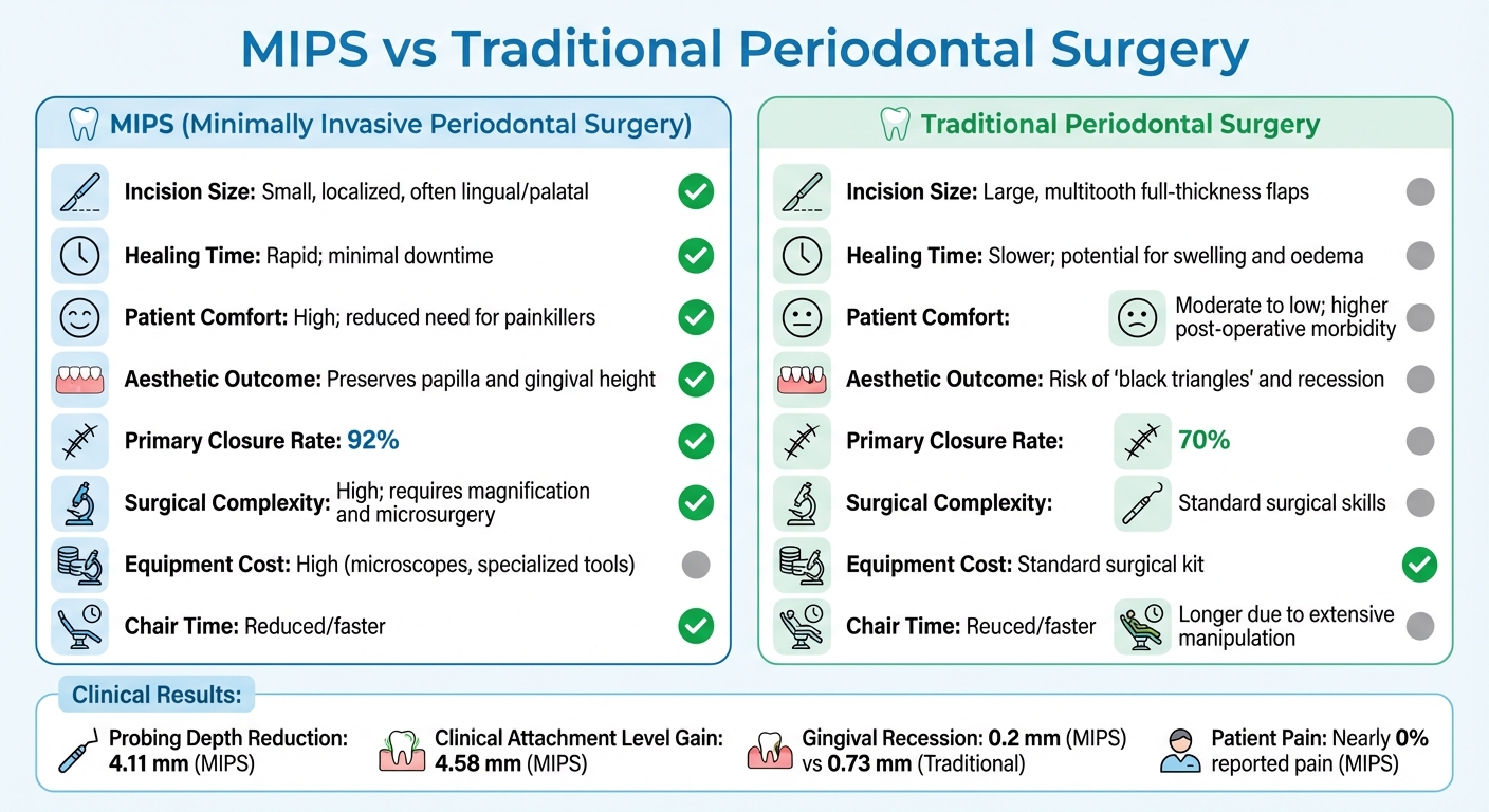
MIPS vs Traditional Periodontal Surgery: Key Differences and Outcomes
When it comes to restoring gum and bone health, MIPS (Minimally Invasive Periodontal Surgery) and traditional periodontal surgery share the same goal. However, they differ significantly in their techniques, recovery processes, and overall outcomes. Below, we explore the distinct advantages and challenges of each approach.
Advantages of MIPS
One of the standout benefits of MIPS is its faster healing and reduced discomfort. According to a Visual Analogue Scale survey, nearly all patients treated with videoscope-assisted MIPS reported no pain on the day of surgery or during follow-ups [5]. In contrast, traditional surgery often leads to noticeable post-operative swelling and discomfort.
Another major advantage is the preservation of soft tissue. Traditional surgery can result in gingival recession and unsightly "black triangles" between teeth. MIPS, on the other hand, maintains papilla height and gingival contour. Research shows that after one year, MIPS patients experienced only 0.2 ± 0.41 mm of gingival recession, compared to 0.73 ± 0.88 mm with standard surgical techniques [1][6].
MIPS also boasts superior wound closure rates. Microsurgical techniques used in MIPS improve primary wound closure from an average of 70% with traditional surgery to an impressive 92% [8]. This better closure protects the blood clot, leading to more effective regenerative outcomes. Dr Stephen K. Harrel highlights the importance of this by stating:
"Maintenance of the blood supply to the flap and minimal trauma of the tissue are critical to the success of all regenerative surgical procedures" [5].
Additionally, MIPS offers reduced surgical time, which benefits both patients and clinicians. While the procedure relies on advanced technology, it often involves shorter chair time due to limited flap reflection and precise handling of tissues [1][2]. Patients typically experience less disruption and require fewer painkillers [8][5].
However, despite these advantages, MIPS is not without its challenges.
Disadvantages of MIPS
A key drawback of MIPS is its high equipment and material costs. The technique demands expensive tools such as operating microscopes, videoscopes, and specialised titanium microsurgical instruments [2][4]. Additionally, biomaterials like Enamel Matrix Derivatives, bone grafts, and bioresorbable membranes contribute to the overall expense [8][5].
Another limitation is the technical complexity of the procedure. MIPS requires a high level of surgical precision, necessitating advanced training for practitioners. Surgeons must master the specialised tools and techniques, which are not universally accessible [4][8].
Finally, MIPS has limited applicability. It is most effective for isolated interproximal defects, particularly three-wall intrabony defects. For cases involving generalised horizontal bone loss or significant tooth mobility, traditional surgery may remain the preferred option [1][5][3].
Comparison Table: MIPS vs. Traditional Surgery
| Feature | MIPS | Traditional Surgery |
|---|---|---|
| Incision Size | Small, localised, often lingual/palatal | Large, multitooth full-thickness flaps |
| Healing Time | Rapid; minimal downtime | Slower; potential for swelling and oedema |
| Patient Comfort | High; reduced need for painkillers | Moderate to low; higher post-operative morbidity |
| Aesthetic Outcome | Preserves papilla and gingival height | Risk of "black triangles" and recession |
| Primary Closure | Approximately 92% | Approximately 70% |
| Surgical Complexity | High; requires magnification and microsurgery | Standard surgical skills |
| Equipment Cost | High (microscopes, specialised tools) | Standard surgical kit |
| Chair Time | Reduced/faster | Longer due to extensive manipulation |
This comparison highlights the advantages of MIPS in terms of healing, aesthetics, and patient comfort, while also acknowledging its higher costs and complexity. On the other hand, traditional surgery remains a more accessible option for a broader range of periodontal conditions.
Clinical Evidence Supporting MIPS
Proven Outcomes in Periodontal Treatment
MIPS has shown consistent results in reducing probing depth (PD) and improving clinical attachment level (CAL), all while maintaining the interdental papilla.
Between 2016 and 2017, Tunnell and Harrel studied 30 patients across 110 sites using Videoscope-Assisted MIPS. Over a follow-up period ranging from 36 to 58 months, they observed an average reduction in probing depth from 6.41 mm to 2.61 mm, alongside a clinical attachment level improvement of 4.16 mm. Additionally, the technique delivered a soft-tissue height gain of 0.36 mm. Impressively, 100% of the patients indicated they would recommend the procedure, further underscoring its effectiveness [5].
Another study focused on NIPSA with Enamel Matrix Derivative (EMD) in treating intrabony defects found a mean PD reduction of 8.25 ± 2.70 mm and a CAL gain of 8.33 ± 2.74 mm after a year [6]. Similarly, AICAST demonstrated CAL gains of ≥6 mm in 88.9% of treated sites, with all cases achieving a residual PD of ≤3 mm [6].
These clinical advancements don’t just stop at measurable outcomes – they also make a noticeable difference in how patients experience the treatment.
Patient-Centred Benefits
Beyond the clinical metrics, MIPS stands out for its patient-friendly approach, offering greater comfort and fewer postoperative issues. From March 2019 to November 2021, researchers at the University Dental Clinic of Ljubljana, Slovenia, conducted a split-mouth study involving 20 patients with Stage III periodontitis. Led by Alja Cmok Kučič and Rok Gašperšič, this study revealed that Minimally Invasive Non-Surgical Therapy (MINST) resulted in significantly less gingival recession in deep molar pockets. The median recession was 0.00 mm for the MINST group, compared to 0.32 mm in the control group. Notably, participants reported no postoperative pain or the need for analgesics [9].
The emphasis on preserving tissue is evident. While the standard MIST technique resulted in a recession increase of 0.73 ± 0.88 mm, the NIPSA method limited recession to just 0.2 ± 0.41 mm [6]. This difference is especially important for the aesthetic zone, where even minor gingival recession can impact appearance. Sylwia Jakubowska from the Medical University of Warsaw highlights the significance of this:
"Preserving papilla integrity and the soft tissue profile is essential for minimising complications, especially in the aesthetic zone" [6].
Conclusion
Minimally invasive periodontal surgery (MIPS) is changing the game in gum disease treatment, offering effective outcomes with less discomfort. Techniques like MIST (Minimally Invasive Surgical Technique), periodontal microsurgery, and non-incisional methods deliver impressive results. They not only reduce probing depth and improve clinical attachment levels but also help preserve the delicate soft tissue structure that traditional surgeries often disrupt.
Clinical evidence strongly supports MIPS. Studies consistently show notable reductions in probing depth, better attachment levels, and minimal gingival recession. On top of that, patients experience less postoperative pain and swelling compared to conventional methods [4][5][6][8].
However, MIPS isn’t a one-size-fits-all solution. Success depends on selecting the right cases, which means considering the bone defect’s morphology and ensuring the patient can maintain excellent oral hygiene [8]. These factors play a crucial role in achieving the best possible outcomes.
Since MIPS requires specialised skills and equipment, it’s important to consult a qualified dental professional. They’ll evaluate your specific periodontal condition and determine if a minimally invasive approach is the right fit for you.
With the right care and guidance, MIPS aims to treat periodontal disease and support tissue regeneration, all while keeping disruption to a minimum [4].
FAQs
What gum issues can be treated with minimally invasive periodontal surgery?
Minimally invasive periodontal surgery is a targeted approach for treating intrabony defects, receding gum lines (recession-type defects), and deep periodontal pockets resulting from periodontitis. These procedures aim to reduce discomfort, encourage quicker recovery, and protect healthy tissue while addressing particular gum concerns.
For personalised advice, it’s important to consult a qualified dental professional to see if this treatment is right for you.
What are the recovery benefits of minimally invasive periodontal surgery compared to traditional methods?
Minimally invasive periodontal surgery is known for offering quicker recovery and reduced post-surgical discomfort compared to traditional approaches. This is largely due to the use of smaller incisions and techniques that limit trauma to the surrounding tissues.
A key advantage of these methods is their emphasis on preserving healthy tissue. By doing so, they promote faster healing, minimise swelling, and often decrease the reliance on pain relief. For many patients, this means a smoother recovery and a quicker return to their usual routines.
Are there any limitations to minimally invasive periodontal surgery for treating generalised gum disease?
Minimally invasive periodontal surgery, while effective in certain cases, does have its limitations when dealing with generalised gum disease. It may not be the best option for addressing all types of gum defects, especially when it comes to deep or advanced lesions. Moreover, the procedure often relies on specialised tools like microscopes and micro-instruments, which aren’t always readily available.
When it comes to generalised gum disease, the results – such as reducing pocket depth or improving attachment levels – may not always outperform those achieved with traditional treatments. To find the most suitable approach for your condition, it’s crucial to discuss your options with a qualified periodontist.
Related Blog Posts
- Advances in Gum Grafting Materials and Techniques
- Minimally Invasive vs. Traditional Gum Surgery: Key Differences
- Minimally Invasive vs Conventional Flap Surgery
- Flapless Surgery: Advances in Periodontal Care
Important Notice: Any surgical or invasive procedure carries risks. Before proceeding, you should seek a second opinion from an appropriately qualified health practitioner.
Individual results may vary. The information provided in this article is for educational purposes only and does not constitute medical advice.
Checkout Related Blogs




Get in touch with us
For more information, call us now to start feeling better. Or fill the form below to make appointment
The Latest News from Complete Smiles
How to Clean Clear Plastic Retainers
Checklist for Choosing Wearable Dental Devices
Checklist for Choosing Cloud AI Platforms in Dentistry

Complete Smiles Bella VistaAccepts All Major Health Funds, Including





