





AI vs. Traditional Oral Cancer Screening
AI is reshaping how oral cancer is detected, offering consistent accuracy, while traditional methods rely on clinician expertise. Here’s what you need to know:
- Traditional Screening (Clinical Oral Examination – COE): Relies on visual inspection and tactile checks during general dental exams. Highly specific (94%-99%) but varies in sensitivity (50%-99%) depending on clinician skill.
- AI-Based Screening: Uses machine learning to analyse images, saliva, and breath samples. Sensitivity averages 87%, specificity 81%, with histopathology analysis reaching 97% sensitivity.
- Key Differences:
- AI is objective and consistent but dependent on quality datasets.
- COE is accessible and cost-effective but subjective and experience-dependent.
- Combined Approach: AI complements COE by acting as a second opinion, improving early detection rates and reducing diagnostic delays.
Quick Comparison:
| Feature | COE | AI-Based Screening |
|---|---|---|
| Sensitivity | 50%-99% | 87%-97% |
| Specificity | 94%-99% | 81%-95% |
| Speed | Manual; time-intensive | Automated; near real-time |
| Cost | Low initial; higher later | Affordable remote options |
| Subjectivity | High; clinician-dependent | Low; data-driven |
AI and COE together offer a balanced, effective approach, especially for early detection in underserved areas.
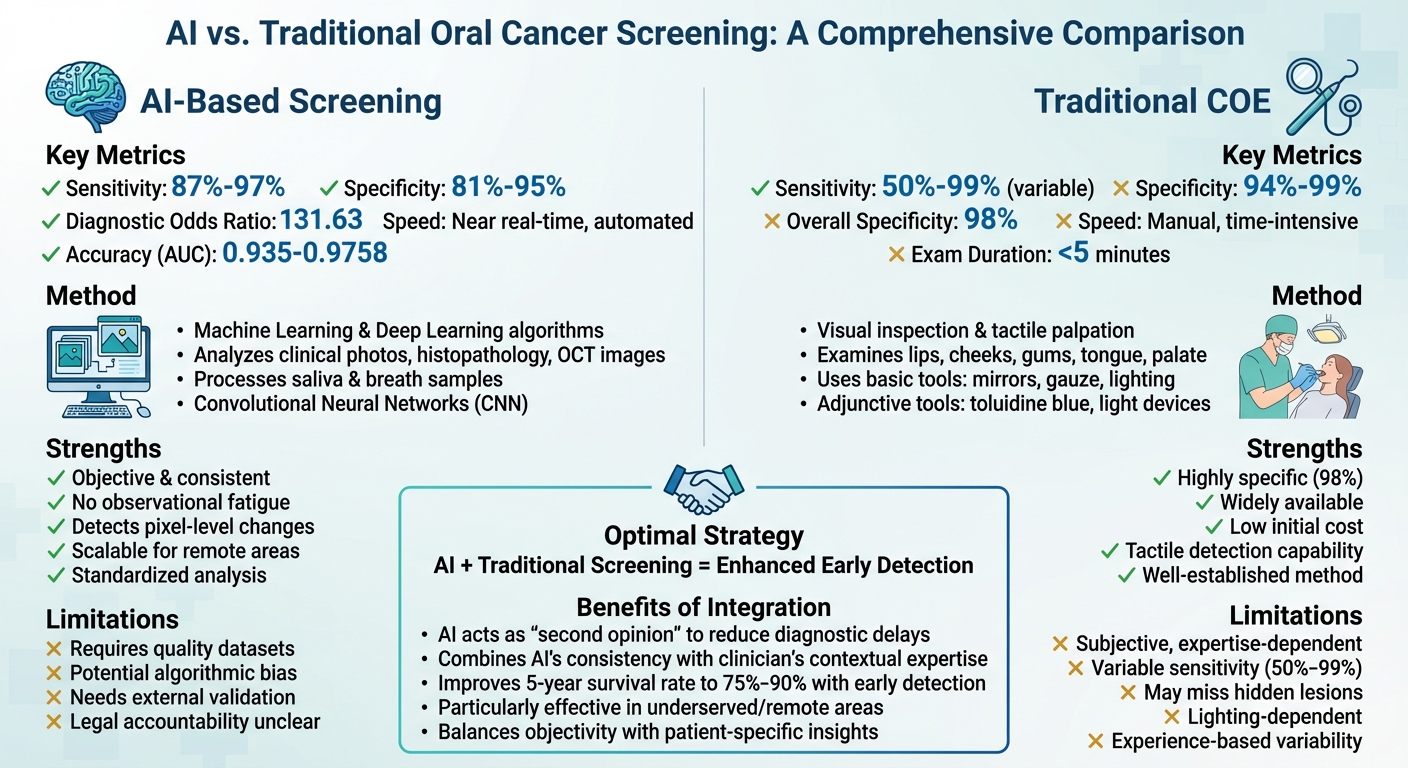
AI vs Traditional Oral Cancer Screening: Performance Comparison
Using AI to Catch Oral Cancer Early | Breakthroughs Can’t Wait
sbb-itb-2be92ed
What Is Traditional Oral Cancer Screening?
Traditional oral cancer screening, also known as the COE (Clinical Oral Examination), is a straightforward procedure involving both visual inspection and tactile palpation of the oral cavity. This examination, which typically takes less than five minutes, is often part of routine dental check-ups [6]. During the process, dentists carefully examine critical areas of the mouth – such as the lips, cheeks, gums, tongue, floor of the mouth, palate, and oropharynx – looking for red patches (erythroplastic), white patches (leukoplakic), or hardened masses. The success of this method relies heavily on the clinician’s expertise.
"The first-line approach to the identification of oral cancer and oral potentially malignant disorders (OPMDs) remains the standard clinical oral examination (COE)."
– IARC Working Group [6]
This process uses basic tools like good lighting, dental mirrors, tongue blades, gauze pads, and gloves [8]. Dentists may also perform bimanual palpation – using fingers inside the mouth and under the chin – to identify abnormalities in the floor of the mouth and submandibular areas. Drying the mucosal surfaces with gauze can help highlight subtle changes in colour and texture [8].
Techniques Used
Adjunctive tools can enhance traditional screening. For example, toluidine blue (1–2% concentration) binds to cells with high DNA content, staining potential lesions a dark blue. This method has demonstrated a sensitivity of 86% and specificity of 68% [6].
Light-based devices are also sometimes employed. These tools use violet-blue light for autofluorescence or acetic acid followed by blue-white light for reflectance. While they can improve detection, they are prone to high false-positive rates, with reflectance specificity as low as 19% [6]. Despite these tools, the strong specificity of traditional screening remains a key advantage.
Strengths of Traditional Screening
The COE boasts an impressive 98% specificity, making it highly effective at identifying patients who are disease-free [6]. It’s a well-established, widely available method that doesn’t require costly equipment, making it accessible in most dental practices. Studies also suggest that dentists are particularly skilled at performing COEs and recognising oral lesions. Early detection of oral cancer through screening significantly improves outcomes, with a five-year relative survival rate of around 90% [1]. It’s worth noting that 75% of all head and neck cancers originate in the oral cavity, with approximately 30% occurring on the tongue, 17% on the lip, and 14% on the floor of the mouth [8].
Limitations of Traditional Screening
Despite its strengths, the COE has some limitations. Its effectiveness is subjective, with sensitivity ranging from 50% to 99%, and it may miss hidden lesions or misidentify benign conditions like geographic tongue or candidiasis [6].
"Errors in diagnosis are most often ones of omission, and therefore, the importance of a systematic approach to the oral, head, and neck cancer examination cannot be overstated."
– Oral Cancer Foundation [8]
Since the COE is primarily a preliminary screening tool rather than a definitive diagnostic method, any suspicious lesion that persists for two to three weeks should lead to a referral for further evaluation or biopsy [9][10]. Unlike AI, which can provide more standardised analysis, traditional screening relies on individual judgement, which can sometimes delay early detection. These challenges underscore the importance of adjunctive methods and open the door for AI-based advancements to refine the process.
What Is AI-Based Oral Cancer Screening?
AI-based oral cancer screening is reshaping how clinicians identify oral potentially malignant disorders (OPMDs) and early-stage cancers. Unlike traditional methods that depend heavily on visual inspections, these systems rely on Machine Learning (ML) and Deep Learning (DL) algorithms to analyse medical data. This approach mimics human decision-making, but without the limitations of fatigue or subjectivity [1][7]. AI processes a variety of inputs, such as clinical photographs, smartphone images, histopathology slides, radiographs, and even molecular data from saliva or breath samples [3][5].
"AI, although a valued tool, should supplement rather than replace healthcare professionals."
– Nature, 2024 [7]
In practice, AI serves as a diagnostic assistant, helping clinicians differentiate between malignant lesions, OPMDs, and healthy tissue [7]. A key technology behind this is the Convolutional Neural Network (CNN), which automatically identifies features in visual data – no manual input required [2][3][5]. Some models even go a step further, analysing molecular and epidemiological data to predict risks like lymph node metastasis or survival rates over five years [3][7].
How AI Works in Oral Cancer Detection
AI systems work by pre-processing images, identifying subtle features like texture and colour changes, and classifying tissue as benign, OPMD, or malignant. These systems can detect minuscule alterations – down to a single pixel – that might escape human observation [2][5][7]. Unlike traditional machine learning, CNNs streamline the process by automating feature extraction [5].
The technology draws from diverse data sources. Imaging options include clinical photographs, Optical Coherence Tomography (OCT), and autofluorescence [2][3][5]. For non-imaging data, AI analyses volatile organic compounds (VOCs) in exhaled breath and salivary exosomes, enabling detection even before symptoms appear [3][5]. In some cases, hybrid systems combine traditional ML with deep learning to improve predictive accuracy [2].
Applications in Clinical Settings
AI-powered tools are already making an impact in clinical settings. For example, the Mobile Mouth Screening Anywhere (MeMoSA) initiative in 2022 used the VGG-19 digital architecture to classify oral lesions from smartphone images. This project aimed to improve screening access in low- and middle-income countries and achieved high accuracy by training on diverse datasets [5].
Another example is a smartphone-based oral cancer probe equipped with 405 nm LEDs for autofluorescence imaging. Paired with a VGG-M deep learning model, this system demonstrated an 86.88% diagnostic accuracy and an AUC of 0.908 in field tests for detecting (pre-)malignant lesions [5]. These tools are especially useful in areas with limited access to specialists. In regions like rural Australia, where healthcare resources can be scarce, AI-based solutions could significantly enhance early detection efforts.
Potential Benefits of AI
AI-based screening offers several advantages. Studies report a pooled sensitivity of 0.87 and specificity of 0.81 for detecting OPMDs and oral cancer [2]. Among different methods, histopathological image analysis by AI stands out, with a sensitivity of 0.97 and specificity of 0.95 [2]. For Optical Coherence Tomography images, AI achieved a sensitivity of 94%, outperforming photographic images (91%) and autofluorescence (89%) [7].
Beyond its accuracy, AI brings consistency and scalability. Unlike human clinicians, it doesn’t suffer from observational fatigue during large-scale screenings [7][1]. AI ensures standardised analysis regardless of lighting or the skill level of the operator, addressing the variability seen in traditional screenings [2][3]. Early detection through AI can dramatically improve survival rates – boosting five-year survival to between 75% and 90%, compared to less than 30% when diagnosed after metastasis [7]. Moreover, it can reduce the reliance on invasive biopsies and the delays involved in waiting for histopathological confirmation [5][7].
These advancements highlight the growing role of AI in transforming oral cancer detection and its potential to complement traditional methods effectively.
Detection Accuracy and Sensitivity Comparison
This section highlights how AI is reshaping the landscape of diagnostic tools by complementing traditional screening methods. A 2024 meta-analysis reveals some telling performance differences: AI-based screening achieved a pooled sensitivity of 87% and a specificity of 81% [2]. In contrast, Traditional Oral Examination (COE) showed sensitivity ranging from 50% to 99%, with specificity between 94% and 99% [11]. This variability in COE stems from the inherent subjectivity of visual assessments [2]. These findings pave the way for a deeper dive into performance metrics.
The type of imaging used also plays a critical role in AI’s diagnostic accuracy. For histopathological images, AI demonstrated its strongest performance, achieving a sensitivity of 97% and a specificity of 95% [2]. Clinical photography yielded slightly lower yet solid results, with sensitivity ranging from 82% to 93.9% [2][12]. Optical Coherence Tomography (OCT) also performed well, with a sensitivity of 94% [7]. The Diagnostic Odds Ratio (DOR) for AI-based screening stands at 131.63, reflecting a high likelihood of accurate diagnosis [2].
"AI algorithms have the potential to function as reliable tools for the early diagnosis of OPMDs and oral cancer, offering significant advantages, particularly in resource-constrained settings."
– Rakesh Kumar Sahoo, School of Public Health, KIIT Deemed to be University [2]
Interestingly, AI’s performance varies by region. In lower-middle-income countries, AI achieved a sensitivity of 95%, significantly outperforming the 82% observed in high-income countries [2]. This underscores AI’s potential to make a meaningful difference in regions with limited access to specialists – a point particularly relevant for Australia’s rural and remote areas.
Comparison Table: AI vs Traditional Screening
| Criteria | Traditional Screening (COE) | AI-Based Screening |
|---|---|---|
| Speed | Manual; clinician-dependent | Rapid; near real-time |
| Accuracy (Pooled) | Variable; expertise-dependent | High (AUC 0.935–0.9758) [2] |
| Sensitivity | 50%–99% [11] | 87%–89.2% [2] |
| Specificity | 94%–99% [11] | 81%–86% [2] |
| Subjectivity | High; prone to variability | Low; objective and data-driven |
| Cost-Effectiveness | Low initial cost; expensive late treatment | Affordable remote screening |
The comparison makes one thing clear: while traditional screening excels in specificity, AI delivers more consistent sensitivity and eliminates human error. Each method has its strengths, and together they form a robust approach to oral cancer detection.
Advantages and Limitations of Each Approach
Advantages of Traditional Screening
The Clinical Oral Examination (COE) remains a cornerstone for detecting oral cancer and potentially malignant disorders [6]. Its key strength lies in its tactile nature, which allows practitioners to detect physical changes that imaging tools might miss. With a specificity rate of 98% in general populations, this method effectively reduces false positives, sparing patients unnecessary worry and avoiding overtreatment. Another major advantage is its accessibility – COE doesn’t require specialised equipment, making it easy to implement across Australia, even in resource-limited settings. Since it’s already a routine part of dental practice, traditional screening continues to be a reliable and familiar method, even as newer technologies emerge.
Advantages of AI-Based Screening
AI offers a fresh perspective by providing consistency and objectivity in diagnostics, addressing the variability that can arise from human interpretation. For instance, AI models have achieved impressive diagnostic accuracy, with an Area Under the Curve (AUC) of 0.9758 [2]. This level of precision is particularly beneficial in remote or underserved areas, where smartphone-based platforms can either relay images to specialists or provide instant automated assessments [3].
"AI technologies are increasingly integrated into accessible tools such as smartphone apps… making them particularly useful for community outreach and in areas where access to specialists is limited."
– Mamata Kamat et al. [3]
AI also empowers non-specialists, like community health workers or general practitioners, by helping them differentiate subtle malignant lesions from benign ones. Beyond visual analysis, AI can process complex data, such as salivary volatile organic compounds and breath samples, further enhancing diagnostic capabilities [3][5]. This makes AI a valuable tool for maintaining high diagnostic standards, even under heavy clinical workloads.
Limitations of Both Methods
Despite their strengths, both traditional and AI-based methods have limitations that affect their practical use. Traditional screening can be subjective, with results influenced by lighting conditions, practitioner expertise, and individual interpretation. These factors can be particularly challenging in Australia’s rural and remote areas, where access to highly trained specialists is limited [2].
AI-based screening comes with its own hurdles. Reliable performance depends on large, well-annotated datasets, and poor data quality can lead to inconsistent outcomes [2]. Issues like algorithmic bias are also concerning, especially when datasets fail to represent diverse populations.
"AI approaches should be standardised, tested longitudinally, and ethical and practical issues related to real-world deployment should be addressed."
– Vineet Vinay, Department of Public Health Dentistry [1]
Another unresolved issue is legal accountability for AI-assisted misdiagnoses, which raises questions about liability and patient safety. These challenges highlight the importance of using AI as a complementary tool rather than a standalone solution. Combining traditional screening with AI can balance their respective strengths, with AI serving as a second opinion to enhance clinical decision-making [7]. Together, they offer the potential for more effective and comprehensive screening.
Study Evidence on Performance
A 2024 meta-analysis of 18 studies revealed that AI models demonstrated a pooled sensitivity of 87% and specificity of 81% across various imaging techniques [2]. When it came to histopathological analysis, the results were even stronger, with sensitivity reaching 97% and specificity at 95%. On the other hand, clinical photography showed lower figures, with sensitivity at 82% and specificity at 73% [2]. These findings highlight AI’s growing ability to rival experienced clinicians in diagnostic tasks.
For example, a study conducted by the Kerman Faculty of Dentistry evaluated 518 intraoral images using the DenseNet-121 AI model. The model achieved an accuracy of 91%, sensitivity of 75%, and specificity of 98%, surpassing the performance of an experienced oral medicine specialist [13]. This suggests that AI could play a crucial role in areas where access to specialists is limited.
The sensitivity of Conventional Oral Examination (COE) varies widely, ranging from 50% to 99%, which heavily depends on the clinician’s expertise [11]. This variability underscores the challenges of relying solely on human judgement. Across multiple studies comparing AI to unaided clinicians, AI’s accuracy in detecting oral mucosal lesions ranged from 74% to 100%, while clinicians’ accuracy ranged from 61% to 98% [4].
However, challenges remain. A 2026 systematic review highlighted that 90.5% of AI studies lacked external validation, raising concerns about how well these models perform in diverse clinical settings [14]. Additionally, the variability between studies is significant, with heterogeneity statistics for sensitivity reaching 98.2% and specificity at 99.2% [2]. These findings suggest that while AI holds promise, it is best utilised as a complementary tool rather than a standalone replacement for traditional screening methods.
"AI algorithms have the potential to function as reliable tools for the early diagnosis of OPMDs and oral cancer, offering significant advantages, particularly in resource-constrained settings."
– Frontiers in Oral Health [2]
Overall, this evidence positions AI as a valuable addition to early detection strategies, especially when used alongside standard screening practices. However, the lack of standardised testing protocols and real-world validation across diverse populations remains a key limitation.
Future Role of AI in Oral Precancerous Detection
AI screening tools are reshaping how clinicians approach oral precancerous conditions. In April 2025, the University of Hong Kong Faculty of Dentistry made waves by opening the first AI-driven clinic dedicated to managing oral potentially malignant disorders. At the heart of this clinic is "OralCancerPredict", a web tool developed by Dr J. Adeoye’s team. With an impressive 94% accuracy rate, it evaluates cancer risk in patients with oral leukoplakia and oral lichenoid mucositis. By incorporating this tool into routine practice, the clinic has reduced unnecessary surgeries for low-risk cases and established consistent six-month surveillance intervals – all without increasing treatment costs [20].
Smartphone-based AI screening is also making diagnostics more accessible. Compact AI models, designed to run on devices with limited processing power, can deliver diagnostic probabilities in under five seconds [19]. This is a game-changer for rural and remote regions in Australia, where access to specialists can be challenging. Non-specialists can now capture clear, centred images of lesions and receive instant risk assessments [19][2].
"This pioneering AI clinic represents a substantial advancement in the prevention, early detection, and management of oral cancer… with the potential to mitigate oral cancer burden, save lives, and improve the quality of life."
– E. Veseli and J. Adeoye, British Dental Journal [20]
These advancements are just the beginning of AI’s role in revolutionising oral disease management.
Future workflows are set to become even more sophisticated. By combining clinical images, histopathology, genomic data, and patient-reported outcomes, AI – using Natural Language Processing – will enable tailored treatment plans [18]. Robotics, like the da Vinci and Versius systems, are already enhancing surgical precision during the removal of precancerous lesions. Meanwhile, IoT-enabled "Smart Oral Devices" are helping monitor hydration and temperature during recovery [18]. These technologies are refining early detection and improving the accuracy of treatments across diverse clinical environments. By 2025, many private insurers had begun covering AI-assisted screenings as part of preventive care [17].
Another key development is the focus on Explainable AI (XAI), which is fostering trust among clinicians. By using heatmaps to highlight specific tissue features flagged by the AI, XAI addresses concerns about "black box" systems. This transparency reassures clinicians that AI serves as a diagnostic aid, complementing rather than replacing their expertise [19][2][21].
Conclusion
AI screening and traditional examinations each bring distinct strengths to the detection of oral cancer. Traditional methods depend on clinical expertise and direct patient interaction, which are vital for understanding a patient’s medical history and lifestyle. On the other hand, AI tools excel at spotting subtle mucosal changes and microscopic abnormalities that can escape human observation, with reported pooled sensitivities ranging between 87% and 89% [2][17].
Together, these methods create a complementary system. AI can act as a "second opinion", highlighting suspicious areas and potentially speeding up the diagnostic process, while clinicians use their expertise to verify findings and make treatment decisions [16][22]. This combined approach maximises the strengths of both – AI’s precision and objectivity paired with the clinician’s ability to interpret context and history. It also helps offset limitations, such as AI’s lack of patient-specific insights and the potential for human fatigue or bias [7][16].
"Machine learning doesn’t just uncover hidden patterns – it equips dentists with a way to prioritise patients who need urgent care, making our interventions more targeted and effective." – Dr. Gus Bal, Bal Dental Centre [16]
The importance of early detection cannot be overstated. When oral cancer is caught in its early stages, the five-year survival rate can reach approximately 90%. However, this drops significantly to around 40% for advanced cases [15][17]. For individuals with oral lesions persisting for more than two weeks, AI screening could help differentiate between benign conditions and early malignancies [17].
As AI continues to advance, its integration with traditional screening methods is likely to improve early detection even further. By leveraging diverse data sources and explainable algorithms, AI has the potential to assist clinicians in delivering quicker, more precise diagnoses across a variety of healthcare settings [2][23].
FAQs
How does AI enhance oral cancer screening compared to traditional methods?
AI is transforming oral cancer screening by using advanced algorithms to analyse imaging data, like photographs and scans, with an impressive level of precision. These systems can spot subtle changes that might go unnoticed during traditional visual exams, boosting early detection rates and minimising the chances of human error.
By processing extensive datasets, AI models can uncover patterns and features associated with oral cancer or precancerous conditions. This capability leads to more accurate and consistent results. It’s particularly useful for identifying early-stage or potentially malignant disorders, allowing for timely interventions and improving outcomes for patients.
What challenges does AI face in detecting oral cancer?
AI has made strides in aiding the detection of oral cancer, but it’s not without its challenges. One major issue is that AI systems can occasionally overlook subtle or unusual signs of oral cancer, leading to missed diagnoses or false negatives. This highlights a critical gap where human expertise remains essential.
The effectiveness of AI tools also hinges on the quality and range of the data they’re trained on. If the training data lacks diversity or carries inherent biases, these tools may struggle to deliver consistent results across different patient groups. This can limit their reliability in real-world applications.
AI’s limitations become even more apparent with complex or rare cases. These situations often require the nuanced understanding of a trained clinician, something AI currently cannot replicate. Additionally, inconsistencies in imaging standards and variations in clinical environments can impact how well AI performs.
While AI has proven to be a helpful addition to diagnostic processes, it works best as a complement to traditional assessments and the expertise of healthcare professionals. It’s a tool, not a replacement.
Can AI-based screening help detect oral cancer in areas with limited access to specialists?
AI-based screening holds great potential for detecting oral cancer, particularly in regions where access to specialists is scarce. These advanced systems have demonstrated impressive diagnostic accuracy, making them a valuable tool for identifying oral precancerous conditions and addressing healthcare gaps in remote or underserved areas.
By examining medical images and patient data, AI tools can aid in early detection and facilitate timely referrals. This can be a game-changer in improving patient outcomes. While these technologies aren’t a substitute for professional care, they serve as a strong complement to traditional methods, broadening the reach of screening efforts where it’s needed most.
Related Blog Posts
- AI Research in Oral Disease Detection: Key Findings
- AI in Oral Cancer Detection: How It Works
- AI vs Radiographs: Caries Detection Accuracy
- AI vs. Traditional Oral Health Risk Assessment
Important Notice: Any surgical or invasive procedure carries risks. Before proceeding, you should seek a second opinion from an appropriately qualified health practitioner.
Individual results may vary. The information provided in this article is for educational purposes only and does not constitute medical advice.
Checkout Related Blogs




Get in touch with us
For more information, call us now to start feeling better. Or fill the form below to make appointment
The Latest News from Complete Smiles
How to Clean Clear Plastic Retainers
Checklist for Choosing Wearable Dental Devices
Checklist for Choosing Cloud AI Platforms in Dentistry

Complete Smiles Bella VistaAccepts All Major Health Funds, Including





