


Stem Cell Differentiation with Nanotechnology
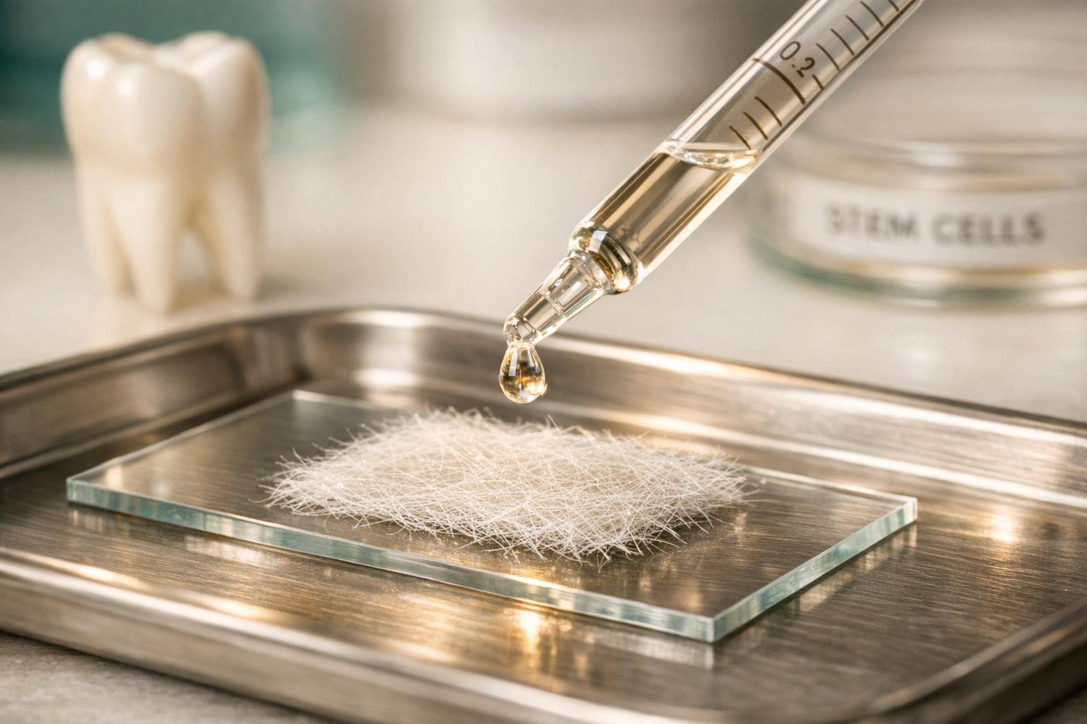
Nanotechnology is transforming dental regeneration by improving how stem cells rebuild damaged teeth and tissues. Here’s a quick breakdown of how it works:
- Dental Stem Cells (DSCs): Found in teeth, these cells can turn into bone, cartilage, and other tissues. They’re easy to collect, especially from wisdom or baby teeth.
- Nanotechnology’s Role: Tiny materials like nanoparticles, nanofibres, and nano-engineered surfaces help guide these stem cells to grow into specific tissues, such as dentin or bone.
- Nanostructured Scaffolds: These 3D frameworks mimic the body’s natural cell support system, encouraging proper cell growth and tissue repair.
- Applications: Used for pulp-dentin repair, bone regeneration, and improving dental implants.
- Safety and Challenges: While promising, proper concentration and biocompatibility of dental nanomaterials are critical to avoid cell damage or toxicity.
Nanotechnology allows precise control over stem cell behaviour, opening new possibilities for repairing dental tissues without relying on traditional materials. However, safety and regulatory approvals remain key steps before these methods become widely used in dental practices.
The Future of Stem Cells | Types, Applications, Grow Human Teeth!
sbb-itb-2be92ed
Nanostructured Scaffolds for Dental Stem Cell Differentiation
Nanostructured Scaffolds for Dental Stem Cell Differentiation: Key Types & Findings
As advancements in nanotechnology continue, nanostructured scaffolds are becoming essential tools in dental tissue engineering. These scaffolds act as temporary 3D frameworks that support cell growth during tissue regeneration. When designed at the nanoscale, they closely replicate the body’s extracellular matrix (ECM) – the natural support system for cells in living tissue. This close resemblance to the ECM makes them particularly promising for dental regeneration. By combining the precision of nanotechnology with the ability to guide stem cell differentiation, these scaffolds offer practical solutions for tissue repair.
Types of Nanostructured Scaffolds
In dental research, several types of nanostructured scaffolds are gaining attention, each with distinct advantages.
Electrospun nanofibrous scaffolds, often made from synthetic polymers like polycaprolactone (PCL) or poly-L-lactic acid (PLLA), are valued for their fine fibre architecture, which mimics the ECM.
"Nanofibrous scaffolds are widely used in dental tissue engineering due to their biomimetic structure, which closely resembles the extracellular matrix (ECM)." – BioMedical Engineering OnLine [1]
However, synthetic polymers like PLLA are naturally hydrophobic, which can hinder cell attachment. To address this, researchers have incorporated graphene oxide (GO) into PLLA scaffolds. GO improves the surface’s hydrophilicity, making it more conducive to cell growth. A study in BMC Oral Health (April 2024) highlighted that a 0.15% GO/PLLA composite scaffold significantly enhanced the expression of differentiation markers RUNX2 and COL1 in human dental pulp stem cells. The study concluded this scaffold could play a key role in oral bone tissue engineering [7].
Another innovative approach involves bioactive coatings. For example, coating PCL nanofibers with Biodentine (a tricalcium silicate cement) creates a hybrid scaffold that combines mechanical support with biological activity. Biodentine releases calcium and silicate ions, which activate signalling pathways to promote stem cell differentiation into odontogenic and osteogenic lineages. Compared to Mineral Trioxide Aggregate (MTA), Biodentine is preferred for its faster dentine bridge formation and easier handling [1].
Nanotopographical surfaces, such as gold nanoparticle gradients, use physical cues to influence stem cell behaviour without the need for biochemical additives. Research from the University of South Australia in 2017 found that 16 nm gold nanoparticle features were the most effective at directing human dental pulp stem cells (hDPSCs) toward osteogenic lineages, outperforming larger nanoparticle sizes (38 nm and 68 nm) [3].
Key Findings from Research
Studies consistently show that nanostructured scaffolds outperform unmodified surfaces in promoting stem cell adhesion and differentiation.
A study published in BioMedical Engineering OnLine (July 2025) demonstrated that PCL scaffolds coated with 0.01% Biodentine achieved peak alkaline phosphatase (ALP) activity by day 14 – a crucial early indicator of mineralisation – and the highest calcium deposition by day 21. However, a higher Biodentine concentration (0.05%) proved cytotoxic, highlighting the importance of precise formulation [1].
"Biodentine/PCL scaffolds demonstrated notable and suitable physical and chemical properties. Furthermore, they enhanced odontogenic/osteogenic differentiation and mineralisation compared to the control group." – BioMedical Engineering OnLine [1]
Nanotopographical surfaces have shown the ability to drive hDPSC self-differentiation toward osteogenic lineages purely through mechanical signals, without the need for soluble growth factors [3].
| Scaffold Type | Key Materials | Key Finding |
|---|---|---|
| Electrospun Nanofibers | PCL, PLLA | High surface area; closely mimics ECM architecture [1][4] |
| Bioactive Composite | PCL + Biodentine (0.01%) | Peak ALP activity day 14; highest calcium deposition day 21 [1] |
| Graphene Oxide Composite | GO/PLLA (0.15%) | Upregulates RUNX2 and COL1 differentiation markers [7] |
| Nanotopographical Gradient | Gold nanoparticles (16 nm) | Induces osteogenic differentiation without biochemical factors [3] |
Applications in Dental Tissue Regeneration
The findings from these scaffolds are now being applied to specific clinical scenarios.
For pulp-dentin complex regeneration, bioactive-coated nanofibrous scaffolds hold significant promise. By stimulating odontogenic markers like DSPP and DMP1, these scaffolds could support the regrowth of dentin-forming cells inside damaged teeth – something traditional restorative materials cannot achieve [1].
In alveolar bone augmentation, GO/PLLA composites and nanotopographical surfaces are particularly effective. Their ability to upregulate bone-forming transcription factors like RUNX2 makes them ideal for regenerating bone around dental implants or in areas of bone loss [7][3].
"hDPSC possess the ability to sense nanoscale geometric cues from their microenvironment." – Clinical and Laboratorial Research in Dentistry [4]
This shift from passive restorative materials to biologically active scaffolds marks a new direction in dental tissue repair, leveraging the cell’s innate ability to react to its environment.
Nanoparticles as Carriers and Differentiation Modulators
Nanoparticles bring a whole new level of precision to controlling stem cell behaviour. Acting as delivery systems, they transport bioactive molecules directly into stem cells. This approach not only shields these molecules from enzymatic breakdown and environmental challenges but also influences the biological mechanisms that guide stem cell differentiation.
Types of Nanoparticles Used in Dental Research
In dental research, a variety of nanoparticles are employed, including metallic ones like gold nanoparticles (AuNPs), ceramic options such as nano-hydroxyapatite (nHA), and carbon-based materials like graphene oxide. Researchers also explore advanced systems like metal-organic frameworks (MOFs) and upconversion nanoparticles (UCNPs). These nanoparticles go beyond merely providing structural support; they actively modulate stem cell behaviour, allowing for precise interventions at the cellular level [2].
How Nanoparticles Work
One of the standout features of nanoparticles is their ability to safeguard fragile biological materials. Growth factors and genetic materials, which typically degrade quickly in biological environments, are protected by nanoparticles until they reach their target cells [6]. Once delivered, many nanoparticles are absorbed by stem cells and accumulate in cellular compartments like endosomes and lysosomes, where they trigger specific cellular responses.
For instance, gold nanoparticles (AuNPs) have been shown to activate transcription factor EB (TFEB) in inflammatory conditions, helping to restore the differentiation potential of stem cells compromised by inflammation [2]. On the other hand, inorganic nanoparticles like nano-hydroxyapatite (nHA) release calcium ions (Ca²⁺), which stimulate the expression of osteogenic genes such as BMP-2 and RUNX-2. This drives stem cells toward bone-forming pathways [6]. These mechanisms have been linked to promising results in recent research.
Research Highlights
A study published in January 2021 in BMC Biotechnology revealed that encapsulating human dental pulp stem cells (hDPSCs) in Alginate-Gelatin/nano-hydroxyapatite (Alg/Gel/nHA) microcapsules significantly enhanced bone-related gene expression within a 3D microenvironment after 28 days compared to controls [6]:
- BMP-2 expression increased by four times
- RUNX-2 expression saw a 5.1-fold rise
Another study, featured in Cell and Tissue Research in March 2025, examined hydroxyapatite nanoparticles (HANPs) sized between 55 and 67 nm. These nanoparticles promoted osteogenic differentiation in dental pulp stem cells (DPSCs) at concentrations of 0.40 mg/mL or lower, enabling calcium deposit formation essential for repairing hard tissues. Although concentrations up to 0.81 mg/mL were tolerated without altering cell morphology, staying at or below 0.40 mg/mL was crucial for preserving the cells’ ability to migrate [5].
"Combining HANPs and DPSCs shows promise for restoring damaged hard tissues, like bone and teeth, and enhancing regenerative therapies." – Mais Emad et al., Faculty of Pharmacy and Medical Sciences, University of Petra [5]
These findings collectively underscore the active role nanoparticles play in driving the regenerative processes crucial for dental tissue repair and periodontal regeneration.
Nanotopography and Surface Modifications in Dental Materials
The texture of a surface at the nanoscale can act as a powerful cue for cellular behaviour. By adjusting nanoscale features like height, spacing, and roughness, researchers can guide stem cells toward specific tissue-forming pathways – without the need for chemical growth factors. This process, known as mechanotransduction, allows stem cells to sense and respond to the surface’s contours, triggering changes in their biochemical activity. Moreover, increasing the surface area provides more anchoring points for dental stem cells, which is crucial for advancing regenerative dental treatments. This technique builds upon earlier innovations like nanostructured scaffolds and nanoparticle carriers, refining how stem cells differentiate.
Nanostructured Implant and Root Surfaces
In 2017, researchers at the University of South Australia explored how nanoscale features influence stem cell behaviour. They created density gradients of gold nanoparticles (16 nm, 38 nm, and 68 nm) on a uniform 5 nm plasma polymer layer. Their findings? The size and spacing of nanofeatures play a critical role. For instance, 16 nm features with interparticle distances under 97 nm were most effective at promoting osteogenic differentiation in human dental pulp stem cells. Meanwhile, 68 nm features encouraged greater cell proliferation [3].
"Carefully tuned surface nanotopography can be used to manipulate and guide the proliferation and differentiation of these cells." – Krasimir Vasilev, School of Engineering, University of South Australia [3]
Similarly, calcium phosphate cements (CPCs) exhibit enhanced performance when scaled down to the nanoscale. Nanotopological CPCs improve cell adhesion, alkaline phosphatase activity, and odontogenic gene expression (e.g., DMP-1 and DSPP). This is achieved through integrin-associated signalling pathways such as FAK, paxillin, Akt, MAPK, and NF-κB [8].
"Nanotopological CPCs significantly enhance the odontogenic differentiation of HDPSCs when compared to conventional micro-CPCs through the integrin-associated signalling pathways." – Hae-Won Kim, Institute of Tissue Regeneration Engineering (ITREN) [8]
These advancements in surface modifications not only influence cellular responses but also lay the groundwork for improved outcomes in endodontics and periodontics.
Applications in Regenerative Endodontics and Periodontics
The principles of nanotopography are now being applied to dental materials used in clinical settings. For example, a study published in BioMedical Engineering OnLine (July 2025) investigated polycaprolactone (PCL) nanofibrous scaffolds coated with 0.01% Biodentine, a tricalcium silicate cement used in root canal treatments. Compared to uncoated scaffolds, the Biodentine-coated version displayed a moderately roughened surface with uniform mineralised deposits. This resulted in the highest calcium deposition by day 21 and peak alkaline phosphatase (ALP) activity by day 14. However, higher Biodentine concentrations (0.05%) were found to be cytotoxic, highlighting the importance of precise dosing [1].
The table below summarises how various nanostructured surface designs affect human dental pulp stem cell (hDPSC) behaviour:
| Material | Modification | Key Outcome |
|---|---|---|
| Gold nanoparticles | 16–68 nm density gradients | Osteogenic differentiation (16 nm) and proliferation (68 nm) [3] |
| Calcium phosphate cement | Nano-crystallite particles | Increased expression of DMP-1 and DSPP via integrin signalling [8] |
| PCL + Biodentine | 0.01% tricalcium silicate coating | Enhanced biomineralisation; peak ALP activity at day 14 [1] |
| PLLA nanofibers | Nanoscale fibrous structure | Improved hDPSC survival and odontogenic/neurogenic potential [4] |
These findings demonstrate that nanoscale surface features can effectively guide dental stem cell behaviour without relying on additional biochemical supplements. This approach complements earlier scaffold and nanoparticle strategies, offering a practical and scalable path for advancing regenerative dental therapies.
Safety, Challenges, and Future Directions
Safety and Biocompatibility Concerns
Nanomaterials hold promise for dental regeneration, but their safety must be carefully managed. One of the key factors is concentration. A study published in Cell and Tissue Research (March 2025) by researchers from the University of Petra and the University of Jordan revealed that hydroxyapatite nanoparticles (HANPs) in the 55–67 nm size range are tolerated by human dental pulp stem cells (DPSCs) up to 0.81 mg/mL without noticeable changes in cell shape or growth. However, their ability to self-renew remains intact only at concentrations below 0.20 mg/mL, while migratory capacity begins to drop past 0.40 mg/mL [5].
Another concern is the role of excessive reactive oxygen species (ROS) in damaged tissue. High ROS levels can trigger cell death through lysosomal and mitochondrial pathways, reducing stem cell survival [2]. Nanoparticle agglomeration further complicates matters by forming surface-connected compartments that disrupt interactions between macrophages and stem cells [5]. To address this, researchers at Chung-Ang University are developing antioxidant nanomaterials, such as manganese-substituted Co₃O₄ nanocrystals, which actively neutralise ROS and protect stem cells in oxidative environments [2].
"Specific types of nanomaterials can be internalized in cells and can accumulate in cellular compartments… which activates autophagy through their biological effects." – Tae-Hyung Kim, School of Integrative Engineering, Chung-Ang University [2]
These safety challenges highlight the importance of strict regulatory measures to ensure the safe application of nanomaterials in clinical settings.
Regulatory and Ethical Considerations
Beyond safety, regulatory frameworks play a critical role in bringing nanomaterials into clinical practice. In Australia, the Therapeutic Goods Administration (TGA) oversees the approval of nanomaterials for dental use, ensuring they meet rigorous safety and biocompatibility standards. Additionally, the Dental Board of Australia requires adherence to evidence-based practices. Innovations like nanotopography-based approaches offer a practical advantage in this context. Unlike traditional methods that rely on soluble factors derived from animals – raising concerns about disease transmission and regulatory scrutiny – these approaches use physical surface cues, simplifying compliance.
"Some of the soluble factors are of animal origin and can cause regulatory problems because of the danger of transmission of diseases." – Akash Bachhuka et al., University of South Australia [3]
The Australian National Fabrication Facility (ANFF) has already supported advancements in this area, providing the tools needed by University of South Australia researchers to create nanostructured gold surfaces, as discussed earlier [3].
The Future of Dental Nanotechnology
Despite its potential, nanotechnology for dental stem cell regeneration remains in the experimental stage. No nanostructured scaffold or nanoparticle-based system has yet become a standard part of Australian dental practice, such as dental implants, highlighting the gap between laboratory research and clinical application.
Still, the future looks promising. The move away from costly biochemical growth factors towards scalable surface engineering offers a more affordable and safer path for clinical use.
"Surface engineering is more cost-effective and safe… it can make cell therapies more affordable." – Krasimir Vasilev, Professor, University of South Australia [3]
Emerging technologies, such as multifunctional nanoparticles, could revolutionise the field. These materials aim to guide stem cell differentiation, reduce oxidative stress, and serve as imaging tools – all in one. Additionally, the concept of point-of-care nano-enabled bioreactors could one day make controlled stem cell expansion more accessible, even outside research labs [2]. While these advancements remain in the research phase, they represent a significant step toward integrating nanotechnology into everyday dental treatments. The foundational work being done now is shaping the future of regenerative dentistry.
Conclusion: Nanotechnology and the Future of Dental Regeneration
Recent studies are paving the way for biologically driven dental regeneration, focusing on the use of patients’ own stem cells to rebuild tissue. By incorporating advanced tools like nanostructured scaffolds, nanoparticle carriers, and specialised nanotopographical surfaces, the field is progressing toward safer and more efficient clinical solutions.
Key discoveries reveal how nanotopography can guide dental pulp stem cells toward bone-forming (osteogenic) pathways using physical cues, which helps cut down on the need for expensive growth factors. For instance, hydroxyapatite nanoparticles sized between 55–67 nm have been shown to encourage osteogenic differentiation at concentrations of 0.40 mg/mL or less [5]. Additionally, hybrid scaffolds combining PCL nanofibers with a 0.01% Biodentine coating demonstrated peak ALP activity by day 14 and maximum calcium deposition by day 21 [9]. These findings highlight the exciting potential of nanotechnology in pushing dental regeneration forward.
Despite these advances, moving from lab research to clinical practice is no small feat. Regulatory hurdles and biosafety concerns still need to be addressed. This includes developing antimicrobial nanocomposites to prevent infection during the healing process. However, the prospect of nano-enabled, point-of-care treatments using a patient’s own dental pulp cells offers hope for faster treatments and potentially lower costs.
As noted earlier:
"To fully realise the therapeutic potential of stem cells in the field of regenerative medicine, precise control of the fate of stem cells should be addressed." – Chang-Dae Kim et al. [2]
While the journey toward routine clinical application continues, each new study builds on this foundation, bringing us closer to a future where dental regeneration becomes a standard part of care.
FAQs
Can nanotechnology regrow tooth pulp or dentine?
Nanotechnology has the potential to transform dental care, especially in regenerating tooth pulp and dentine. Studies indicate that nanoparticles such as hydroxyapatite, bioactive glass, and tricalcium silicate can impact stem cell differentiation and essential cellular activities involved in the regeneration process. These developments could pave the way for groundbreaking advancements in dental treatments.
Are dental nanomaterials safe for people?
Research shows that some nanomaterials in dental care, like bioactive glass and hydroxyapatite nanoparticles, are biocompatible and can aid in the growth and differentiation of stem cells. This suggests they could be safe for use, provided they undergo proper evaluation and are applied correctly.
When will these treatments be used in Australian dental clinics?
As research and technology continue to evolve, these treatments are likely to make their way into Australian dental clinics. Since 2016, studies have been investigating nanotechnology-based regenerative therapies, and further progress is expected soon. These advancements are set to influence how such therapies are applied in dental care.
Related Blog Posts
- Stem Cells in Periodontal Regeneration: Current Trials
- Antimicrobial Nanocomposites for Tooth Regeneration
- Nanocomposites for Dental Stem Cells: Key Insights
- Future of Stem Cells in Tooth Regeneration
Important Notice: Any surgical or invasive procedure carries risks. Before proceeding, you should seek a second opinion from an appropriately qualified health practitioner.
Individual results may vary. The information provided in this article is for educational purposes only and does not constitute medical advice.
Checkout Related Blogs


Get in touch with us
For more information, call us now to start feeling better. Or fill the form below to make appointment
The Latest News from Complete Smiles
How to Clean Clear Plastic Retainers
Checklist for Choosing Wearable Dental Devices
Checklist for Choosing Cloud AI Platforms in Dentistry

Complete Smiles Bella VistaAccepts All Major Health Funds, Including


