


How 3D Dental Models Improve Patient Understanding

3D dental models are transforming how dentists explain oral health to patients. By replacing flat, hard-to-interpret X-rays with detailed, interactive visuals, these models make it easier for patients to understand their dental conditions and treatment options. Here’s how they work and why they’re effective:
- Clear Visualisation: Patients can see their teeth and gums in 3D, making it simpler to grasp issues like cavities, bone loss, or fractures.
- Improved Communication: Dentists can explain complex conditions and procedures more effectively, bridging the gap between technical terms and patient understanding.
- Enhanced Decision-Making: With visual aids, patients feel more confident about their treatment choices and are more likely to proceed with recommended care.
- Efficient Technology: Intraoral scanners and CBCT imaging capture precise 3D data in minutes, replacing uncomfortable traditional impressions.
- Practical Applications: From orthodontics to implants, 3D models help simulate outcomes, show treatment options, and even create physical replicas for hands-on explanations.
This technology empowers patients to take an active role in their dental care, improving understanding, trust, and satisfaction. Whether through digital visuals or 3D-printed models, these tools are reshaping the dental experience for both patients and practitioners.
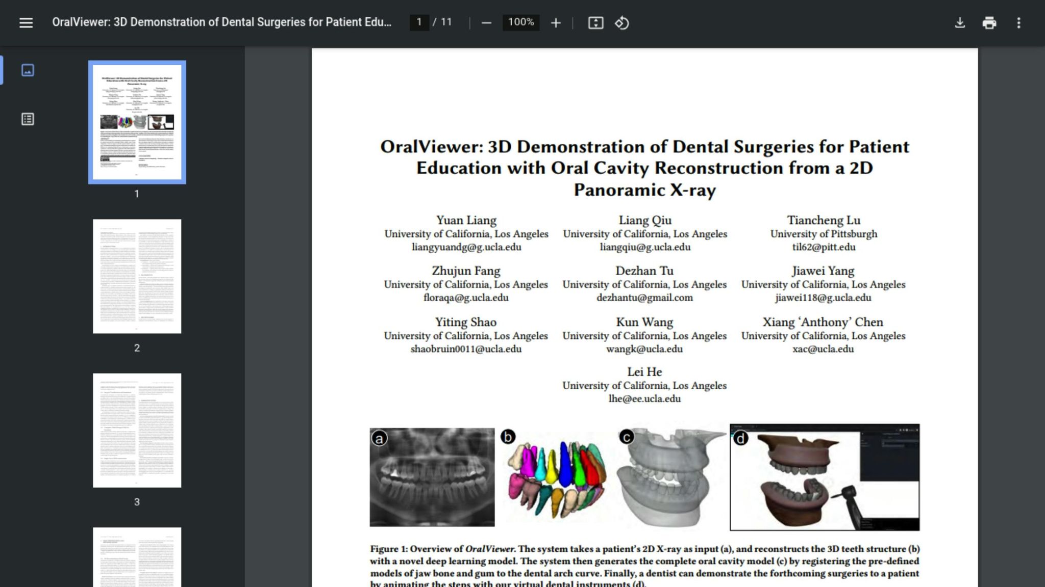
OralViewer: 3D Demonstration of Dental Surgeries for Patient Education with Oral Cavity …
How 3D Dental Models Work
How 3D Dental Models Are Created: From Scan to Patient Communication
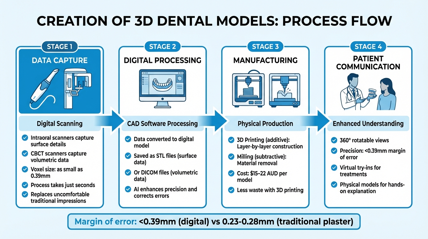
The journey to creating 3D dental models starts with data capture. Dentists use intraoral scanners to take digital impressions of teeth and gums, or CBCT scanners to gather volumetric data from deeper structures. CBCT scanners work by rotating around the patient’s head, snapping multiple images from various angles, which are then combined into a detailed 3D model [4]. This process takes just a few seconds, sparing patients the discomfort of traditional impression materials. Once the data is collected, it moves seamlessly into digital design.
Next, the captured data is processed using CAD software, which converts it into a digital model. These models are typically saved as STL files for surface data or DICOM files for volumetric information. AI plays a crucial role by enhancing scanning precision, correcting errors, and eliminating interference.
"Every physical impression that comes into our dental lab is digitised and converted into a 3D model ready for manufacture on our state-of-the-art computer aided manufacturing (CAM) systems" – Richard Salter, Managing Director at Avant Dental [2]
Creating 3D Dental Models from Patient Scans
There are multiple ways to create these digital replicas. Intraoral scanners are used for capturing surface details directly, while CBCT scans provide highly detailed volumetric data with voxel sizes as small as 0.39 mm [8]. Once a digital model is created, it can be physically manufactured through 3D printing (additive manufacturing) or milling (subtractive manufacturing). 3D printing, which builds objects layer by layer, is becoming the go-to method for complex structures because it produces less waste and is more economical.
Advantages of 3D Models Over 2D Images
With 3D digital models, dentists gain a tool that far surpasses the capabilities of traditional 2D images. Standard 2D X-rays offer flat, single-angle views, often causing anatomical structures to overlap and obscure important details. In contrast, 3D models provide a fully rotatable, in-depth view, allowing dentists to examine the oral cavity from any angle [4]. This makes it possible to uncover hidden root canals, measure bone density in Hounsfield units, and evaluate the precise relationship between teeth and jaw – details that would be invisible on a standard X-ray.
The precision of these models is another major leap forward. Digital models typically have a margin of error of less than 0.39 mm, while traditional plaster models show variations between 0.23 and 0.28 mm [8].
"3D imaging captures detailed, volumetric data that allows for a comprehensive view of the oral cavity" – Helix Dental [4]
Improving Communication Between Dentists and Patients
Technical jargon and flat, 2D X-rays can leave patients feeling lost when it comes to understanding their dental health. By transforming these abstract ideas into clear, 3D visualisations, dentists can close the communication gap. Here’s how 3D models make it easier for patients to grasp dental conditions and treatment options.
Explaining Dental Conditions with 3D Models
When a dentist points out a dark spot on a traditional X-ray, it’s often hard for patients to understand what it means. Is it serious? Where exactly is the issue? That’s where 3D models shine. They allow patients to see their teeth from every angle, clearly showing the location of a cavity, the depth of a fracture, or the extent of bone loss around a tooth.
This technology is especially helpful for revealing complex internal structures. Hidden root canals, periapical lesions, or unusual anatomy – like dens invaginatus (a developmental anomaly) – can be brought to life, making them easier for patients to comprehend.
Dentists can even compare current 3D scans with older ones to visually demonstrate changes over time. Whether it’s decay progression or damage from teeth grinding, these side-by-side comparisons can highlight why a certain treatment is necessary.
Showing Treatment Options and Expected Results
3D models don’t just explain problems – they also help patients see solutions. For example, in restorative dentistry, patients can preview the size, shape, and arrangement of proposed crowns or dentures through a "virtual try-in." This feature removes much of the uncertainty surrounding major procedures.
Orthodontic patients can benefit too. Using simulations, they can see how their teeth will shift and align over time. This is particularly reassuring for those starting clear aligner therapy, which often requires 20–30 aligners [9]. Seeing the projected outcome helps build confidence in the process. Similarly, for dental implants, specialised software can simulate how the implant will fit within the jawbone, showing how the final crown will look and function in relation to surrounding teeth and bone structure.
"The ability to visualise and understand their treatment through 3D images helps reduce anxiety and build trust in the care they receive."
– Helix Dental [4]
For an even more hands-on approach, physical 3D-printed models provide a tangible way to explain complicated procedures. These models are especially useful for cases that are hard to communicate via a screen. And with material costs ranging between $15–22 AUD per model [3], they offer an affordable way to improve patient understanding and confidence in their treatment plans.
sbb-itb-2be92ed
Helping Patients Make Better Decisions
Thanks to the use of 3D visuals, patients are now better equipped to make confident and well-informed decisions about their dental care. By transforming complex dental conditions into interactive, easy-to-understand visual models, these tools offer a clear pathway for patients to grasp their treatment options.
Supporting Informed Consent with Visual Tools
For informed consent to be meaningful, patients need to fully understand the procedures they are agreeing to. This is where 3D models shine. They provide a detailed view of the exact areas of concern in the mouth and visually demonstrate how proposed treatments will address these issues [11][4]. This not only simplifies the consent process but also encourages patients to take an active role in their healthcare decisions.
Increasing Patient Involvement and Satisfaction
When patients can see and even interact with models that represent their specific dental conditions, they become more involved in their care journey [10]. This marks a shift from the traditional "Guild Model", where decisions were primarily clinician-led, to an "Interactive Model", where patients are actively engaged. With clear visual evidence, patients can make decisions based on more than just verbal explanations [12].
In one study involving postgraduate students, participants reported an average improvement score of 8.38 ± 0.82 (on a 10-point scale) in their understanding of procedures after using 3D models [3]. This improved comprehension often leads to greater confidence in the treatment process and higher satisfaction with outcomes.
"3D printed models are easier for the patients to understand and serve as an effective treatment planning tool for the clinician."
– Ankit Arora, MDS, Reader at KM Shah Dental College [7]
The growing demand for this technology is evident in the dental industry. Over half of dental laboratories now use 3D scanners, and 20% have adopted 3D printing technology [10]. These advancements are reshaping how care is delivered and understood, benefiting both patients and clinicians alike.
Using 3D Models in Your Dental Practice
Incorporating 3D models into your dental practice doesn’t mean upending your entire workflow. With careful timing, thoughtful customisation, and proper staff training, these tools can seamlessly enhance patient consultations.
When to Use 3D Models During Appointments
3D models are incredibly useful during diagnostics, helping to visually identify issues like cavities or bone loss. For treatment planning, they allow for virtual try-ins, giving patients a preview of prosthetic designs. Position a monitor so patients can see the intraoral scan in real-time – this not only keeps them engaged but also helps them understand exactly where problems lie [4].
When it comes to orthodontics and implants, 3D models shine. They can simulate tooth movement or show precisely how an implant will fit within the jawbone, taking into account nerve placement and bone density.
"Digital dentistry involves a small wand camera – called an intra-oral scanner – that’s connected to a computer… If you’re curious to see a detailed picture of your mouth and teeth your dentist can position the screen so you can see what’s going on, as it happens."
– Keppel Dental [5]
Tailoring 3D Models for Each Patient
Every patient is unique, and customisation is key to making 3D models effective. High-resolution scans paired with CAD software allow you to isolate specific anatomical areas [13][14]. These models, which cost between $15–$22 AUD per unit, can be crafted with materials that mimic the properties of tissue [1].
To ensure accurate scans, it’s important to keep teeth dry using a dental syringe and use lip and cheek retractors to give the scanner unobstructed access to oral structures [4]. Additionally, storing patient data digitally means prosthetics can be easily reproduced or adjusted when necessary [6].
Equipment Needs and Staff Training
To get started, you’ll need an intraoral scanner like the Medit i700, Primescan, or iTero, along with a computer. If you’re planning on printing in-house, a 3D printer with post-processing units for washing and curing is also required [15][2].
Dental assistants play a critical role in this workflow. They handle patient scans, manage file uploads, oversee the print queue, and take care of post-processing. Training your team for these tasks allows dentists to focus on treatments while ensuring a reliable digital record is maintained [2]. In Australia, dental assistants earn an average salary of $60,000 to $70,000 per year as of 2025, while a Certificate III in Dental Assisting costs around $2,490 upfront [16].
"The greatest ROI for 3D printing for a general dentist is for night guards and for in-house aligners."
– Dr. Usa Bunnag, Bunnag Comprehensive Dentistry [15]
Dr. Usa Bunnag of Bunnag Comprehensive Dentistry, based in North Bethesda, Maryland, adopted a 3D printing system in 2023 using SprintRay equipment. By using printed surgical guides, he reduced implant surgery times to just 15 minutes. His practice also cut night guard production costs to about $29 AUD (roughly $25 for design and $4 for processing), enabling same-day or next-day delivery with no adjustments needed in 95% of cases [15].
If you’re just starting, begin with intraoral scanning before committing to full 3D printing hardware. This allows your team to become familiar with digital impressions first [2]. Ensure that your scanner software is compatible with your dental lab’s systems for smooth file transfers, whether through open STL files or specific portals. Finally, keep in mind that resin printer tanks typically need replacing every 6 to 10 months, so factor this into your budget [15].
Conclusion
3D dental models are transforming the way dentists and patients communicate, turning complex dental data into clear, visual representations. These models make it easier to explain issues like hidden bone loss, implant placements, and orthodontic outcomes, bridging the gap between technical expertise and patient understanding. This visual clarity not only simplifies discussions but also builds trust and makes treatment planning smoother.
When patients can visually grasp what their dentist is explaining, they feel more confident and informed, which often leads to higher treatment acceptance rates. As Ankit Arora, MDS, Reader at KM Shah Dental College, puts it:
"3D printed models are easier for the patients to understand and serve as an effective treatment planning tool for the clinician." [7]
But the benefits of 3D models go beyond education. They actively engage patients in their care, fostering a collaborative approach to dental treatment. By promoting transparency and helping set realistic expectations, these tools enhance the overall patient experience. Plus, they improve comfort by replacing traditional, often uncomfortable, impressions with digital scans, and they can even reduce the need for multiple appointments.
For dental practices aiming to improve communication and strengthen patient relationships, 3D dental models represent more than just a tech upgrade. They mark a shift towards a more collaborative, patient-friendly approach to dental care, seamlessly integrating into the digital dentistry landscape.
FAQs
How do 3D dental models help patients better understand their dental health?
3D dental models offer an in-depth, three-dimensional perspective of your teeth, gums, and surrounding areas, providing a clearer picture of dental conditions and treatments. Unlike traditional 2D X-rays that produce flat images, these models deliver a more lifelike and detailed view of your oral health.
They also make communication easier between you and your dentist. By using these models, dentists can explain diagnoses and treatment options in a way that’s easier to grasp. For patients, this means they can visualise procedures and potential outcomes more clearly, boosting both understanding and confidence. Plus, 3D imaging enables tailored treatment plans, ensuring your care is customised to suit your specific needs.
How do 3D dental models enhance patient understanding and treatment planning?
3D dental models offer patients a clear and engaging way to see their dental issues and understand proposed treatments. These models provide a detailed look at the complex structures of the mouth, making it easier for patients to grasp their oral health situation and the care process.
For dental professionals, these models bring precision and accuracy to treatment planning. They allow for personalised care by addressing each patient’s specific needs. On top of that, 3D models can simplify workflows, often cutting down treatment times and improving communication between dentists and patients. This collaborative process helps patients feel more informed and confident about their dental care choices.
How do 3D dental models help patients better understand their dental health?
3D dental models offer patients a clear and interactive way to grasp their oral health. Instead of relying on traditional explanations, these models visually showcase issues like cavities, misaligned teeth, or gum disease, making complex dental conditions much easier to understand.
When patients can see detailed representations of their teeth and the suggested treatments, they feel more informed and confident about their choices. This method enhances communication between the dentist and the patient, paving the way for a more collaborative and tailored treatment journey.
Related Blog Posts
- AR in Dental Care: Patient Education Tools
- How 3D Imaging Improves Orthodontic Treatment Outcomes
- CBCT Technology: Mechanisms Behind 3D Imaging
- 3D Imaging in Orthodontics: How It Works
Important Notice: Any surgical or invasive procedure carries risks. Before proceeding, you should seek a second opinion from an appropriately qualified health practitioner.
Individual results may vary. The information provided in this article is for educational purposes only and does not constitute medical advice.
Checkout Related Blogs


Get in touch with us
For more information, call us now to start feeling better. Or fill the form below to make appointment
The Latest News from Complete Smiles
How to Clean Clear Plastic Retainers
Checklist for Choosing Wearable Dental Devices
Checklist for Choosing Cloud AI Platforms in Dentistry

Complete Smiles Bella VistaAccepts All Major Health Funds, Including


