


Ultimate Guide to AI in Dental Implant Radiology

Artificial intelligence (AI) is transforming dental implant radiology by automating complex tasks, improving accuracy, and supporting clinicians in decision-making. From analysing CBCT scans to detecting peri-implant issues, AI tools are becoming crucial for pre-implant assessments, surgical planning, and post-operative monitoring. These systems help identify anatomical landmarks, measure bone quality, and monitor complications like bone loss, making them particularly useful for busy clinics and regional practices in Australia. However, clinicians must oversee AI outputs to ensure accuracy and address challenges like dataset bias and regulatory compliance.
Key Highlights:
- Imaging Methods: AI supports panoramic, periapical, and CBCT imaging for precise evaluation and implant planning.
- Pre-Implant Assessments: AI measures bone dimensions, detects pathologies, and generates standardised reports.
- Surgical Planning: Tools assist in implant positioning, surgical guide design, and avoiding critical structures.
- Post-Operative Monitoring: Systems flag complications like bone loss early, aiding in consistent follow-up care.
- Benefits: Improved diagnostic precision, reduced variability, and streamlined workflows.
- Limitations: Dataset bias, false readings, and integration challenges.
- Regulations: Must comply with TGA standards and privacy laws in Australia.
AI is a support tool, not a replacement for clinical judgement. Gradual implementation, staff training, and ongoing oversight are key to leveraging its potential responsibly.
Big data and artificial intelligence: The future in implant dentistry?
How AI is Used in Dental Implant Diagnostics
Artificial intelligence (AI) is transforming the field of dental implantology by simplifying processes at every stage – from initial assessments to ongoing monitoring. By employing deep learning models to analyse CBCT, panoramic, periapical, and intraoral scans, AI automates many manual tasks and enables more efficient workflows [1][3]. This technology not only enhances diagnostic accuracy but also improves surgical planning and post-operative care.
AI for Pre-Implant Assessment
AI tools play a crucial role in pre-implant evaluations by analysing CBCT scans to measure ridge dimensions and assess bone quality based on grey values and trabecular patterns [1][3]. These systems can pinpoint anatomical landmarks and identify conditions like cysts, tumours, or periodontal disease, helping clinicians tailor treatment plans. For instance, if AI flags underestimated bone height, it may lead to the selection of shorter implants. Similarly, detecting asymptomatic maxillary sinus lesions could prompt referrals before procedures like grafting [1][3][6][8].
Clinics such as Complete Smiles Bella Vista, which frequently use CBCT for implant planning, benefit significantly from AI integration. These systems generate rapid, standardised reports that align with Australian diagnostic standards, streamlining the pre-operative process and ensuring consistent care.
AI for Surgical Planning
When it comes to surgical preparation, AI combines CBCT and intraoral data to recommend the best implant positions and dimensions. It also aids in designing surgical guides and, when paired with robotic systems, can adjust drill trajectories in real time to avoid critical structures [1][3][6]. By processing pre-operative CBCT data and tracking anatomy with markers, these tools ensure precise placement. Research shows that AI-assisted or robot-guided techniques achieve greater accuracy and reduced deviations at the implant entry and apex compared to freehand methods, though much of this data stems from controlled environments [1][6].
Additionally, AI models can identify implant systems and components from radiographs with an impressive accuracy rate of 92–95%, far surpassing the approximately 37% accuracy reported for periodontists working with lower-quality images [2][7]. This capability is particularly valuable in saving time when ordering parts for repairs or restorations, especially in cases where patient records are incomplete [2][5][7].
AI for Post-Operative Monitoring
AI continues to provide value after surgery by comparing new radiographs to baseline images, identifying changes like marginal bone loss or complications such as abutment loosening and implant fractures [2][3][7][8]. By quantifying bone loss rates, these systems can detect peri-implantitis early – often before symptoms appear [3]. For practices with structured recall programmes, integrating AI into routine radiographic reviews ensures consistent monitoring and timely interventions. This is especially beneficial for high-risk patients, such as smokers or individuals with diabetes or a history of periodontitis [4].
Benefits and Limitations of AI in Dental Implant Radiology
Clinical Benefits of AI
AI is transforming dental implant radiology by improving diagnostic precision in detecting peri‑implant bone loss, anatomical landmarks, and pathologies on cone‑beam computed tomography (CBCT) and periapical radiographs [1]. Studies show that deep learning models outperform clinicians in identifying dental implant systems, even from low‑quality radiographs [2]. By automating tasks like bone-level measurements, nerve canal tracing, and CBCT segmentation, AI simplifies routine processes, giving clinicians more time to focus on patient care and treatment planning.
Survey results reveal that 68% of specialists appreciate AI’s ability to detect subtle details, while 64% see its value in supporting less experienced dentists [1]. AI also enhances diagnostic accuracy, reduces radiation exposure through advanced image reconstruction and denoising, and provides consistent monitoring of marginal bone loss. This early detection capability is particularly helpful in addressing peri‑implantitis before it progresses.
Limitations and Risks of AI
Despite its advantages, AI in dental implant radiology has its challenges. One major issue is dataset bias. Models trained on images from specific regions or manufacturers may struggle to generalise to the diverse implant systems, bone types, and radiographic equipment commonly found in Australian practices [1]. Additionally, performance can be compromised by factors like metal artefacts, scatter, or variations from low‑dose imaging protocols.
False readings are another concern, emphasising the need for thorough clinical validation. Many AI systems function as "black boxes", meaning their decision-making processes are not easily understood by practitioners. This lack of transparency can make it harder for clinicians to trust or interpret AI recommendations. Integration with existing practice management and imaging systems also poses challenges, potentially disrupting established workflows. For these reasons, AI should be viewed as a supportive tool, with clinicians maintaining full responsibility for final diagnostic and treatment decisions.
Regulatory and Ethical Considerations
In Australia, AI tools used for diagnostics or treatment planning are classified as medical devices and regulated by the Therapeutic Goods Administration (TGA). To be listed on the Australian Register of Therapeutic Goods, manufacturers must demonstrate robust analytical and clinical validation [6]. Additionally, under the Privacy Act 1988, dental radiographs and CBCT scans are classified as health information, requiring secure storage, transmission, and proper de‑identification, particularly when cloud‑based processing is involved. Practices must obtain explicit consent if patient images are used for purposes beyond direct care or ensure they meet de‑identification standards.
Ethically, patients should be informed when AI is involved in their implant diagnosis or treatment planning. They should also be reassured that a registered dentist remains responsible for all clinical decisions. Practices, such as Complete Smiles Bella Vista, that incorporate AI into their workflows should review and update their privacy policies, consent forms, and vendor agreements. They should also establish protocols for ongoing validation, performance monitoring, and clinician oversight. These measures ensure that AI integration is not only effective but also aligns with regulatory and ethical obligations, maintaining trust and accountability as the technology evolves.
sbb-itb-2be92ed
How to Integrate AI into Dental Practices
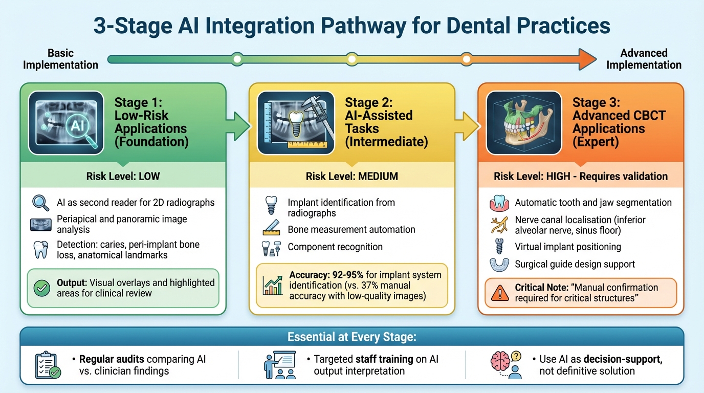
AI Integration Stages in Dental Implant Radiology: From 2D Analysis to Advanced CBCT Planning
Gradual Implementation of AI Tools
When introducing AI into dental practices, it’s best to start small and expand gradually. Begin with low-risk applications like using AI as a second reader for 2D radiographs, such as periapicals and panoramic images. These tools can help detect caries, peri-implant bone loss, and anatomical landmarks by providing visual overlays and highlighting areas for clinical review [1][3]. Once your team becomes comfortable with these basic tools, you can move to Stage 2, which involves AI-assisted tasks like implant identification and bone measurement on radiographs. Research indicates that AI performs these tasks with high accuracy, even when working with lower-quality images [2][7].
The next step, Stage 3, involves more advanced applications with CBCT imaging. These include automatic tooth and jaw segmentation, nerve canal localisation, and virtual implant positioning. At this stage, it’s essential to use AI outputs as decision-support tools rather than definitive solutions. For example, critical structures like the inferior alveolar nerve and sinus floor should always be manually confirmed before finalising surgical plans [1][6]. Regular audits comparing AI-generated suggestions with clinician findings help ensure accuracy and build trust in the system. By following this phased approach, you can integrate AI tools safely and effectively without overwhelming your team. It’s also crucial to provide targeted training so all staff members can correctly interpret AI outputs.
Training Requirements for Clinicians
For AI to be effective in dental practices, clinicians need to understand how it works, including its training data, supported image types, and limitations. For instance, AI may struggle with unusual anatomy or poor-quality scans [1][2]. Training should focus on interpreting heatmaps, which highlight areas where the model detects potential issues or important structures. Staff need to remember that these highlighted areas indicate probability, not certainty, and that non-highlighted regions could still contain subtle findings [1][3].
When it comes to structured AI reports – such as bone measurements, implant identification, or peri-implant bone loss scores – clinicians should double-check critical values against manual measurements or alternative image slices. Any discrepancies should be reconciled with clinical findings to ensure accuracy [1][2].
Hands-on workshops using anonymised images from your own practice can be an excellent way to demonstrate how to interpret AI outputs correctly. These sessions can also highlight common errors and reinforce safe usage practices. Additionally, radiographers and dental assistants should be trained on optimising image acquisition for AI. Consistent field of view, resolution, and patient positioning are key factors that improve accuracy [2]. Proper training ensures that your team can use AI effectively without relying too heavily on its automated outputs.
Working Across Dental Specialties
Once AI tools are fully integrated and the team is trained, they can enhance collaboration across different dental specialties. AI generates standardised, visual reports, which include annotated radiographs or CBCT slices, segmented anatomical structures, and detailed measurements like bone height, bone density, and proximity to critical structures. These reports can be shared electronically among general dentists, implant specialists, and radiologists, reducing misunderstandings and aligning expectations during case discussions [1][3]. Cloud-based AI platforms connected to electronic dental records also allow specialists to comment directly on AI-generated findings, enabling faster consultations and smoother workflows [2][3].
For more complex cases, such as full-arch or multi-implant procedures, AI-driven CBCT analysis can provide automatic jaw and tooth segmentation, identify anatomical constraints, and suggest initial virtual implant positioning [1][6]. This data allows for seamless collaboration among specialists. For example, a periodontist can assess bone quality and grafting needs, a prosthodontist can evaluate prosthetic space and occlusion, and an oral surgeon can plan flap design and implant angulation – all using the same AI-annotated images.
Practices like Complete Smiles Bella Vista (https://completesmilesbv.com.au), which offer a range of implant and specialist services, use AI-powered tools for tracking bone levels. This enables general dentists and periodontists to coordinate peri-implant maintenance and intervene early if bone loss is detected [8]. By creating a shared, structured data environment, AI fosters transparency and supports cohesive, multidisciplinary care.
The Future of AI in Dental Implant Radiology
Over the next few years, AI is poised to transform how dental practices in Australia approach implant planning and monitoring. This technology promises to improve diagnostic precision, particularly in areas like assessing bone quality, identifying nerve locations, and detecting potential complications. By automating tasks such as pre-reading, risk flagging, and integrating reports, AI aims to streamline workflows and bring greater consistency to dental diagnostics [1].
Beyond diagnostics, AI is expected to play a key role in personalised treatment planning. By combining radiographic data with patient history, it can help customise implant size, placement, and timing for each individual [1]. For dental practices in regional and remote areas, AI-annotated imaging could be a game-changer, enabling real-time specialist reviews and making expert advice accessible even across vast distances [1].
While AI offers many benefits, it is important to remember that it serves as a support tool rather than a replacement for human expertise. Decisions that involve patient preferences, complex medical histories, or aesthetic considerations still require the nuanced judgement of a skilled clinician [1][9]. Australian dentists will need to refine their ability to interpret and validate AI-generated insights, using the technology as a helpful "second opinion" rather than the final authority [1].
However, there are challenges to address. Many AI models are trained on limited datasets, which may not fully reflect Australian populations or imaging protocols without local validation [1]. Issues like regulatory compliance, privacy concerns, and integration hurdles also need careful attention [1]. Additionally, smaller or regional clinics may face barriers such as high initial costs and the need for staff training, which could delay adoption [1].
Advanced imaging centres like Complete Smiles Bella Vista (https://completesmilesbv.com.au) are already well-positioned to incorporate AI for more precise planning and better communication with patients [1]. As AI becomes embedded in CBCT units and practice management software, the role of clinicians is likely to evolve. Dentists may shift their focus toward oversight and shared decision-making, with algorithms handling repetitive analyses while they concentrate on more complex problem-solving and patient care [1]. For AI to truly enhance dental practice, its integration must be done responsibly, with ongoing training and collaboration across disciplines to support – not replace – clinical judgement [1][9].
FAQs
How is AI enhancing precision in dental implant radiology?
Artificial intelligence (AI) is transforming the field of dental implant radiology, offering a new level of precision and efficiency in diagnostics. With AI-powered tools, radiographic images can be analysed with incredible detail, uncovering crucial information like bone density, nerve locations, and the suitability of specific areas for implants – details that might otherwise go unnoticed.
By automating the interpretation of these images, AI minimises the risk of human error and provides valuable support for making well-informed decisions. This advancement not only streamlines treatment planning but also improves the overall experience and outcomes for patients receiving dental implants.
What are the potential limitations and risks of using AI in dental implant radiology?
AI has undeniably brought progress to dental implant radiology, but it’s not without its challenges and risks. One of the main concerns is the accuracy and dependability of AI algorithms. These systems rely heavily on the quality and variety of the data they’re trained with. If the data is incomplete or skewed, it can result in diagnostic errors or flawed treatment plans.
Another challenge lies in the absence of human intuition. While AI can process vast amounts of data, it cannot replicate a dentist’s clinical judgement or adapt to the unique nuances of individual patients. On top of that, introducing AI into dental practices can come with steep upfront costs and ongoing maintenance expenses, which may not be practical for every clinic.
There’s also the matter of ethical and privacy concerns. AI systems often require access to large volumes of patient data, making it essential to comply with data protection laws and ensure patient confidentiality throughout the process. Balancing these considerations is key to successfully integrating AI into dental care.
How can dental clinics effectively adopt AI technology into their workflows?
Integrating AI into dental clinic workflows offers a way to make diagnostics and treatment planning more efficient, ultimately improving both the clinic’s operations and patient care. Start by pinpointing areas where AI can make a difference – think analysing radiographs for quicker diagnoses, predicting treatment success rates, or automating routine tasks like scheduling appointments.
For a smooth implementation, clinics should prioritise investing in specialised AI software designed for dental practices and ensure staff receive the necessary training to use it effectively. Partnering with dental technology experts and keeping up with the latest AI advancements can help clinics stay ahead. It’s also essential to comply with Australian health regulations and ethical standards when introducing AI into your practice.
Related Blog Posts
- How AI Improves Diagnostic Consistency in Dentistry
- AI-Powered Radiology: What Dentists Need to Know
- AI vs Radiographs: Caries Detection Accuracy
- AI in Tooth Replacement: Economic Benefits
Important Notice: Any surgical or invasive procedure carries risks. Before proceeding, you should seek a second opinion from an appropriately qualified health practitioner.
Individual results may vary. The information provided in this article is for educational purposes only and does not constitute medical advice.
Checkout Related Blogs


Get in touch with us
For more information, call us now to start feeling better. Or fill the form below to make appointment
The Latest News from Complete Smiles
How to Clean Clear Plastic Retainers
Checklist for Choosing Wearable Dental Devices
Checklist for Choosing Cloud AI Platforms in Dentistry

Complete Smiles Bella VistaAccepts All Major Health Funds, Including


