


Top 5 Benefits of Endodontic Microscopes
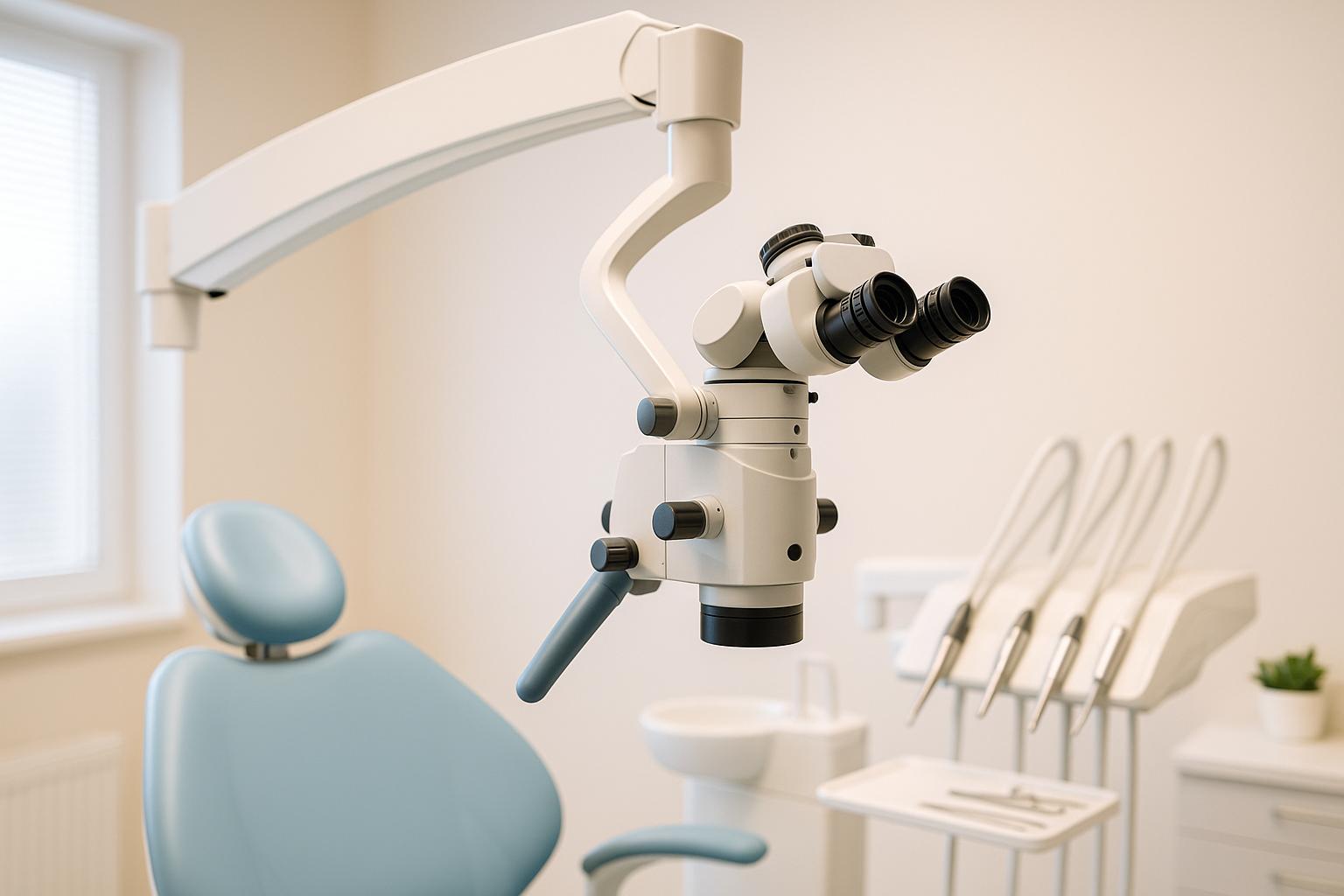
Endodontic microscopes are game-changers in dental care. They provide better visibility, more accurate diagnoses, and precise treatments, ensuring improved patient outcomes and comfort. Here’s why they matter:
- Better Visualisation: Magnifies up to 25× with bright, shadow-free illumination, revealing tiny details like hidden canals or fractures.
- More Accurate Diagnosis: Detects issues as small as 0.006 mm, improving identification of cracks, decay, and complex anatomy.
- Precise Treatment: Enables careful cleaning and shaping of root canals, reducing risks of complications and retreatments.
- Efficiency and Ergonomics: Speeds up procedures while promoting better posture for practitioners, reducing fatigue.
- Improved Patient Outcomes: Minimally invasive techniques preserve natural tooth structure, reduce discomfort, and enhance recovery.
Quick Comparison:
| Feature | Without Microscope | With Microscope |
|---|---|---|
| Magnification | 2.5×–6× (loupes) | 4×–25× |
| Resolution | 0.2 mm | 0.006 mm |
| Illumination | Overhead lighting with shadows | Bright, coaxial, shadow-free |
| Posture | Awkward, strain-inducing | Neutral, upright |
| Success Rate | Lower | Up to 94% |
Endodontic microscopes enhance every step of dental procedures, making them a vital tool for both dentists and patients.
1. Better Visualisation
Magnification and Illumination Quality
Endodontic microscopes bring a new level of clarity to root canal procedures with their advanced magnification capabilities. These devices typically offer magnification levels between 4× and 25×, far surpassing the 2.5×–6× range of traditional loupes [2].
What makes these microscopes stand out even more is their combination of high magnification and bright, coaxial illumination. This powerful pairing creates the perfect conditions for viewing even the tiniest details during treatment. With such a clear and detailed view, dentists can make more accurate diagnoses and treatment plans.
"For the differentiation of canal anatomy based on endodontic principles, it is essential to have a clear, deep view allowing to see everything down to the smallest detail."
– Dr. Fabio Gorni [6]
Impact on Diagnostic and Treatment Accuracy
With these crystal-clear images, clinicians can diagnose and treat dental issues with an unmatched level of precision. The improved visualisation helps identify even the smallest canals, fractures, or anatomical variations that might have been missed with traditional methods. This is especially crucial for locating hidden or accessory canals during root canal therapy [3][7].
Another standout feature is the large depth of field, which keeps different levels of the tooth structure in focus. This makes it easier to navigate and understand complex root canal anatomy [8]. The result? More precise cleaning, shaping, and filling of root canals, reducing the likelihood of treatment failures or postoperative issues [7].
Patient and Practitioner Benefits
The benefits of enhanced visualisation extend beyond just technical improvements – it positively impacts both patients and practitioners. For dentists, the improved visibility facilitates minimally invasive procedures, preserving more of the natural tooth structure while achieving better treatment outcomes [3].
"The greatest indicator of long-term retention of teeth is the volume of healthy natural dental tissue that remains after we finish treating a tooth."
– David Clark, Founder of the Academy of Dental Microscopy [2]
Patients also gain a clearer understanding of their dental health. Real-time images from the microscope can be shared with them, making it easier to explain their condition and the recommended treatment plan. This transparency often leads to greater trust and acceptance of the proposed procedures [3].
From a clinical standpoint, the enhanced visibility reduces risks during treatment. For instance, practitioners can observe instrument movements more accurately and create better root canal access, which significantly lowers the chances of complications like instrument breakage [8].
"The microscope lowers the risk of instrument breakage as you can create much better root canal access and observe the instruments’ movement more reliably."
– Dr. Dean Raicov, Endodontist in Germany [8]
2. More Accurate Diagnosis
Impact on Diagnostic and Treatment Accuracy
Endodontic microscopes take diagnosis to a whole new level by offering incredibly detailed visualisation. With the ability to detect details as small as 0.006 mm, these tools significantly improve diagnostic accuracy, compared to the previous limit of 0.2 mm [5]. This leap in resolution makes it possible to identify complex dental conditions that might otherwise go unnoticed.
Thanks to magnification up to 25 times greater than the naked eye, practitioners can uncover subtle issues like caries and tiny cracks that, if ignored, could lead to serious complications [10]. These microscopes also excel at revealing hidden anatomy and managing sclerosed canals, both of which are notoriously challenging in traditional dental practices [10].
A key feature is the coaxial lighting system, which eliminates shadows and ensures even illumination across the field of view [11]. This shadow-free lighting allows dentists to thoroughly examine the tooth’s internal structures, ensuring no detail is overlooked. This level of precision supports a more systematic and thorough diagnostic process.
Magnification and Illumination Quality
The combination of high magnification and superior illumination is what makes endodontic microscopes indispensable for accurate diagnosis. Unlike the limited capabilities of the naked eye or low-magnification loupes, these microscopes provide the clarity required for intricate dental procedures [11].
This clarity becomes even more critical as practitioners age, as visual acuity naturally declines after 40 years of age [10]. Endodontic microscopes help overcome this limitation, enabling dentists to detect enamel cracks, fractures, and subtle changes in dentin colour or texture, which are crucial for diagnosing conditions like internal resorption [9].
Patient and Practitioner Benefits
The precision offered by endodontic microscopes benefits both patients and practitioners in meaningful ways. For patients, the ability to detect dental problems early means they can receive prompt treatment, reducing the risk of complications [3]. Early diagnosis often leads to less invasive treatments and better long-term oral health outcomes.
For practitioners, the microscope’s capabilities reduce uncertainty during diagnosis and improve treatment planning. By revealing issues like hidden decay or damage, dentists can address problems before they escalate into more serious conditions [3]. Research highlights the positive impact of magnification on treatment success, reinforcing the value of this technology [1].
The widespread adoption of microscopes by nearly 90% of endodontists by 2008 underscores their transformative role in improving diagnostic accuracy [9].
Importance of Dental Microscope in Endodontic Treatment
3. More Precise Treatment
Endodontic microscopes take diagnostic accuracy a step further by enabling highly precise treatment. With magnification levels ranging from 4× to 25×, these tools provide an exceptional view of the tooth’s internal structures, such as root canals and microfractures [12]. The bright, coaxial illumination eliminates shadows, ensuring consistent lighting that allows for precise navigation through even the most intricate root canals [8]. This combination of enhanced visibility and clarity supports more accurate treatment execution.
Impact on Diagnostic and Treatment Accuracy
The improved visualisation offered by these microscopes ensures the complete removal of infected tissue and careful shaping of root canals [12]. Their ability to reveal tiny fractures or longitudinal cracks – often missed during standard procedures – helps practitioners address issues that could otherwise lead to treatment failure [8].
With superior magnification, dentists can also effectively manage complex anatomical variations, such as three-rooted premolars or C-shaped canals [14]. These often-challenging features are made visible and can be treated with greater precision [8]. Additionally, obstacles like calcifications or canal obliterations can be identified and removed, clearing the way for appropriate care [8].
Microscopes are particularly valuable for locating hidden or accessory root canals that may be overlooked during conventional treatments. Addressing these ensures a higher success rate and reduces the likelihood of retreatment [16][13].
Patient and Practitioner Benefits
The precision afforded by endodontic microscopes delivers significant advantages for both patients and professionals. For patients, this accuracy allows for minimally invasive treatments that preserve more of the natural tooth structure and reduce discomfort [3]. The detailed imagery also helps practitioners create tailored treatment plans that align with each patient’s unique dental anatomy [3]. These precise methods enhance the earlier diagnostic improvements, contributing to better overall outcomes.
"The greatest indicator of long-term retention of teeth is the volume of healthy natural dental tissue that remains after we finish treating a tooth." – David Clark, Founder of the Academy of Dental Microscopy [2]
For practitioners, the ergonomic design of these microscopes reduces fatigue during lengthy procedures and improves focus [17]. The ability to work with such precision often translates to faster and more effective treatments [15]. Additionally, the built-in documentation features support thorough record-keeping and clearer communication with patients [17].
Endodontic microsurgery performed with magnification boasts a success rate of 94%, compared to just 59% without it [10]. This significant improvement in outcomes minimises the need for retreatment or corrective procedures, benefitting both patients and dental professionals [3].
sbb-itb-2be92ed
4. Better Efficiency and Ergonomics
Endodontic microscopes bring a dual advantage to the table: they make procedures more efficient and create a more comfortable working environment for practitioners. This combination not only speeds up treatment but also allows dentists to work with less physical strain.
Efficiency in Procedure Time and Workflow
With enhanced visualisation, identifying issues becomes quicker, cutting down chair time for patients. The integration of improved four-handed dentistry techniques also simplifies instrument handling, ensuring a smoother workflow for practitioners [18]. Additionally, strategic use of magnification helps keep procedures on track and efficient [18].
These improvements don’t just save time – they also contribute to a better experience for both the dentist and the patient.
Patient and Practitioner Benefits
The ergonomic design of endodontic microscopes directly supports more accurate and dependable outcomes. Statistics show that over 90% of endodontists rely on dental operating microscopes in their practice, largely due to the physical comfort they provide [19].
"The clinician’s position when working with the scope allows for a more favorable upright working environment, helping to alleviate occupational neck, back and shoulder issues." [19]
By encouraging a neutral seated posture and working in the 12–11 o’clock position, these microscopes help maintain body symmetry and ease pressure on joints [18]. As Juan Carlos Ortiz Hugues, DDS, CEAS explains:
"The microscope calls for the operator to work in the neutral seated work posture." [18]
This approach significantly reduces postural strain compared to traditional methods, making a noticeable difference in daily practice [2][18].
Dentists using these tools frequently report less eye fatigue, fewer musculoskeletal issues, and even reduced psychological fatigue [2]. Features like binocular extenders and multifocal lenses with working distances of 200 to 400 millimetres ensure proper posture without sacrificing visibility [18]. Plus, the effective use of mirrors allows practitioners to work on all tooth surfaces and quadrants while maintaining a neutral posture [18].
5. Improved Patient Outcomes and Comfort
The ultimate aim of any dental treatment is to ensure the best possible results for patients while keeping them comfortable throughout the process. Endodontic microscopes help achieve both by enabling greater precision and less invasive procedures.
Magnification and Illumination Quality
Endodontic microscopes significantly enhance patient outcomes by offering unparalleled visual clarity. Their magnification capabilities allow dentists to see details the human eye simply cannot. While the human eye can distinguish objects 0.2 millimetres apart, a microscope can identify details as small as 0.006 millimetres [5]. This level of precision helps practitioners spot tiny canals or fractures that might otherwise go unnoticed.
Additionally, advanced lighting systems in these microscopes eliminate shadows that often hinder traditional dental procedures. As Global Surgical Corporation explains:
"When a light source isn’t parallel to your line of sight, you will encounter shadows in the deep, dark holes you find during procedures like root canals or delivering a broken root tip. Microscopes eliminate these shadows by unifying the light source and your line of sight within the body of the microscope. Many doctors we speak with describe the final product (the vision you receive through a microscope) as a ‘new realm’, giving them the ability to see in fine details unlike ever before." [22]
This enhanced visibility reduces uncertainty during diagnosis, making even the smallest cracks, early decay, or microleakage clearly visible [10]. As a result, dentists can plan and execute treatments with greater accuracy.
Impact on Diagnostic and Treatment Accuracy
The precision provided by microscopic visualisation leads to more predictable treatment outcomes. With enhanced visibility, practitioners can achieve consistency and reliability in their procedures.
Higher magnification also improves fine motor skills, allowing dentists to perform delicate movements required for navigating complex root canal systems [10]. This precision ensures a more thorough cleaning and shaping of canals, directly benefiting the patient.
Patient and Practitioner Benefits
The use of microscopes in dentistry offers a range of advantages for both patients and practitioners. For patients, the precision of these tools often means faster healing times and fewer complications after procedures [14].
Minimally invasive approaches made possible by microscopes help preserve more of the healthy tooth structure, reducing discomfort [20][21]. As David Clark, Founder of the Academy of Dental Microscopy, highlights:
"The greatest indicator of long-term retention of teeth is the volume of healthy natural dental tissue that remains after we finish treating a tooth." [2]
By minimising trauma to surrounding tissues, microscopes also help reduce post-operative pain, swelling, and recovery time [20]. This means patients can return to their daily lives more quickly and with less discomfort.
Another advantage is the ability to personalise treatment. Microscopes allow dentists to tailor their techniques to each patient’s unique anatomy, rather than relying on standardised methods [14]. This individualised approach ensures a more effective and comfortable experience.
Studies back up these benefits, with research showing that treatments performed with magnification yield better results than those without [1]. For example, a study tracking long-term outcomes found that 12 to 30 months after endodontic microsurgery, the combined rate of complete and partial healing was 90.5% [23].
For patients seeking high-quality endodontic care, choosing an endodontist who uses microscopic techniques can lead to a more comfortable experience and better long-term results [20]. This technology not only improves comfort but also helps preserve the health of teeth for years to come, showcasing its transformative impact on modern dental care.
Comparison Table
Microscopes have revolutionised endodontics by offering better visualisation, improved diagnostic capabilities, and greater precision in treatment. The table below compares traditional methods with microscope-assisted endodontics, showing why dental operating microscopes are now used by over 90% of endodontists [19].
| Aspect | Traditional Methods | Microscope-Assisted Endodontics |
|---|---|---|
| Magnification | Limited to 2.5x to 4x with loupes [24] | Variable magnification from 5x to 20x or more [24] |
| Visual Detail Resolution | 0.2 millimetres between distinguishable objects [11] | 0.006 millimetres between distinguishable objects [11] |
| Illumination | Standard overhead lighting with shadows | Bright, focused illumination that eliminates shadows [25] |
| Diagnostic Accuracy | Relies on visual and tactile skills [4] | Better detection of fractures, decay, and hidden canals [24] |
| Root Canal Detection | Tiny canals and obstructions may go unnoticed [14] | Identifies minute root canals and cracks [14] |
| Treatment Precision | Limited by naked eye capabilities [24] | Greater accuracy in cavity preparations and crown margins [24] |
| Practitioner Posture | Often requires awkward positioning | Promotes neutral, upright posture, reducing strain [18] |
| Procedure Efficiency | Standard workflow limitations | Improved efficiency through enhanced visualisation [14] |
| Tissue Preservation | Higher risk of removing healthy tissue | Minimally invasive, preserving more tooth structure |
These comparisons highlight how microscopes bring significant advancements in diagnostic depth, treatment precision, and overall efficiency.
The ergonomic benefits stand out as a major advantage. Dr. Juan Carlos Ortiz Hugues, DDS, CEAS, emphasises the importance of posture:
"The microscope calls for the operator to work in the neutral seated work posture." [18]
This upright, comfortable positioning contrasts with the awkward angles often required by traditional methods, which can lead to fatigue and long-term musculoskeletal issues.
Dr. Carlos Murgel sheds light on the precision offered by microscopes:
"If the objective is to be sure that everything is excellent, any doubts of working with the naked eye or limited magnification become certainties when the same work is performed under a microscope, which provides adequate light and maximum magnification. The details ‘lost’ in one are details ‘gained’ visually in another." [18]
Beyond individual procedures, microscopes enhance the overall workflow. They are particularly effective in four-handed dentistry, making the process more seamless and benefiting both clinicians and patients.
Conclusion
The use of endodontic microscopes has revolutionised the field of dental care, improving every stage of treatment – from accurate diagnosis to faster patient recovery. Studies reveal that procedures performed with the aid of microscopes achieve healing rates exceeding 90% and successfully handle more than 70% of complex cases [23][26]. These numbers highlight how far this technology has advanced compared to traditional methods.
"Dental microscopes have redefined the field of endodontics, offering unmatched precision, improved outcomes, and enhanced patient experiences." – Hammond Pond Dental Group [14]
The enhanced visualisation provided by these microscopes allows dentists to perform minimally invasive procedures, preserving more of the natural tooth structure. This not only reduces patient discomfort but also shortens recovery times. Additionally, the ergonomic design of these tools helps practitioners avoid fatigue and physical strain, even during lengthy or intricate procedures.
Modern dental practices increasingly recognise the importance of adopting cutting-edge technology to provide the highest standard of care. Clinics like Complete Smiles Bella Vista, led by Dr James Hanna, incorporate these advanced tools into their treatment protocols, ensuring their patients benefit from the latest advancements in endodontics.
For patients, selecting a dental practice equipped with microscopic technology can make all the difference in preserving natural teeth. Procedures that were once considered too challenging are now routine, redefining what is possible in modern endodontic care.
FAQs
What are the benefits of using endodontic microscopes in dental procedures?
Endodontic microscopes bring a range of advantages to dental procedures, particularly during root canal treatments. With magnification up to 25x and exceptional illumination, these tools enable dentists to spot intricate details like hidden canals, fractures, or calcifications that might otherwise go unnoticed. This level of precision allows for more thorough cleaning and shaping of root canals, lowering the chances of reinfection and improving overall treatment success.
Beyond just enhancing accuracy, these microscopes also help dentists manage anatomical variations with greater ease. This leads to better success rates and improved long-term outcomes. For patients, the benefits are clear: treatments that are not only more effective but also more comfortable and efficient.
What are the ergonomic advantages of using endodontic microscopes for dentists?
Endodontic microscopes provide notable ergonomic advantages for dentists, helping them maintain better posture, reduce physical strain, and ease eye fatigue. By encouraging a neutral seated position where the head, shoulders, and hips are properly aligned, these tools help dentists avoid forward bending and awkward movements that can lead to discomfort or even long-term physical issues.
On top of this, the improved visibility and magnification allow dentists to work from a comfortable distance, enhancing focus and efficiency during procedures. This combination of ergonomic support not only safeguards the dentist’s physical health but also enables more precise and effective treatments for patients.
How do endodontic microscopes improve patient comfort and recovery?
Endodontic microscopes play a key role in improving patient care by enabling treatments that are both precise and less invasive. Their advanced magnification and lighting capabilities allow dentists to spot and address even the tiniest details within a tooth’s structure. This level of accuracy helps minimise the chances of missing canals or leaving behind any infection, which are common causes of discomfort after treatment.
Because these microscopes support less invasive techniques, they help preserve more of the natural tooth and reduce trauma to nearby tissues. As a result, patients often experience shorter recovery periods, less pain, and a quicker return to their daily lives.
Related Blog Posts
- How X-Rays Help in Dental Treatment Planning
- What Is Minimally Invasive Cavity Preparation?
- Flapless Implant Surgery: Benefits and Process
- Periodontal Pocket Depth: Measurement Techniques
Important Notice: Any surgical or invasive procedure carries risks. Before proceeding, you should seek a second opinion from an appropriately qualified health practitioner.
Individual results may vary. The information provided in this article is for educational purposes only and does not constitute medical advice.
Checkout Related Blogs


Get in touch with us
For more information, call us now to start feeling better. Or fill the form below to make appointment
The Latest News from Complete Smiles
How to Clean Clear Plastic Retainers
Checklist for Choosing Wearable Dental Devices
Checklist for Choosing Cloud AI Platforms in Dentistry

Complete Smiles Bella VistaAccepts All Major Health Funds, Including


