





Machine Learning for Periodontal Disease Staging
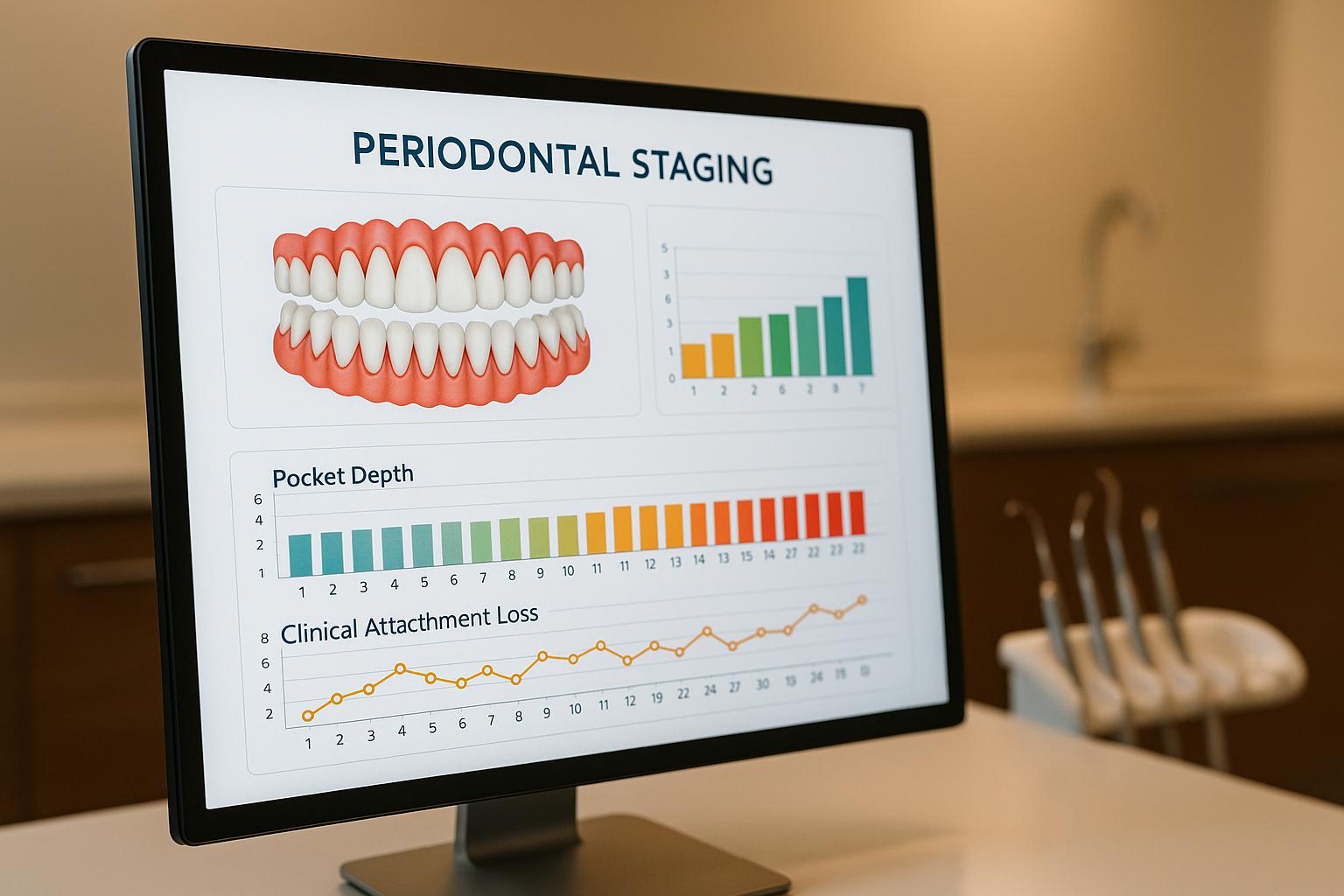
Machine learning is transforming how we diagnose and stage periodontal disease, offering more accurate and consistent tools for dental professionals. By analysing clinical data and radiographic images, these systems improve diagnostic precision and reduce variability in assessments. Here’s what you need to know:
- Clinical Data Models: Algorithms like decision trees and random forests achieve up to 98.6% accuracy using structured data, such as bone loss, smoking, and diabetes status.
- Deep Learning Models: Tools like CNNs analyse radiographs directly, automating bone loss detection with accuracies ranging from 77% to 88%.
- Hybrid Approaches: Combining deep learning with machine learning classifiers offers staging accuracy of 88.2%, balancing image analysis with clinical data insights.
For Australian practices, these technologies streamline periodontal care, especially in busy or remote clinics. They align with 2017 World Workshop guidelines, helping clinicians make data-driven decisions while saving time. However, challenges like data quality, integration with existing systems, and interpretability require careful consideration.
Key Takeaway: Machine learning enhances periodontal staging accuracy, but its success depends on the quality of clinical data, radiographs, and seamless integration into dental workflows.
[IPCAI 2021] Oral: Automating Periodontal Bone Loss Measurement via Dental Landmark Localisation
1. Standard Machine Learning Models
Standard machine learning (ML) models, such as decision trees, random forests, kNN, SVMs, and shallow neural networks, are highly effective in staging periodontitis when working with structured clinical data. Unlike deep learning systems that analyse raw images, these models rely on clinical measurements and radiographic data extracted from patient records. They strike a balance between accuracy, ease of use, and interpretability, making them practical for clinical settings.
Accuracy and Performance
When trained on carefully chosen clinical attributes, these models deliver impressive results. For instance, a decision tree algorithm using just 10 clinical and radiographic attributes achieved an accuracy of 97.2% for staging periodontitis based on the 2017 World Workshop classification [2]. Random forest models performed slightly better, achieving 97.9% accuracy for grading using the same dataset [2].
Interestingly, even when the input was reduced to four key attributes – percentage bone loss relative to age, diabetes status, radiographic bone loss, and smoking status – random forest and kNN models maintained high grading accuracy [2]. This highlights the potential to streamline data collection without compromising diagnostic performance.
In a study of 282 patients, seven ML algorithms were tested for staging and grading accuracy [3]. Most models, except AdaBoost, achieved over 85% training accuracy for staging and around 90% for grading [3]. On a separate validation cohort of 50 patients, the models maintained approximately 80% accuracy for staging and 90% for grading [3]. Among these, the shallow neural network stood out, achieving 81% accuracy for staging and 94% for grading, and was later incorporated into a web-based decision-support tool [3].
When applied to image-derived features extracted from panoramic radiographs by convolutional neural networks (CNNs), these models showed lower accuracy compared to structured clinical data. For example, a hybrid ResNet50 + SVM model achieved 88.2% staging accuracy, while other CNN-standard ML combinations, such as DenseNet121+SVM and VGG16+SVM, ranged between 80.6% and 85.4% accuracy [2]. These results underscore that structured clinical inputs currently provide better outcomes for standard ML models than image-based approaches.
Inputs and Data Requirements
These models rely heavily on clinical measurements and systemic risk factors routinely collected during periodontal exams. Inputs typically include clinical attachment loss, probing depth, bleeding on probing, plaque index, tooth mobility, furcation involvement, number of teeth lost to periodontitis, percentage radiographic bone loss, and patient factors like smoking status, diabetes, and age [2][3]. The quality and consistency of this data significantly affect model performance.
For optimal results, data must adhere to the 2017 World Workshop classification guidelines [2]. This requires comprehensive periodontal charting with multiple measurements per tooth, standardised radiographic protocols, and accurate documentation of systemic health conditions.
Research suggests that robust models can be trained with datasets of around 280 to 300 well-characterised cases [2][3]. In Australia, where single-centre datasets may be smaller, pooling anonymised data across clinics or using cross-validation techniques can help avoid overfitting. Ensuring high-quality radiographic data is also critical to minimise errors [2][4].
Clinical Integration and Practicality
One major advantage of standard ML models is their seamless integration into existing clinical workflows. These systems can be embedded into practice management or electronic dental record software, enabling automatic staging and grading suggestions once clinical data is entered [2][3].
In everyday practice, this offers chairside decision support for general dentists, flagging cases for specialist referral and standardising documentation for insurance and maintenance schedules. These tools can also support remote or teledentistry applications, where structured data is uploaded for a preliminary assessment before an in-person visit [6].
For Australian clinics like Complete Smiles Bella Vista, these systems align well with local guidelines and funding models. They help clinicians quickly classify disease severity, prioritise complex cases, and maintain efficiency in busy environments. The ability to work with compact datasets – sometimes as few as four attributes – makes these tools practical for real-time use [2].
Interpretability and Transparency
Among standard ML models, decision trees are particularly valued for their interpretability. They use simple if-then rules – such as thresholds for bone loss, smoking status, or diabetes – to mirror established periodontal classification criteria [2]. This makes them easy for clinicians to understand and explain to patients.
While models like random forests, kNN, and SVMs often deliver higher accuracy, they function more as "black boxes" unless paired with tools like feature importance plots to improve transparency [2][3]. In situations requiring clear explanations, such as patient discussions or medico-legal documentation, decision trees or simplified ensembles are often preferred, even if it means a slight trade-off in accuracy.
A practical approach involves training a high-accuracy model (e.g., random forest or SVM) to identify key clinical variables, then using these variables to create a simplified decision tree or rule set [2][4]. This method combines the performance of advanced models with the clarity of a transparent system. Hybrid pipelines can also highlight the specific factors influencing a staging decision, helping clinicians align algorithm outputs with established guidelines. This balance of accuracy and interpretability is critical as the field moves towards more complex deep learning solutions.
2. Deep Learning and Combined Models
Deep learning, particularly through tools like CNNs and YOLOv8, has revolutionised periodontal staging by analysing raw radiographic images. These models learn to detect patterns of bone loss and disease severity without needing manual feature selection. While traditional clinical-data models remain highly accurate, deep learning provides an image-focused alternative. When combined with traditional machine learning classifiers, these hybrid approaches offer a robust solution for image-based diagnosis. This evolution in data utilisation is shaping how we evaluate model performance.
Accuracy and Performance
Deep learning models trained on panoramic radiographs generally achieve staging accuracies between 77% and 88%, which, while slightly below the best clinical-data models, are still clinically useful [2][4]. Hybrid approaches that merge deep learning feature extraction with traditional classifiers often outperform standalone methods. For instance, ResNet50 + SVM achieved an 88.2% staging accuracy, while combinations like DenseNet121, EfficientNetB0, InceptionV3, and VGG16 paired with SVMs or random forests ranged between 80.6% and 85.4% [2]. Key to these results is standardised preprocessing, including contrast normalisation, noise reduction, and image resizing.
One notable hybrid framework, published in Nature Scientific Reports, used deep learning to identify the cemento-enamel junction (CEJ) and alveolar bone crest. It then applied conventional computer-aided diagnosis (CAD) methods to calculate percentage bone loss and assign stages based on the 2017 World Workshop criteria [1]. This system demonstrated a Pearson correlation of 0.73 with radiologists’ staging and an intraclass correlation coefficient (ICC) of 0.91 [1].
Meanwhile, YOLOv8 models trained on 2,000 panoramic radiographs achieved a mean average precision (mAP50) of 0.995 for detecting bone loss regions, automating radiographic assessment workflows [4]. These models can segment teeth, detect landmarks, quantify bone loss, and assign periodontitis stages – tasks traditionally requiring significant clinician time.
Despite these advances, clinical-data models still achieve 97–99% accuracy for staging [2]. This suggests that traditional machine learning remains highly effective for practices with detailed clinical records. However, deep learning shines in scenarios where imaging data is abundant or where automated radiographic analysis can complement clinical decision-making.
Inputs and Data Requirements
Deep learning models primarily rely on radiographic images, with panoramic radiographs being the most common, though some models also use periapical or bitewing films [1][2][4]. These inputs often include expert annotations of key landmarks, such as the CEJ and alveolar crest.
Hybrid models have shown strong performance using only a few clinical variables alongside image-derived features [2]. Standardised preprocessing – contrast normalisation, noise reduction, and resizing – along with expert annotations, remains essential [1][2][4]. Object-detection models like YOLOv8 also require detailed bounding-box and segmentation annotations, typically provided by periodontists or radiologists [4].
For Australian practices, consistent digital radiography workflows and high image quality are critical. Pooling anonymised datasets from multiple practices – such as centres like Complete Smiles Bella Vista – can help create the diverse training sets needed for robust models, provided ethical and legal guidelines are followed. Research indicates that datasets of around 2,000 well-annotated radiographs are sufficient for training high-performing systems [4].
Once the data needs are met, attention must turn to how these models can be seamlessly integrated into clinical workflows.
Clinical Integration and Practicality
Incorporating deep learning tools into everyday practice is becoming increasingly feasible with the rise of web-based and chairside applications. For example, one neural network-based system was deployed as a web application, allowing clinicians to input clinical parameters and receive staging predictions. This system achieved 81% test accuracy for staging and 94% for grading in an independent cohort of 50 patients [3].
In Australia, integrating AI into digital radiography systems can provide real-time support for periodontal staging [4]. These tools are particularly beneficial for less experienced clinicians, helping them apply the 2017 classification and identifying cases that may require specialist referral [2].
Practical implementation, however, requires strict adherence to data security and privacy laws, especially if images and clinical data are processed via cloud-based platforms [3][6]. AI systems should be designed to act as decision-support tools rather than replacements for clinical judgement, aligning with professional standards and medico-legal requirements. Clinics like Complete Smiles Bella Vista, which already leverage advanced digital workflows, are well-positioned to trial these tools, provided they undergo proper validation and governance.
Hybrid models also offer flexibility by separating feature extraction from classification. This allows clinics to adjust staging thresholds without needing to retrain the entire CNN [1][2].
Interpretability and Transparency
A common criticism of deep learning models is their "black box" nature, where the reasoning behind their decisions is unclear [5]. To address this, interpretability strategies are being employed.
CNN-based models are increasingly using visual explanation methods like saliency maps, Grad-CAM, and heatmaps. These tools highlight image regions that influenced the model’s decision, such as areas of bone loss or CEJ-to-crest distance, helping clinicians verify that the system is focusing on relevant structures [4][5].
Hybrid models further enhance transparency by combining deep learning with rule-based logic. For instance, a framework that used deep learning to locate CEJ and bone crest, followed by explicit percentage bone loss calculations tied to the 2017 World Workshop criteria, provides a clear, traceable process for clinicians to review [1]. Similarly, decision-tree components allow practitioners to see how specific risk factors contribute to stage assignments [2].
Emerging recommendations include performance dashboards and regular calibration to maintain model reliability [4][5]. For Australian practitioners, adopting models with case-level explanations and auditable decision paths will be essential to meet medico-legal standards and maintain trust.
Training clinicians to interpret AI outputs, understand confidence scores, and recognise when manual review is necessary – such as with poor-quality images or unusual anatomy – is key to successful integration [5]. Ultimately, these tools should enhance, not replace, the expertise and judgement of clinicians.
sbb-itb-2be92ed
Advantages and Disadvantages
When it comes to periodontal staging, selecting the right machine learning approach is all about finding the right balance between accuracy, practicality, and the resources available in your practice. Whether you’re considering standard machine learning models, deep learning systems, or hybrid approaches, each option offers its own strengths and challenges.
Standard machine learning models – like decision trees, random forests, support vector machines (SVMs), and k-nearest neighbours (k-NN) – work exceptionally well with structured clinical data. These models have demonstrated staging accuracies of up to 98.6% when using key clinical variables such as percentage bone loss adjusted for factors like age, smoking status, diabetes, and radiographic bone loss measurements [2]. For Australian practices that already collect detailed periodontal charting and patient histories, this approach is practical and efficient. These models require smaller datasets, train quickly on standard computers, and offer results that are easy for clinicians to interpret. For example, decision trees clearly outline which variables influence staging decisions, making them transparent and defensible in medico-legal situations.
However, these models aren’t without limitations. They rely on manual measurements, which can be time-consuming and prone to variability. Additionally, if your practice’s patient demographics or charting protocols differ from the data used to train the model, its performance may decline. Standard models also can’t pick up on subtle image patterns, such as early bone architecture changes visible on radiographs but not yet reflected in percentage measurements.
To overcome these challenges, deep learning models offer an alternative by analysing raw imaging data. Systems like convolutional neural networks (CNNs) and object-detection frameworks like YOLOv8 excel at identifying patterns of bone loss directly from panoramic radiographs, without needing manually crafted features. YOLOv8, for instance, achieves a high localisation precision with a mAP₅₀ of 0.995 [4]. These systems can automate tooth-level assessments, quantify bone loss percentages, and assign stages based on the 2017 World Workshop criteria – tasks that traditionally demand significant clinician time.
But deep learning comes with its own trade-offs. These models require large datasets with expert annotations, as well as GPU resources and technical expertise for training. For smaller Australian practices, building and maintaining such datasets can be impractical without external support. Deep learning models are also less interpretable; while tools like heatmaps can highlight which parts of an image influenced a decision, the reasoning isn’t as clear as it is with decision trees. This "black box" nature can make it challenging to build trust, comply with regulations, or explain decisions to patients. Performance-wise, deep learning models typically achieve staging accuracies between 77% and 88% [2][4], which, while clinically useful, falls short of the accuracy achieved by standard clinical-data models. They also struggle with grading disease severity, where variables like probing depth and attachment loss provide richer information than images alone [2].
Hybrid models aim to combine the strengths of both approaches. By using deep learning for feature extraction and classical machine learning for classification, these models strike a balance. For example, ResNet50 paired with SVM achieved an 88.2% staging accuracy on preprocessed panoramic radiographs, while other combinations like DenseNet121 + SVM and EfficientNetB0 + SVM showed accuracies ranging from 80.6% to 85.4% [2]. One hybrid framework, published in Nature Scientific Reports, used deep learning to identify key landmarks and conventional methods to calculate bone loss and assign stages. This system demonstrated a Pearson correlation of 0.73 with radiologists’ staging and an intraclass correlation coefficient of 0.91, indicating strong reliability [1].
Hybrid models are particularly appealing for practices that can work with moderately sized, well-curated datasets. They are more efficient than pure deep learning models and allow clinics to adjust staging thresholds without retraining the entire neural network. They also tend to be more interpretable, as the classical classifier component can reveal feature importance or decision boundaries. However, hybrid systems still require substantial datasets and computational resources, adding complexity to their implementation.
For Australian practices, the choice of approach will largely depend on their existing workflows and resources. Clinics like Complete Smiles Bella Vista, which already utilise advanced digital imaging and maintain comprehensive clinical records, may find hybrid models an attractive option. On the other hand, practices with strong periodontal charting but limited imaging infrastructure might lean towards standard machine learning models. Smaller clinics could explore deep learning tools through cloud-based platforms or vendor partnerships rather than attempting to develop in-house systems.
It’s important to note that all machine learning approaches share some common risks. Models trained on narrow datasets – whether limited by demographics, specific radiographic equipment, or patient populations – may underperform when applied to broader or more diverse groups. Dataset bias can particularly affect accuracy in Australia’s diverse population or in patients with high rates of systemic conditions. Moreover, reported accuracies often come from controlled research environments and may not fully reflect real-world performance. Ongoing validation, calibration, and using AI outputs as a supplement to, rather than a replacement for, clinical judgement are essential for any model.
Here’s a summary of the key trade-offs:
| Model Type | Key Advantages | Key Disadvantages | Typical Use Case in Australian Practice |
|---|---|---|---|
| Standard ML (decision trees, random forest, SVM, k-NN) | High accuracy with structured data (up to 97.2–98.6% for staging) [2]; low computational demand; easy to implement and interpret; works well with existing charting data | Limited ability to analyse raw images; performance drops with noisy or incomplete data; relies on manual measurements | Practices with strong periodontal charting and limited imaging infrastructure; chairside decision support; integration with practice management software |
| Deep Learning (CNNs, YOLOv8) | Analyses raw radiographs directly; automates assessments and bone loss quantification; reduces reliance on manual measurements; high detection precision (mAP₅₀ up to 0.995) [4] | Requires large, expertly labelled datasets; high computational costs; less interpretable; staging accuracy (77–88%) often lower than clinical-data models [2][4]; struggles with grading | Practices with high-quality digital radiography and large image repositories; research settings; automated screening and triage; integration with imaging systems |
| Hybrid Models (deep learning + classical ML) | Combines deep learning’s feature extraction with traditional classifiers; staging accuracy up to 88.2% (ResNet50 + SVM) [2]; strong reliability (ICC 0.91) [1]; more efficient with moderately sized datasets; better interpretability than pure CNNs | Requires substantial datasets and GPU resources; more complex to develop and maintain; inherits some data dependency of deep learning | Multidisciplinary clinics with advanced digital workflows; practices combining imaging and clinical data; research collaborations; vendor-supplied AI platforms |
Conclusion
Machine learning is opening up new possibilities for periodontal staging in Australian dental practices, offering tools tailored to various clinical needs and resource levels. Traditional machine learning models – like decision trees, random forests, and support vector machines – boast impressive staging accuracies of 97.2% to 98.6%. These models rely on structured clinical data, including variables such as percentage bone loss relative to age, diabetes status, smoking history, and radiographic measurements [2]. For practices already maintaining detailed periodontal charts, these systems provide reliable, efficient chairside decision support without requiring significant computational resources.
On the other hand, deep learning systems take an image-based approach, analysing panoramic radiographs with staging accuracies typically between 77% and 88% [2][4]. These systems automate tasks like measuring bone loss and can handle large image volumes consistently. For instance, a hybrid model combining deep learning feature extraction with traditional machine learning classifiers achieved an intraclass correlation coefficient of 0.91 when compared to expert radiologists [1]. This level of reliability makes these tools particularly useful in settings like radiology hubs, university clinics, and screening programs, where access to high-quality imaging and IT infrastructure is a given.
For many practices, clinical data–based machine learning tools are an accessible starting point. These systems can flag high-risk patients, support evidence-based recall scheduling, and help less experienced clinicians apply the 2017 World Workshop classification consistently. Specialist periodontal clinics – such as Complete Smiles Bella Vista – may find hybrid deep learning systems especially beneficial for standardising complex case assessments, creating visual aids for patient education, and integrating with advanced services like implant planning.
In rural or regional settings, where teledentistry and remote triage are becoming more common, image-based models can assist in referral decisions. These systems are particularly helpful when only panoramic radiographs and limited clinical data are available [2][4][7]. However, implementing such tools requires reliable connectivity and robust data security. Smaller clinics concerned about costs might consider a phased approach: starting with low-cost structured-data tools integrated into existing periodontal charting software, then expanding later to include cloud-based radiograph analysis services once their clinical value has been demonstrated.
Regulatory and clinical governance considerations are just as important as performance metrics. AI tools should act as decision support systems, not replacements for clinical judgment. Even highly accurate models can fail in specific cases or when applied to populations or imaging equipment that differ from their training data. To ensure patient safety, Australian practices should validate AI tools locally, using representative patient samples and assessing performance across different age groups and among patients with conditions like diabetes [2][4][5]. Clear protocols for using AI outputs and documenting clinician reasoning when overriding recommendations can help patients understand that AI serves as an adjunct, not a standalone decision-maker.
Data quality and seamless workflow integration are critical for success. Practices should regularly audit their records to ensure that probing depths, clinical attachment loss, bone loss percentages, and systemic factors are consistently documented, as these are essential inputs for machine learning tools [2][4]. Imaging and practice management software must be capable of securely exporting structured data or DICOM radiographs, and staff should be trained on both the strengths and limitations of AI systems. Models may struggle with unusual anatomy, radiographic artefacts, extensive restorations, or implants [7][5], so clinicians must be prepared to exercise their judgment in such cases. Any AI tool used must comply with TGA regulations and be incorporated into informed consent discussions when it influences diagnosis or treatment planning. Regular audits comparing AI outputs with periodontist assessments can help identify performance issues and maintain safety standards. Collaboration between universities, specialist centres, and larger group practices can further enhance the development of Australian training datasets, ensuring AI tools are reliable and equitable across diverse communities.
FAQs
How does machine learning enhance the staging of periodontal disease compared to traditional methods?
Machine learning models are transforming how periodontal disease is staged, offering a more efficient way to analyse complex data patterns compared to traditional methods. These models can handle vast datasets, including clinical records and imaging results, to pick up on subtle signs of disease progression that might escape manual evaluation.
Using algorithms like decision trees, support vector machines, and neural networks, machine learning delivers precise, reliable, and timely diagnoses. This empowers dental professionals to create more personalised treatment plans, which could lead to better patient outcomes and help minimise long-term oral health issues.
What challenges arise when using machine learning to stage periodontal disease in dental practices?
Integrating machine learning tools into dental practices to assess and stage periodontal disease comes with its share of hurdles. One of the biggest obstacles is the need for high-quality, standardised data to train these models properly. Differences in patient records, imaging methods, and clinical documentation can create inconsistencies, potentially affecting the accuracy and dependability of these tools.
Another critical issue is ensuring these systems fit seamlessly into existing clinical workflows. Dentists and other dental professionals might need extra training to understand and effectively use the insights these tools provide. On top of that, safeguarding patient data is non-negotiable. Compliance with privacy laws and ethical practices is essential when introducing such technologies into dental care.
Even with these challenges, machine learning advancements bring exciting possibilities. They could transform the way periodontal disease is diagnosed and managed, paving the way for more precise and tailored treatment options for patients.
How do hybrid models improve the accuracy of periodontal disease diagnosis by combining deep learning and traditional machine learning techniques?
Hybrid models bring a new level of precision to diagnosing periodontal disease by blending the strengths of deep learning with traditional machine learning techniques. Deep learning shines when it comes to detecting intricate patterns in medical images like X-rays, while traditional machine learning is adept at analysing structured data such as patient history and clinical metrics. When combined, these methods offer a more thorough analysis, boosting the accuracy of diagnoses and disease staging.
For instance, deep learning algorithms can pinpoint subtle changes in bone structure or detect signs of tissue inflammation from imaging. At the same time, traditional models evaluate risk factors such as age, smoking habits, and gum health indicators. This combined approach provides a well-rounded view of periodontal conditions, enabling dental professionals to make more tailored and informed decisions for their patients.
Related Blog Posts
- AI Research in Oral Disease Detection: Key Findings
- AI in Dentistry: Predicting Periodontal Disease
- AI vs Radiographs: Caries Detection Accuracy
- How AI Predicts Periodontal Bone Loss
Important Notice: Any surgical or invasive procedure carries risks. Before proceeding, you should seek a second opinion from an appropriately qualified health practitioner.
Individual results may vary. The information provided in this article is for educational purposes only and does not constitute medical advice.
Checkout Related Blogs




Get in touch with us
For more information, call us now to start feeling better. Or fill the form below to make appointment
The Latest News from Complete Smiles
How to Clean Clear Plastic Retainers
Checklist for Choosing Wearable Dental Devices
Checklist for Choosing Cloud AI Platforms in Dentistry

Complete Smiles Bella VistaAccepts All Major Health Funds, Including





