





5 Implant Surface Modifications for Less Inflammation
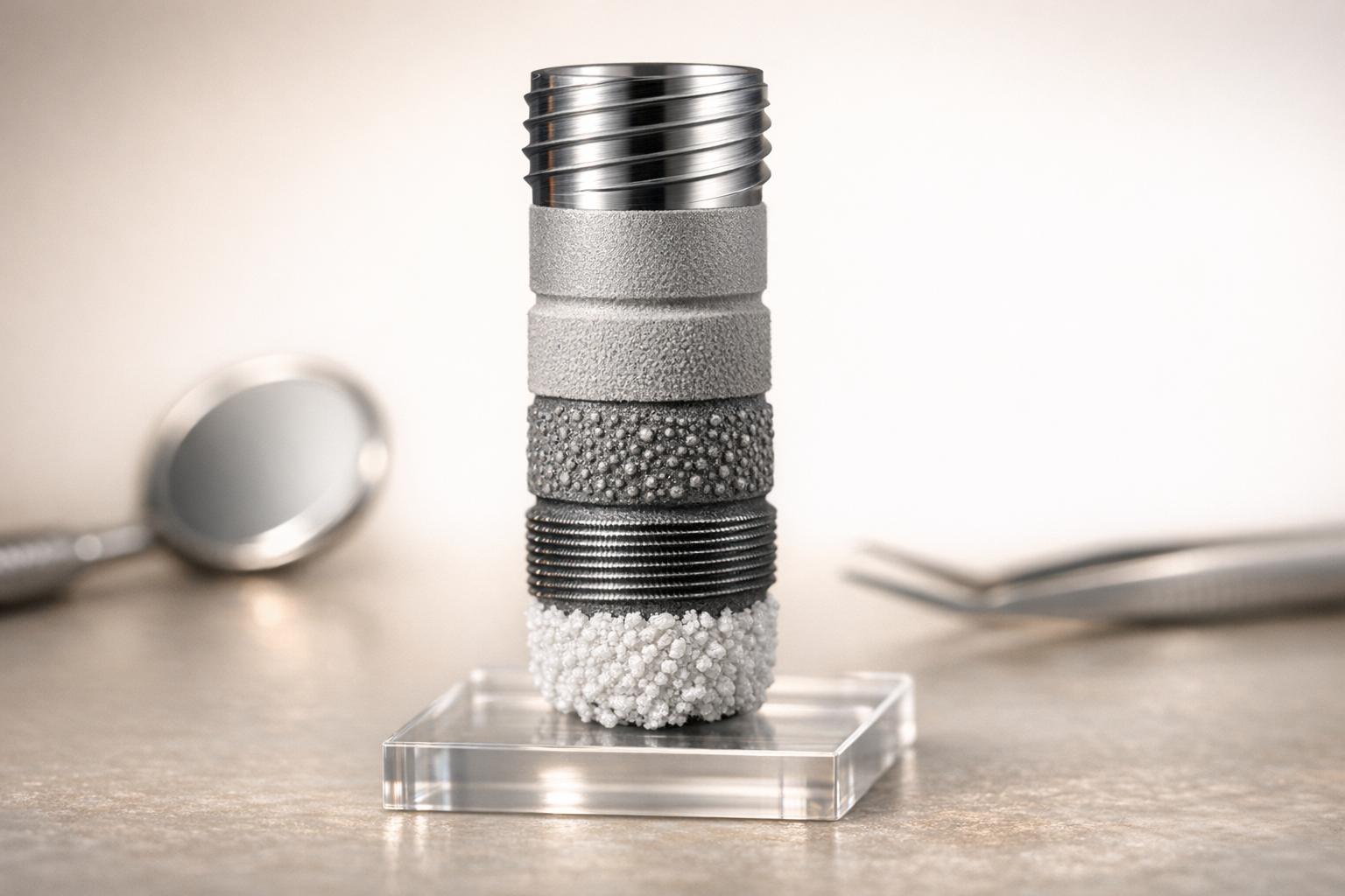
Dental implants work well for most people, with success rates between 90% and 98%, but inflammation and long-term stability can still be challenges. The surface of the implant plays a key role in healing and reducing risks like infections or bone loss. New surface modification techniques aim to improve healing and osseointegration while lowering inflammation. Here’s a quick summary of the five methods covered:
- Nanostructured Topographies: Tiny surface patterns encourage bone cell growth and reduce inflammation by shifting immune cells to a healing state.
- Hydrophilic Modifications: Water-attracting surfaces improve early healing and reduce bacterial adhesion.
- Bioactive Coatings: Special coatings release drugs or growth factors to manage inflammation and speed up healing.
- Hydroxyapatite Coatings: Mimics natural bone, promoting faster bonding and reducing inflammation.
- Surface Charge Optimisation: Electrically charged surfaces attract bone-forming proteins and repel bacteria.
Each method targets inflammation, infection risks, and osseointegration differently. While promising, some require further human trials to confirm long-term results.
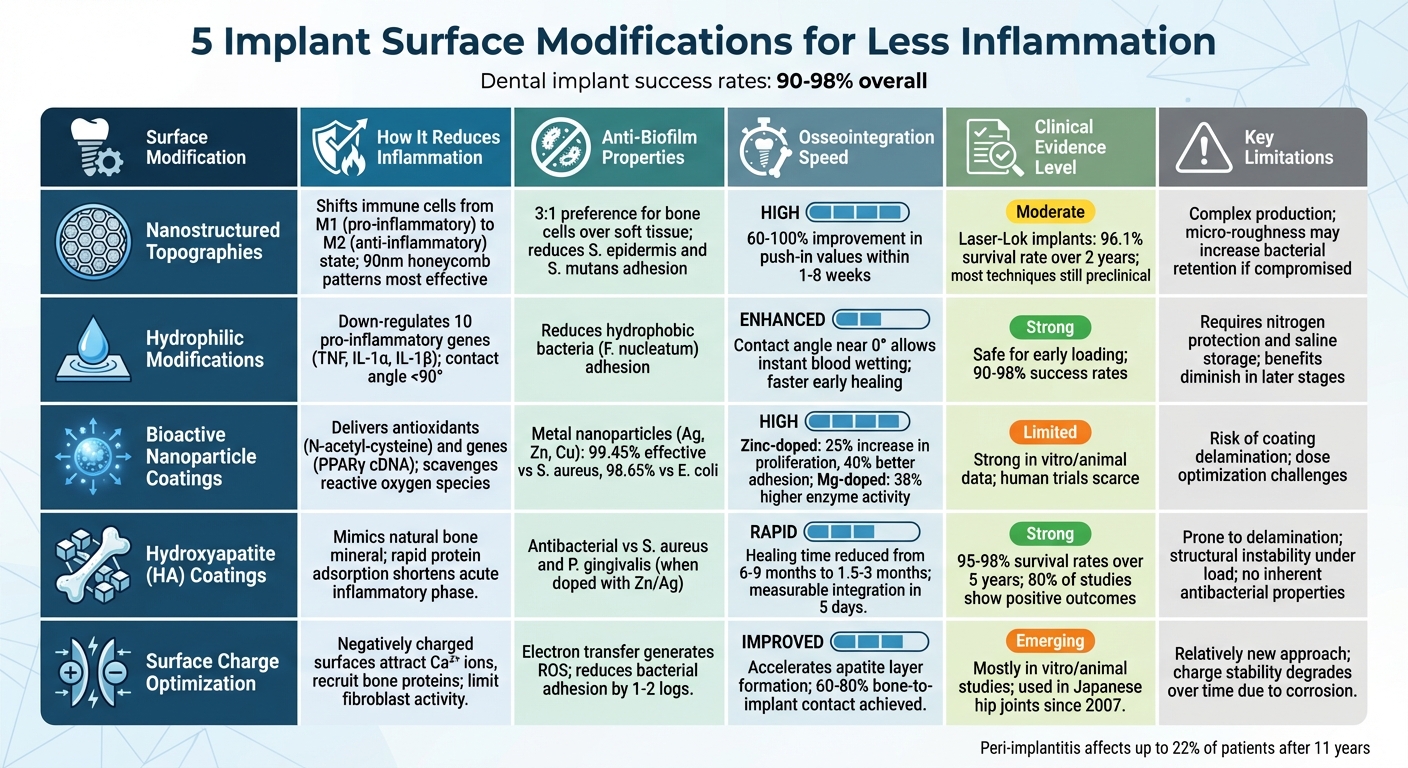
Comparison of 5 Dental Implant Surface Modifications for Inflammation Reduction
Recent Advances in Implants- Part 2
sbb-itb-2be92ed
1. Nanostructured Topographies
Nanostructured surfaces, ranging from 10–100 nm, are designed to replicate natural bone proteins and collagen. This helps improve cell communication and directs the healing process towards quicker integration while minimising inflammation.
Inflammation Reduction Mechanism
A key player in reducing inflammation is osteoimmunomodulation. Nanostructures – especially 90 nm honeycomb TiO₂ patterns – encourage macrophages (immune cells) to transition from a pro-inflammatory M1 state to an anti-inflammatory M2 state. This shift is essential for enhancing osseointegration.
"Achieving a balanced immune response – transitioning from pro-inflammatory (M1) to anti-inflammatory (M2) macrophage phenotypes – is critical for successful integration." – Li Yue, Tianjin Stomatological Hospital [9]
When cells interact with nanostructured surfaces, they activate the RhoA/ROCK signalling pathway, which plays a role in regulating immune responses [9].
Anti-biofilm Efficacy
Beyond modulating the immune response, nanostructured surfaces promote the adhesion of osteoblasts (bone-forming cells) while discouraging bacterial colonisation. These surfaces demonstrate a 3:1 preference for osteoblasts over fibroblasts (soft tissue cells), compared to the 1:1 ratio observed on smooth surfaces [7]. Additionally, they significantly reduce the adhesion of bacteria such as Staphylococcus epidermis and Streptococcus mutans, both of which are common causes of implant infections [6][7].
Osseointegration Speed
Nanorough surfaces provide more binding sites for proteins like vitronectin and fibronectin, which speeds up mechanical interlocking with bone. Research shows that micro-nano composite surfaces can improve biomechanical "push-in" values by 60–100% within the first 1–8 weeks after implantation [6]. Among these, disordered 15 nm high nanotube arrays stand out, as they enhance osteoblast activity while reducing bone resorption by osteoclasts [6].
Clinical Evidence Level
While laboratory and animal studies highlight promising outcomes, human clinical evidence is still evolving. One exception is Laser-Lok implants, which feature nanoscale microchannels that create biological seals. These implants achieved a 96.1% survivability rate over two years in clinical trials [8]. Such results underscore the potential of nanostructured surfaces for improving long-term implant stability. However, most nanostructuring techniques remain in preclinical stages, requiring further human trials before becoming widely available [9].
This approach opens the door to further innovations aimed at enhancing implant performance.
2. Hydrophilic Chemical Modifications
Hydrophilic surfaces, defined by a contact angle below 90 degrees, naturally attract water and blood. This interaction creates conditions that promote faster healing and help reduce inflammation. By modifying standard titanium implants, these surfaces are designed to actively encourage positive biological responses.
Inflammation Reduction Mechanism
The way hydrophilic surfaces reduce inflammation stems from their interaction with immune cells at a genetic level. For instance, Straumann‘s modSLA surface, which is treated under nitrogen and stored in saline to preserve high surface energy, has been shown to down-regulate 10 pro-inflammatory genes, including TNF, IL-1α, and IL-1β, compared to standard SLA surfaces [12].
"An attenuated pro-inflammatory response may be an important molecular mechanism for faster and/or improved wound healing." – Mohammed A. Alfarsi, School of Dentistry and Oral Health, Griffith University [12]
Super-hydrophilic TiO₂ coatings, produced through techniques like microarc and thermal oxidation, also play a role. Interestingly, treating titanium in ambient air rather than pure oxygen improves hydrophilicity and cell attachment by avoiding the formation of a rapid oxide layer that could hinder beneficial diffusion [4].
Osseointegration Speed
Ultra-hydrophilic surfaces, with a contact angle nearing 0 degrees, allow blood to instantly wet the surface [10]. This immediate blood spreading supports early cell adhesion and osteoblastic differentiation. Nobel Biocare‘s TiUltra implant surface demonstrated notable improvements in soft tissue outcomes, such as increased keratinised tissue height, during a two-year clinical study compared to traditional machined surfaces [10].
"Based on clinical trials, early implant loading of hydrophilic surfaces is considered safe and predictable." – Gabriel Bosch, University of Zurich [11]
These early biological benefits highlight the potential for hydrophilic surfaces to enhance implant performance, though further research is encouraged.
Clinical Evidence Level
While hydrophilic surfaces clearly benefit early healing phases, their advantages often diminish in later clinical stages [11]. Current evidence supports faster osseointegration and stable early loading, with dental implants generally achieving success rates between 90% and 98% [3]. However, long-term performance data, especially in challenging conditions like poor bone quality or in preventing peri-implantitis, remains limited [11]. Since peri-implantitis affects up to 22% of patients after 11 years [2], ongoing studies aim to better understand these surfaces’ long-term protective effects.
3. Bioactive Nanoparticle Coatings
Bioactive nanoparticle coatings take implant technology a step further by actively delivering therapeutic agents to manage inflammation. Unlike passive surfaces, these coatings interact directly with the healing process. Materials like titania nanotubes and mesoporous silica act as reservoirs, enabling the controlled release of anti-inflammatory drugs, growth factors, and even genetic material [13].
Inflammation Reduction Mechanism
Nanoparticles loaded with antioxidants, such as N-acetyl-cysteine, help reduce oxidative stress by scavenging reactive oxygen species (ROS) after implantation [13]. Some systems incorporate chitosan-gold nanoparticles to deliver genes like PPARγ cDNA, which lower inflammatory molecules and encourage bone remodelling – an approach especially helpful for individuals with diabetes [13]. Additionally, biopolymers like chitosan come with natural anti-inflammatory properties, modulating the immune response without requiring extra drug loading [13].
"Nanostructured surfaces are shown to have an impact on both indirect and direct cell interactions, guiding specific molecular events."
– Chrysa Marasli, Laboratory of Biology, National and Kapodistrian University of Athens [13]
These coatings not only reduce inflammation but also promote osteoblast adhesion over fibroblasts, enhancing bone integration. This dual capability strengthens their antibacterial performance as well.
Anti-Biofilm Efficacy
Metal nanoparticles, including silver, zinc, and copper, play a significant role in reducing inflammation by preventing bacterial biofilm formation – a major cause of peri-implantitis [13] [2]. Copper-doped coatings, for instance, have shown antibacterial effectiveness of 99.45% against Staphylococcus aureus and 98.65% against E. coli [14]. Similarly, zinc-doped surfaces can increase osteoblast proliferation by 25% and improve cell adhesion by 40% [14]. These nanoparticles disrupt bacterial cell walls and generate ROS, eliminating pathogens before they can form colonies [13] [14].
Osseointegration Speed
Coatings like nano-hydroxyapatite and BMP-2–loaded titania nanotubes accelerate the transition from initial stability to firm osseointegration by providing numerous cell-binding sites [13] [7]. By combining drug delivery with enhanced binding capacity, these coatings improve both mechanical stability and biological integration. Magnesium-doped titanium surfaces, for example, have been found to increase alkaline phosphatase activity by 38% and cell proliferation by 4.5 times [14]. Additionally, titanium implants with titania nanotubes loaded with ibandronate demonstrated significantly higher removal torque values and improved bone density after just four weeks compared to standard anodised implants [7].
Clinical Evidence Level
Although in vitro and animal studies consistently show reduced inflammation and faster healing, human clinical trials remain scarce [13] [2]. While the technology holds great potential under controlled conditions, broader commercial application will require further clinical validation. As Chrysa Marasli from the National and Kapodistrian University of Athens highlights:
"Future research is necessary to assess their clinical behaviour in humans and proceed to widespread commercialisation" [13]
The current evidence supports the biological mechanisms of these coatings, but long-term human data – particularly for patients with systemic conditions like diabetes, where implant survival rates can drop to around 80% – is still under development.
4. Hydroxyapatite (HA) Coatings
Hydroxyapatite (HA) coatings stand out for their ability to create a bioactive interface that mimics the natural composition of bone. As the main inorganic component of bone, HA replicates the mineral environment, encouraging a faster transition from inflammation to bone formation.
How HA Reduces Inflammation
HA coatings act as a bioactive scaffold, directly supporting bone formation [2][15]. Their hydrophilic nature ensures immediate interaction with blood, leading to rapid protein adsorption. This process kickstarts healing and shortens the acute inflammatory phase [15]. João Vicente Calazans Neto, writing for The Journal of Prosthetic Dentistry, highlights:
"Hydroxyapatite surface treatment is effective in the osseointegration of titanium dental implants because it favors the absorption of proteins, adhesion, and proliferation of bone cells." [16]
These coatings not only promote cell attachment but also serve as reservoirs for therapeutic agents like curcumin. They avoid issues like residual blasting particles, which can hinder healing or prolong inflammation [2][3][15]. This combination of biochemical and structural benefits lays the groundwork for faster and more effective osseointegration.
Accelerating Osseointegration
The chemical properties of HA enable it to bond directly with bone, speeding up osseointegration far beyond what physical attachment alone can achieve [2]. Modern HA coatings have reduced healing times from 6–9 months to just 1.5–3 months by facilitating rapid scaffold formation [15]. In some experimental models, drug-loaded HA coatings have shown measurable osseointegration within just five days [2].
Innovations like HA–carbon nanotube composites and strontium-doped HA further enhance osteoblast activity, accelerating bone growth [2]. Heat treatments at 500°C optimise the balance between mechanical strength and biocompatibility, while gradient HA layers help minimise thermal expansion mismatches that can otherwise cause the coating to delaminate [2]. This dual approach not only reduces inflammation but also ensures the implant’s long-term stability.
Evidence from Clinical Studies
Clinical data supports the effectiveness of HA coatings, with survival rates exceeding 95% over five years [15][17]. Modern dental implants with HA coatings report survival rates between 95% and 98% over the same period [15]. A systematic review from July 2025 examined 15 studies on HA coatings, finding that 80% of them reported positive osseointegration outcomes. However, three long-term studies noted no significant differences between coated and uncoated implants after several years [16].
One concern is the potential for coating delamination, where the HA layer separates from the titanium base. This can cause localised inflammation and affect implant stability [2][15]. While HA itself lacks antibacterial properties, doping it with elements like zinc or silver can combat biofilm formation from pathogens such as S. aureus and E. coli, all without hindering bone growth [2].
5. Surface Charge Optimisation
Surface charge optimisation takes implant–tissue interaction to the next level by introducing ‘smart’ surfaces that actively influence healing. The electrical properties of an implant’s surface play a key role in determining which proteins, ions, and cells attach to it first. This process is crucial in speeding up inflammation resolution and improving overall outcomes.
Inflammation Reduction Mechanism
Negatively charged surfaces have a unique ability to attract Ca²⁺ ions, which act as bridges to cell-adhesion proteins like integrins, fibronectin, and osteonectin[18]. This targeted recruitment encourages osteoblast growth while limiting fibroblast activity, reducing the risk of fibrous encapsulation – a common cause of chronic inflammation[18]. As Cecilia Yan Guo from the Department of Dental Materials Science at the University of Hong Kong points out:
"A negatively charged surface is essential to obtain a bioactive material with good osseointegration properties." [18]
While standard sandblasting techniques can create these beneficial negative charges on titanium surfaces, the effect tends to diminish over time, limiting its long-term effectiveness[18].
Anti-Biofilm Efficacy
Surface charge modifications also show promise in preventing bacterial colonisation. For instance, silver-nanoparticle-doped surfaces created through plasma immersion ion implantation can store electrons transferred from bacterial membranes. This electron transfer triggers the production of reactive oxygen species (ROS), which cause oxidative stress and inhibit bacterial growth, including strains like S. epidermidis, without relying on antibiotics[2]. Considering that antibiotic-resistant implant infections in the US cost an estimated US$55–70 billion annually[2], these advancements could provide a much-needed alternative.
Osseointegration Speed
Charged surfaces also speed up the development of a bone-like apatite layer, which is essential for forming a strong chemical bond between the implant and the surrounding bone[18]. Techniques such as NaOH etching combined with heat treatment create a negatively charged sodium titanate layer. This method has been successfully applied in artificial hip joints in Japan since 2007[18]. However, analysis of retrieved titanium implants shows that even the most successful ones, in place for up to 17 years, typically achieve only 60%–80% bone-to-implant contact[18]. This highlights the potential for further improvement through charge optimisation.
Clinical Evidence Level
Surface charge optimisation, alongside advancements like nanostructuring and bioactive coatings, holds great potential to reduce complications. However, it remains a "relatively new" approach[18]. Most evidence for its anti-biofilm and osseointegration benefits comes from in vitro and animal studies, with limited large-scale human trials conducted so far. While orthopaedic applications provide some validation, dental-specific clinical evidence is still developing.
Comparison Table
Each surface modification technique comes with its own strengths and challenges when it comes to reducing inflammation around dental implants. The table below summarises the key methods, benefits, and limitations of these approaches, drawing on the detailed mechanisms discussed earlier.
| Surface Modification | Inflammation Reduction Method | Anti-Biofilm Properties | Osseointegration Rate | Clinical Evidence | Primary Limitations |
|---|---|---|---|---|---|
| Nanostructured Topographies | Minimises infection-driven inflammation by reducing bacterial adhesion [19]. | Less biofilm formation compared to machined or grit-blasted surfaces [19]. | High; significantly increases bone-to-implant contact and torque removal [19]. | Shows improved tissue and bone integration in vivo [19]. | Complex to produce; micro-roughness may lead to higher bacterial retention if nano-structure is compromised [19]. |
| Hydrophilic Modifications | Prevents pathogenic attachment and encourages rapid soft tissue sealing [19][21]. | Reduces adhesion of hydrophobic bacteria like F. nucleatum [19]. | Enhanced; particularly beneficial during early osseointegration stages [19]. | Strong evidence of increased bone-to-implant contact and density [19]. | Requires controlled storage conditions, such as nitrogen protection and saline, to retain activity [19]. |
| Bioactive Coatings | Speeds up healing through growth factor signalling, such as BMP-2 [19]. | Variable; some peptides (e.g., GL13K) show bactericidal effects [19]. | High; significantly improves bone-to-implant contact (average increase of 7.29) [20]. | Meta-analyses confirm better longevity and integration [20]. | Risk of coating delamination and challenges in dose optimisation [20][22]. |
| Hydroxyapatite (HA) Coatings | Encourages rapid mineralisation, shortening the "vulnerable" healing period [19][20]. | Shows antibacterial effects against S. aureus and P. gingivalis [19]. | Rapid; suitable for immediate loading scenarios [19]. | Proven to enhance osseointegration, especially when combined with BMP-2 [20]. | Prone to structural instability under load-bearing conditions, with potential degradation risks [22]. |
| Surface Charge Optimisation | Promotes osteoblast adhesion while limiting fibroblast proliferation [19]. | Reduces bacterial adhesion by 1–2 logs [19]. | Improves osteoblast adhesion and proliferation [19]. | Higher success rates for immediate loading compared to machined implants [19]. | Long-term stability of the charge or oxide layer may degrade due to corrosion [19][22]. |
While hydrophilic modifications and hydroxyapatite coatings excel in promoting rapid osseointegration, bioactive coatings and surface charge optimisation stand out for their anti-biofilm properties. These benefits are often linked to mechanisms like reactive oxygen species generation and electron transfer [2][19].
However, there is a trade-off. Modified surfaces that enhance initial integration also show higher rates of peri-implantitis recurrence. Rough surfaces, for instance, have a recurrence rate of about 52%, compared to just 17% for smooth, machined surfaces [5]. This highlights the importance of balancing surface properties with long-term maintenance.
Clinical evidence also varies across these techniques. Hydrophilic modifications and bioactive coatings are supported by robust human trials and meta-analyses [19][20], whereas surface charge optimisation is still relatively new, with most data coming from laboratory and animal studies rather than large-scale clinical trials [18].
This comparison underscores the need for careful selection of surface modification techniques, considering both inflammation control and osseointegration for optimal outcomes.
Conclusion
Surface modifications play a crucial role in reducing inflammation and improving the long-term success of implants. The five advanced techniques discussed – ranging from nanostructured topographies to surface charge optimisation – show how titanium can be transformed from a passive material into "smart surfaces" that encourage faster healing and lower infection risks [2]. These advancements are paving the way for the next generation of implant technology.
"The surface of the titanium implant directly interfaces with blood, cells, and tissues in vivo, and the surface properties can have profound influences on protein- and cell-based interactions that then promote or impede osseointegration." – Guang Zhu, Researcher, Shenzhen Institute of Advanced Technology [2]
While early results are encouraging, there’s still a need for long-term clinical studies extending beyond 5–10 years to validate these benefits [3][1]. Much of the current evidence is derived from laboratory and animal studies, which don’t fully replicate the complexities of the human body – especially in individuals with conditions like diabetes. These limitations highlight the need for further research.
The future of implant technology lies in multifunctional hybrid surfaces designed to tackle antibacterial resistance, enhance osseointegration, and control inflammation. With the rising prevalence of peri‐implantitis and the growing challenge of antibiotic-resistant infections, advancements in nanotechnology, additive manufacturing, and smart biomaterials are critical to improving the outcomes for Australians increasingly relying on dental implants.
FAQs
Which implant surface option is best for reducing peri-implantitis risk?
Biomimetic surfaces with calcium phosphate coatings are regarded as a top choice for lowering the risk of peri-implantitis. These surfaces can carry osteogenic agents, improve osseointegration, and may help reduce inflammation, ultimately decreasing the chances of peri-implantitis.
Do these surface modifications change healing time or when an implant can be loaded?
Surface treatments on implants can improve how well they integrate with the bone, which might reduce the time needed for healing before the implant is ready for use. However, the results can vary based on the method applied. It’s best to speak with your dentist to see how these developments could affect your recovery and treatment schedule.
Are these modified implant surfaces proven safe for long-term use in humans?
Some implant surface changes, like biomimetic coatings and surface engineering, have proven safe for long-term use in humans. Studies show they can improve osseointegration – helping implants bond better with bone – while ensuring safety. These developments focus on boosting implant integration and minimising inflammation, all without negatively affecting patient outcomes.
Related Blog Posts
- Preventing Implant Infections with Surface Engineering
- How Hybrid Coatings Improve Osseointegration
- Surface Modifications for Better Osseointegration
- Electrochemical Surface Modification for Implants
Important Notice: Any surgical or invasive procedure carries risks. Before proceeding, you should seek a second opinion from an appropriately qualified health practitioner.
Individual results may vary. The information provided in this article is for educational purposes only and does not constitute medical advice.
Checkout Related Blogs




Get in touch with us
For more information, call us now to start feeling better. Or fill the form below to make appointment
The Latest News from Complete Smiles
How to Clean Clear Plastic Retainers
Checklist for Choosing Wearable Dental Devices
Checklist for Choosing Cloud AI Platforms in Dentistry

Complete Smiles Bella VistaAccepts All Major Health Funds, Including





