


How Occlusal X-rays Help Detect Oral Issues
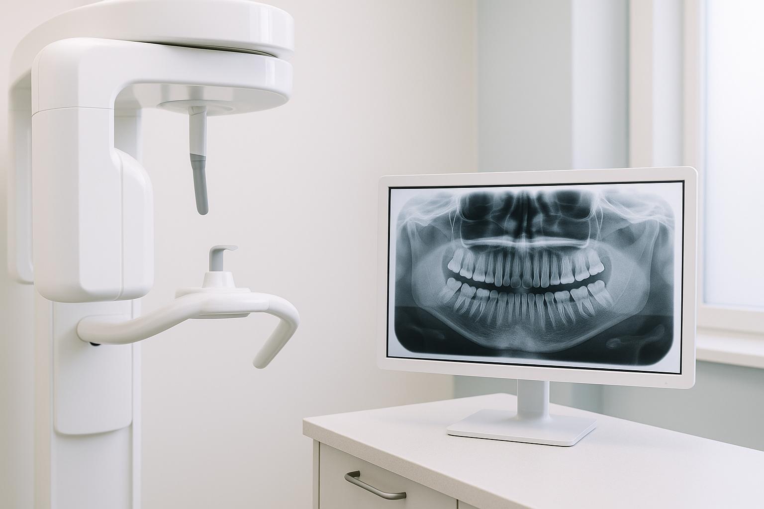
Occlusal X-rays provide a detailed image of your entire dental arch and surrounding structures in one shot. They’re particularly useful for identifying oral health problems that might not be visible during a standard dental exam, such as impacted teeth, jaw fractures, cysts, and infections. These X-rays are also commonly used in paediatric dentistry to monitor tooth development and eruption.
Here’s what you need to know about occlusal X-rays:
- What They Show: Full dental arch, jaw structures, and tissues beneath the gums.
- Key Uses: Detecting impacted teeth, jaw injuries, cysts, abscesses, and monitoring tooth growth in children.
- Comparison to Other X-rays: Broader view than bitewing or periapical X-rays but less detailed for small-scale issues.
- Safety: Modern digital systems reduce radiation exposure by up to 90% compared to older methods and comply with Australian safety standards.
Whether diagnosing fractures, tracking dental growth, or spotting hidden issues, occlusal X-rays are a practical tool for dentists to ensure early detection and effective care.
Main Uses of Occlusal X-rays
Monitoring Tooth Growth and Eruption
Occlusal X-rays play a key role in keeping track of tooth development, especially in children and teenagers. These images let dentists examine how teeth are forming, making it easier to spot any potential issues early on. Early detection means dentists can intervene before minor problems turn into major orthodontic challenges later on [1][5][8].
In paediatric dentistry, these X-rays are particularly useful for checking if teeth are growing as they should and for evaluating the position of teeth that haven’t erupted yet. When children are undergoing orthodontic treatment, occlusal X-rays provide a detailed look at the entire dental structure. This helps dentists predict how teeth are likely to erupt and whether there’s enough room for all the permanent teeth to fit properly [8].
These X-rays are also incredibly helpful for assessing dental trauma.
Finding Jaw Fractures and Trauma
For patients with suspected jaw injuries – whether from accidents, sports, or other incidents – occlusal X-rays offer a clear view of the damage. They can reveal fractures or cracks in the jawbone and surrounding structures that might not be noticeable during a standard clinical exam, especially in cases where patients report jaw pain after an injury [1].
This imaging allows dentists to evaluate the severity of the trauma and plan the appropriate treatment.
Detecting Cysts, Tumours, and Infections
Occlusal X-rays are also effective at uncovering serious conditions like cysts, abscesses, tumours, and infections that lie beneath the gum line. These are often missed during routine check-ups, making X-rays a critical tool for identifying hidden issues [1][3].
While a physical exam focuses on surface-level problems, occlusal X-rays provide a deeper look into the bone and surrounding tissues. For preventive care, most people benefit from dental X-rays every six to eighteen months. However, those with specific risk factors – such as a history of jaw injuries or bone health conditions – might need them more frequently [6].
Benefits and Drawbacks of Occlusal X-rays
Benefits
Occlusal X-rays capture a full-arch image in a single shot, making them an efficient tool for initial dental assessments [1][7].
They are particularly useful in paediatric dentistry, where they help monitor the development of primary and permanent teeth while identifying any irregularities [1][5][11]. Additionally, they expose patients to minimal radiation levels, a benefit further enhanced by modern digital imaging systems [1][7].
These X-rays are also effective for detecting jaw fractures, cysts, abscesses, and unerupted teeth [3][6]. Beyond dental structures, they can provide images of the hard and soft palate as well as the mucous membranes beneath the tongue [10].
Drawbacks
While occlusal X-rays are valuable, they lack the detailed three-dimensional imaging that cone-beam computed tomography (CBCT) provides, which can limit their accuracy in complex cases [1][7]. They may struggle to clearly capture posterior or overlapping areas, potentially leading to incomplete visualisation.
Fine details like small interproximal cavities, early bone loss, or hairline root fractures may go unnoticed due to their lower resolution and absence of three-dimensional capability [1][7]. For these specific concerns, bitewing or periapical X-rays – or even CBCT – might be better suited. As a result, while occlusal X-rays are excellent for broader evaluations, they are less effective in diagnosing subtle or small-scale dental issues.
X-ray Type Comparison
To choose the most appropriate imaging method, it’s helpful to compare occlusal X-rays with other dental X-ray types. Here’s a quick breakdown:
| X-ray Type | Field of View | Typical Usage | Radiation Exposure |
|---|---|---|---|
| Occlusal | Full upper or lower arch | Monitoring tooth development, assessing jaw fractures, detecting cysts | Moderate (low overall) |
| Bitewing | Small section (crowns only) | Identifying cavities between teeth, detecting bone loss | Low |
| Periapical | Single tooth and root area | Diagnosing root infections, abscesses, and bone health | Low |
Occlusal X-rays provide a broader perspective but lack the fine detail of bitewing or periapical X-rays, which are better suited for close-up imaging of specific areas [1][7]. Experts recommend occlusal X-rays for focused diagnostic purposes, such as monitoring dental development in children, evaluating jaw trauma, or identifying large lesions or cysts [3][6][5]. However, they are not typically used for routine cavity detection in adults.
Selecting the right type of X-ray depends on the clinical question and patient requirements, always adhering to Australian safety standards set by ARPANSA [7]. These guidelines prioritise minimising radiation exposure and ensuring X-rays are only performed when clinically necessary, with extra caution for children. In the following section, we’ll explore the procedure and safety measures that highlight the role of occlusal X-rays in comprehensive dental care.
How the Procedure Works and Safety Measures
Patient Setup and Positioning
When it comes to capturing a full-arch image with occlusal X-rays, getting the patient’s position right is key. Typically, the patient sits upright to stay comfortable and steady. A larger film or digital sensor is placed inside their mouth – either on the roof (for maxillary occlusal images) or the floor (for mandibular occlusal images), depending on which area needs to be examined. The patient bites down gently to hold the sensor in place, ensuring the entire dental arch and surrounding jaw structures are visible in one shot. The X-ray machine is set up outside the mouth, and the actual imaging process takes just a few seconds [1][3][7]. Once the patient is positioned correctly, strict safety protocols are followed to minimise radiation exposure.
Radiation Safety Steps
Modern dental clinics in Australia prioritise radiation safety by following strict protocols. Patients are typically provided with lead aprons and thyroid collars, especially if they are children or pregnant women [1][2][4]. The use of digital X-ray technology plays a big role in reducing radiation exposure – by up to 90% compared to traditional film-based systems – while still delivering high-quality images. To put it into perspective, the radiation dose from an occlusal X-ray is less than 0.01 millisieverts (mSv), which is just a small fraction of the average daily background radiation Australians are exposed to [9]. Digital systems also allow for immediate image capture and review, which not only speeds up the diagnostic process but also aids in precise treatment planning. For more vulnerable patients, extra precautions are taken, and the procedure is only performed when absolutely necessary [4][5].
Australian Standards and Guidelines
All these safety measures are backed by Australia’s stringent regulatory standards. Dental clinics operate under the guidelines set by the Australian Radiation Protection and Nuclear Safety Agency (ARPANSA) and the Dental Board of Australia. These standards are rooted in the ALARA principle (As Low As Reasonably Achievable), ensuring that radiation exposure is kept to the minimum needed for accurate diagnosis. This involves regular calibration of equipment, continuous staff training, and thorough documentation of procedures. Clinics also undergo regular audits and inspections to confirm that protective gear is in good condition, staff training is up-to-date, and recorded radiation levels stay within safe limits [4][5].
sbb-itb-2be92ed
How Occlusal X-rays Support Complete Dental Care
Early Detection and Treatment
Occlusal X-rays play a key role in preventive dental care by spotting potential problems before they escalate into serious conditions. These detailed images help dentists identify issues like cysts, abscesses, jaw fractures, and infections early on, when treatments are typically simpler and less invasive [3].
Addressing these concerns early not only reduces the complexity of treatment but also helps patients maintain better oral health without breaking the bank. For instance, catching an impacted tooth early allows for planned preventive measures, avoiding emergency extractions that often require more complicated surgery and longer recovery.
Use Across Different Dental Fields
The insights gained from occlusal X-rays extend across multiple areas of dentistry. Their diagnostic value makes them indispensable in various specialties. In paediatric dentistry and orthodontics, these images are used to track tooth development, monitor eruption patterns, and assess jaw alignment to guide treatment planning [1] [5] [8].
In oral and maxillofacial surgery, as well as emergency dental care, occlusal X-rays are critical for evaluating injuries, such as jaw fractures or displaced teeth, enabling swift and effective treatment [1] [3] [6]. Meanwhile, in periodontics and endodontics, they are instrumental in detecting bony defects, cysts, and complications related to root canals [6].
Modern Imaging at Complete Smiles Bella Vista

At Complete Smiles Bella Vista, advanced imaging technology, including digital occlusal X-rays, enhances the quality of care provided. The clinic’s state-of-the-art digital radiography systems meet Australian standards, delivering high-quality images quickly and with minimal radiation exposure.
Digital X-rays outperform traditional film-based systems by offering sharper images, faster processing times, and reduced radiation levels [7]. This technology supports a proactive approach to dental care, enabling early diagnosis and effective management of oral health issues. Whether it’s assessing unerupted teeth for orthodontic purposes, analysing bone structure for implants, or identifying abnormalities that could affect treatment, occlusal X-rays provide the detailed information needed for safer and more precise treatment plans [3].
Occlusal Radiography
Conclusion
Occlusal X-rays play a vital role in modern dentistry, providing a detailed view of the entire dental arch that can’t be achieved through a simple visual check. They are especially helpful in identifying issues like impacted teeth, jaw fractures, cysts, and abscesses – problems that might otherwise go unnoticed until they become more severe [1][3].
In paediatric cases, early detection through occlusal X-rays can help prevent complications down the road [2][3]. This makes them an essential tool for proactive dental care.
With advancements like digital occlusal X-rays, patients are exposed to up to 80% less radiation compared to traditional film, while also benefiting from sharper images delivered more quickly [9]. In Australia, dental practices adhere to strict radiation safety standards, guided by the ALARA (As Low As Reasonably Achievable) principle. This ensures that every X-ray is carefully considered and optimised to protect patient health.
Occlusal X-rays are indispensable across various dental fields, including paediatric dentistry, orthodontics, oral surgery, and emergency care. Routine X-rays, typically recommended every 6 to 18 months based on individual risk factors, allow patients to stay ahead of potential issues and maintain their oral health.
FAQs
What are occlusal X-rays, and how do they help detect oral health issues?
Occlusal X-rays are a specialised type of dental imaging that offers a broad view of either the upper or lower jaw. They’re especially helpful in spotting issues like impacted teeth, jaw fractures, cysts, or other abnormalities that might not be detected during a routine dental check-up.
These X-rays provide detailed visuals of the jaw and nearby structures, enabling dentists to diagnose conditions and develop treatment plans with greater precision. This imaging technique plays a key role in keeping oral health on track and catching potential problems before they escalate.
How is radiation exposure minimised during occlusal X-rays, especially for children?
When it comes to occlusal X-rays, particularly for children, safety is a top priority. Modern dental X-ray machines are built to use minimal radiation, and protective gear like lead aprons and thyroid collars are employed to safeguard sensitive areas.
Dentists also adhere to the ALARA principle (As Low As Reasonably Achievable). This means X-rays are taken only when truly needed, and the lowest possible radiation dose is used. These careful steps ensure that the diagnostic benefits far outweigh any risks, making it a reliable way to support oral health.
When would a dentist recommend occlusal X-rays instead of other dental X-rays?
Occlusal X-rays are often recommended when a dentist needs a detailed image of the entire arch of either your upper or lower teeth. These X-rays are especially helpful in spotting issues like impacted teeth, jaw fractures, cysts, or irregularities in tooth development. They can reveal problems that might not be noticeable during a routine dental check-up or with other types of X-rays.
With their ability to offer a broad view of your oral structures, occlusal X-rays are an important tool for identifying issues early and planning effective treatments. This ensures you receive the care you need to maintain healthy teeth and gums.
Related Blog Posts
- How X-Rays Help in Dental Treatment Planning
- AI vs. Traditional Methods: Impacted Teeth Detection
- How Full-Mouth X-Rays Aid Diagnosis
- CBCT in Endodontics: Accuracy and Limitations
Important Notice: Any surgical or invasive procedure carries risks. Before proceeding, you should seek a second opinion from an appropriately qualified health practitioner.
Individual results may vary. The information provided in this article is for educational purposes only and does not constitute medical advice.
Checkout Related Blogs


Get in touch with us
For more information, call us now to start feeling better. Or fill the form below to make appointment
The Latest News from Complete Smiles
How to Clean Clear Plastic Retainers
Checklist for Choosing Wearable Dental Devices
Checklist for Choosing Cloud AI Platforms in Dentistry

Complete Smiles Bella VistaAccepts All Major Health Funds, Including


