





Diagnostic Tools for Periodontal Surgery Planning
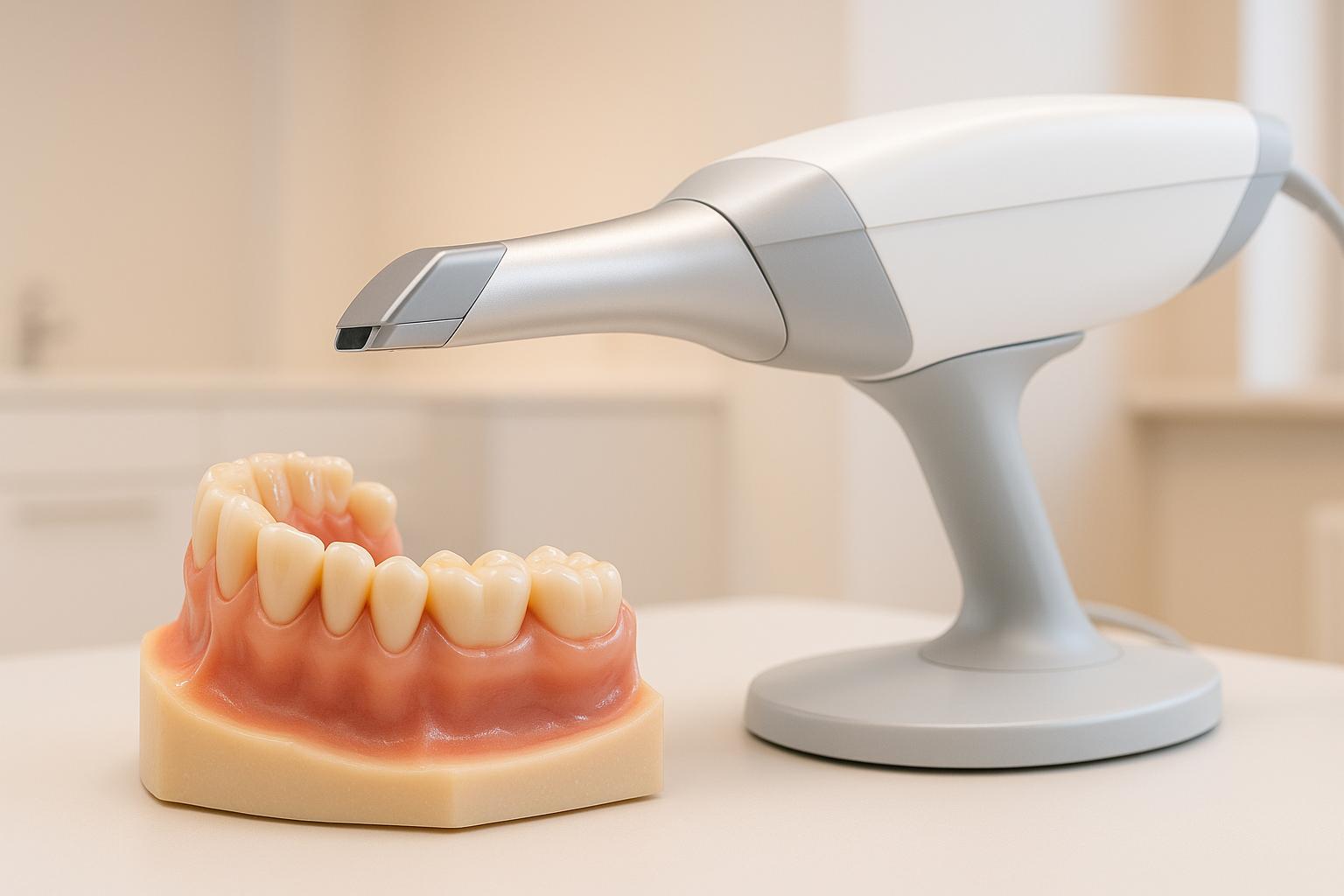
Australian dental practices combine clinical examinations, manual probing, and advanced technologies like CBCT imaging, digital probing systems, and AI tools. These methods assess gum health, bone defects, and disease severity to guide treatment plans.
Key tools include:
- Manual Probing: Measures gum pocket depths and clinical attachment levels.
- Digital Probing Systems: Automate measurements with higher precision (e.g., Florida Probe®).
- 2D X-rays: Detect bone loss and periodontal defects.
- 3D Imaging (CBCT): Provides detailed views of bone and soft tissue for complex cases.
- AI Integration: Analyses imaging and clinical data for personalised care.
Challenges include high costs (e.g., CBCT equipment ranges from AUD 50,000–150,000), compliance with Australian regulations, and staff training. However, these tools improve planning accuracy, reduce risks, and enhance outcomes. Smaller clinics often refer patients to specialist centres for advanced diagnostics.
Emerging methods like MRI and ultrasonography offer non-radiation alternatives but remain in early development. As technology evolves, Australian practitioners must balance cost, safety, and precision to deliver optimal care.
Periodontics | Diagnosis & Periodontal Exam | INBDE, ADAT
Clinical Examination and Periodontal Probing
While advanced imaging and AI diagnostics are reshaping the dental landscape, clinical examination and periodontal probing remain indispensable in Australian practices. These methods provide crucial baseline data that guide surgical planning and help assess gum health and tissue attachment levels [3][7].
The process starts with a comprehensive examination of the periodontal tissues. Practitioners look for signs like inflammation, bleeding, and pocket formation. This hands-on evaluation not only identifies periodontal issues but also lays the groundwork for determining whether surgical intervention is needed [3][7].
Periodontal probing takes the assessment further by delivering measurable insights into tissue health. Using calibrated probes, practitioners measure pocket depths and clinical attachment levels, creating a detailed periodontal map for each tooth. This data plays a direct role in shaping treatment plans and estimating outcomes.
Manual Periodontal Probing
In Australian dental practices, manual periodontal probing is still considered the gold standard for evaluating gum health. This method involves using a calibrated probe to measure the depth of the gingival sulcus or periodontal pocket at multiple points around each tooth [3][6].
The process is straightforward: the practitioner inserts the probe alongside the tooth, measuring from the gingival margin to the pocket base. Results are recorded in millimetres, and the clinical attachment level is calculated using a simple formula:
Attachment level (mm) = Probing depth (mm) – Gingival margin (mm)
For instance, if a tooth has a probing depth of 6mm and the gingival margin is 2mm above the cemento-enamel junction, the clinical attachment level would be 4mm.
Manual probing also tracks bleeding on probing, an indicator of active inflammation, and evaluates furcation involvement in multi-rooted teeth. These measurements help pinpoint areas requiring surgical attention and monitor the progression of periodontal disease over time [3][6].
That said, manual probing isn’t without its challenges. Factors like probing force, angulation, operator technique, and patient discomfort can all influence the accuracy of measurements. Despite these limitations, it remains a cornerstone in periodontal diagnostics, providing the essential data needed for surgical planning [6].
While manual methods are effective, digital systems are stepping in to add more precision and efficiency.
Digital and Electronic Probing Systems
Digital probing systems are making a big impact in periodontal diagnostics by addressing many of the limitations of manual techniques. Tools like the Florida Probe®, InterProbe™, and pa-on Parometer® are gaining traction in Australian practices for their ability to enhance precision and streamline workflows [6].
Take the Florida Probe®, for example. It delivers measurements with 0.2mm precision using a constant force of 0.15N, minimising variability caused by operator technique [6]. Plus, it automatically records and transfers data into digital charts, eliminating transcription errors and saving time.
Here’s how electronic and manual probing compare:
| Feature | Manual Probing | Electronic Probing |
|---|---|---|
| Measurement Precision | ≈1mm | 0.2mm (Florida Probe®) |
| Force Control | Operator-dependent | Constant (0.15N) |
| Data Recording | Manual charting | Automated charting |
| Workflow Efficiency | Slower, prone to errors | Faster, reduces errors |
Modern electronic systems often come equipped with additional sensors, allowing them to detect calculus, monitor temperature changes, and even measure sulfide levels. These advanced features go beyond the traditional scope of pocket depth measurements [6].
The benefits are clear: improved precision, reduced operator variability, and enhanced data management. However, adopting these systems requires careful consideration. Factors such as the upfront investment (in AUD), staff training, and compatibility with existing digital systems should be weighed, especially for smaller clinics.
Ultimately, these digital tools don’t replace traditional methods – they complement them. By integrating manual probing with digital systems, along with radiographic imaging and other diagnostic technologies, Australian dental practices can gather the comprehensive data needed to plan effective treatments and achieve the best surgical outcomes.
X-rays and Radiographic Imaging for Periodontal Diagnosis
Once clinical data is collected through probing, radiographic imaging sharpens the diagnostic picture by examining the underlying bone structures. This step is essential in periodontal diagnosis as it reveals bone loss, defects, and disease progression that may not be apparent during clinical assessments.
In Australian dental practices, strict safety measures are in place to minimise radiation exposure. X-rays are typically recommended only after a thorough clinical evaluation, and the use of digital radiography systems helps lower radiation doses compared to traditional film-based methods. This careful approach ensures effective use of both 2D and 3D imaging for accurate diagnosis.
2D X-rays: Insights and Limitations
Intraoral periapical and bitewing X-rays remain the primary tools for periodontal diagnosis in Australia. These imaging techniques provide detailed views of the jawbone, helping to estimate bone loss and identify periodontal defects.
Periapical X-rays are particularly effective at detecting early-stage bone loss. A study involving 100 patients with moderate to severe periodontitis examined 2,536 teeth and 5,072 proximal surfaces using radiographic and surgical measurements. The findings revealed that periapical X-rays detected small osseous destruction in 3.81% of cases, compared to just 0.81% with panoramic X-rays – making periapical X-rays 4.7 times more effective in identifying minor defects [3].
While panoramic X-rays offer a broader view of the jaw, they are less sensitive to smaller defects. A key limitation of 2D imaging is its inability to provide a three-dimensional view of periodontal structures. This can lead to underestimating bone loss in milder cases and complicates the assessment of defect morphology, especially around multi-rooted teeth. Additionally, buccal or lingual surface defects may go unnoticed due to the projection’s limitations.
Such challenges are particularly significant when planning complex surgical procedures that require an in-depth understanding of bone architecture.
Comparing 2D and 3D Imaging
While 2D imaging provides valuable baseline information, 3D techniques add a new level of precision, especially for complex cases. The introduction of cone-beam computed tomography (CBCT) has been transformative in periodontal diagnosis and surgical planning. CBCT offers highly accurate three-dimensional views of bone destruction, allowing clinicians to map periodontal pockets and defects with precision – an essential feature for surgical planning and implant placement.
| Imaging Feature | 2D X-rays | 3D Imaging (CBCT) |
|---|---|---|
| Bone Loss Detection | Good for general assessment | Detailed 3D mapping |
| Defect Morphology | Limited detail | Complete 3D visualisation |
| Surgical Planning | Basic guidance | Comprehensive roadmap |
| Radiation Exposure | Lower dose | Higher dose |
| Cost Consideration | More affordable | Higher investment |
| Availability | Widely accessible | Increasingly available |
CBCT can even visualise both bone and soft tissue using radio-opaque agents, enabling precise, non-invasive mapping of periodontal pockets. This capability opens the door to less invasive yet highly accurate diagnostic methods.
The choice between 2D and 3D imaging depends on the clinical scenario. For routine check-ups and monitoring, 2D X-rays are cost-effective and efficient. However, 3D imaging is preferred for cases involving complex bone defects, multi-rooted teeth, advanced periodontitis, or dental implant planning. Australian practitioners must weigh the diagnostic benefits of advanced imaging against factors like cost and radiation exposure – particularly for younger patients or those needing repeated imaging.
Emerging technologies such as ultrasonography and MRI may soon provide safer diagnostic alternatives, offering comprehensive imaging of both hard and soft tissues without ionising radiation. These advancements could transform periodontal diagnosis in the future.
sbb-itb-2be92ed
3D Imaging and New Technologies
Modern 3D imaging technologies are transforming periodontal diagnostics, offering a level of detail and precision that goes far beyond traditional radiography. These advancements not only enhance diagnostic accuracy but also help clinicians create more targeted treatment plans while addressing concerns about radiation exposure.
Cone-Beam Computed Tomography (CBCT)
CBCT has become a game-changer in periodontal care, providing detailed three-dimensional images of bone structures that standard X-rays simply cannot achieve. This technology allows for precise bone mapping and the assessment of pathologies, reducing uncertainty when dealing with complex periodontal issues.
Research has shown that CBCT delivers more accurate visualisation of bone damage compared to traditional radiography, making it an essential tool for diagnosing and planning treatments for periodontitis [8]. For example, it clearly displays furcation involvement and root morphology, offering a full picture of the extent and severity of bone loss. This detailed imaging is particularly useful for planning surgical interventions, such as guided bone regeneration. In these cases, CBCT helps map defects and design customised barrier membranes or grafts tailored to the patient’s unique anatomy, ensuring more predictable outcomes.
However, adopting CBCT technology comes with challenges. The equipment costs range between AUD 50,000 and AUD 150,000, and dental practices must also invest in specialised training and comply with strict radiation safety standards. Ongoing maintenance is another factor to consider, making CBCT a significant but valuable investment for advanced periodontal care.
Intraoral Scanners (IOS) Integration
Intraoral scanners (IOS) are another powerful tool that complements CBCT by accurately capturing the contours of soft tissues and dental surfaces. These scanners create detailed digital impressions, which are particularly useful for precise surgical planning. When combined with CBCT data, they enable clinicians to build a complete virtual model of both hard and soft tissues. This integration supports highly accurate treatment plans and customised surgical approaches.
One case study from the Journal of Periodontology illustrated the combined use of CBCT and intraoral scanning in a complex bone grafting procedure. The CBCT images provided detailed measurements of the bone defect, while the intraoral scan mapped the soft tissue contours. Together, these technologies allowed for the creation of a customised surgical guide and graft, leading to successful bone regeneration and improved outcomes [6].
Intraoral scanners also stand out for their accessibility. While CBCT systems require a significant financial commitment, IOS technology is more affordable, with costs typically ranging from AUD 10,000 to AUD 30,000. This makes them a practical option for many dental practices looking to enhance their diagnostic and surgical capabilities.
New Non-Radiation Imaging Technologies
Emerging non-radiation imaging methods are pushing the boundaries of periodontal diagnostics. Techniques like MRI and ultrasonography, often paired with artificial intelligence, offer exciting possibilities for improving diagnostic precision while eliminating radiation exposure.
MRI, for instance, is showing promise in visualising periodontal ligaments, gingival tissues, and bone marrow changes. This capability goes beyond what traditional imaging methods can achieve, offering deeper insights into inflammatory processes and helping monitor treatment progress over time.
Ultrasonography, on the other hand, provides real-time imaging that can dynamically assess soft tissue inflammation and pocket depth. This is particularly valuable for patients who need frequent monitoring, such as those undergoing long-term periodontal therapy or management of peri-implant diseases.
Despite their potential, these technologies are still in the research phase for periodontal applications. Current limitations include lower spatial resolution compared to CBCT, as well as high costs and limited availability. MRI systems, in particular, require significant investment and specialised facilities. However, their radiation-free nature makes them appealing for use with young patients, pregnant women, and others who need repeated imaging. As these technologies evolve, they could become more accessible and practical for routine periodontal care.
The integration of artificial intelligence with these imaging methods is also on the horizon. AI algorithms are being developed to analyse imaging data automatically, potentially improving diagnostic accuracy and streamlining treatment planning. This combination of advanced imaging and AI could make cutting-edge periodontal diagnostics more widely available in the future.
Currently, the use of advanced imaging tools like CBCT and intraoral scanners varies across Australian dental practices. Specialist periodontal clinics are more likely to adopt these technologies to enhance surgical planning and improve patient outcomes.
Digital Systems and Artificial Intelligence in Periodontal Diagnosis
Artificial intelligence (AI) is reshaping how periodontal diseases are diagnosed and treated, offering more consistent and reliable assessments by integrating diverse diagnostic data. With advancements in 3D imaging, these tools enhance surgical planning by combining clinical insights with imaging data for a more precise approach.
AI Integration in Periodontal Surgery Planning
AI systems bring together data from various sources like intraoral scans (IOS), cone-beam computed tomography (CBCT) images, patient demographics, clinical examination results, and even salivary biomarkers. This combination creates detailed diagnostic profiles, offering a clear picture of both soft and hard tissue health while factoring in individual risk elements and disease progression patterns [2].
By standardising measurements, AI tools reduce variability that often depends on the clinician’s judgment. They also improve patient comfort by limiting the need for invasive diagnostic procedures [2].
Take PerioAI, for example. This system uses IOS and CBCT data to measure gingiva-bone distances, providing 3D visualisations of tissue relationships that go beyond the capabilities of traditional 2D imaging [2]. This approach aligns with the growing emphasis on precision and personalised care in periodontal practices across Australia.
Another key development is the Periodontal Risk Scoring System (PRSS), which boasts up to 70% accuracy in predicting high-risk patients [4]. By analysing a combination of patient demographics, clinical data, and imaging results, these AI tools help clinicians make informed decisions and craft tailored care plans.
AI also plays a critical role in early detection. By analysing imaging data in detail, these systems can spot subtle changes in tissue structure or bone density that signal the onset of periodontal disease. Early detection allows for timely intervention, potentially improving long-term outcomes [2].
Future AI Applications in Australian Dental Practices
The future of AI in periodontal care is promising. Imagine real-time AI analysis of diagnostic images providing immediate treatment recommendations, enabling practitioners to discuss options with patients during the same visit. Predictive analytics could also become a game-changer, with systems forecasting long-term disease progression or implant success rates based on individual patient factors.
Incorporating genetic, microbiome, and lifestyle data into AI systems could elevate personalised care to new levels, revolutionising how periodontal disease is prevented and managed [1]. For rural and remote Australian communities, AI-powered tools could enable general practitioners to conduct advanced assessments and refer patients for specialised care when necessary [4].
However, adopting these technologies involves several practical considerations. For accurate AI analysis, Australian dental practices need standardised imaging data from IOS and CBCT systems. Seamless integration with existing clinical workflows and electronic health records is crucial, and compliance with Australian health regulations, such as the Privacy Act 1988 (Cth), must be ensured.
Training dental staff is equally important. Practitioners need to understand how to interpret AI-generated insights and incorporate them into their decision-making processes without compromising their clinical judgment. While challenges remain, the potential benefits of AI-driven diagnostic systems – greater precision, personalised care, and improved efficiency – make them a valuable addition to modern periodontal practices. As these technologies become more refined and accessible, they are likely to become indispensable tools in dental care across Australia.
Summary and Considerations for Australian Dental Practices
Key Points for Effective Diagnosis and Planning
Planning for periodontal surgery requires combining various diagnostic tools to create a thorough and accurate assessment. At the heart of this process are clinical examinations and periodontal probing, with manual probing recognised as the gold standard for evaluating tissue health across six sites per tooth [3][5]. The most reliable diagnoses come from integrating these clinical insights with radiographic imaging and advanced digital technology.
Radiographic imaging is indispensable for confirming periodontitis and determining the extent of bone loss. While periapical radiographs excel in detecting early defects, panoramic imaging is less precise in this regard [3]. For even greater accuracy, 3D imaging tools like cone-beam computed tomography (CBCT) provide detailed visualisations of bone structure and periodontal pockets, offering a level of detail that 2D techniques cannot match [6].
Digital probing systems, such as the Florida Probe® and InterProbe™, further enhance diagnostic accuracy by automating measurements and charting, which reduces the risk of human error and improves workflow efficiency [6]. When used together, these diagnostic methods offer a comprehensive view that supports better treatment planning and improved outcomes for patients. These tools are essential for addressing the specific challenges faced by local practices.
Local Considerations for Australian Practitioners
Beyond the technical aspects, Australian dental practitioners must navigate local regulatory and cost-related factors. Compliance with the Dental Board of Australia guidelines and the Australian Radiation Protection and Nuclear Safety Agency (ARPANSA) standards is non-negotiable for ensuring patient safety and the lawful use of radiographic equipment.
Strict infection control protocols are another critical consideration, especially when using diagnostic tools across multiple patients. Staying updated with the Australian Dental Association‘s (ADA) evolving recommendations on imaging and data management is key to maintaining best practices as technology advances.
For smaller practices, the high upfront costs of advanced imaging equipment can be a significant barrier. Establishing referral relationships with larger specialist centres that already have these technologies may offer a practical solution. This approach allows smaller clinics to provide patients with access to advanced diagnostics without the financial burden of purchasing the equipment themselves.
Patient consent is another important factor. Australian healthcare standards require that patients fully understand the benefits and risks associated with radiographic imaging. As non-radiation imaging methods like ultrasonography and MRI continue to develop, they may offer safer and more accessible alternatives in the future [6].
Specialist Clinics and Advanced Care
Specialist clinics across Australia demonstrate how advanced diagnostic tools can transform periodontal care. A prime example is Complete Smiles Bella Vista, under the leadership of Dr. James Hanna. This clinic showcases how cutting-edge technology and personalised treatment plans can work hand in hand to deliver exceptional care.
By integrating tools like digital X-rays, CBCT, and intraoral scanners, these clinics can thoroughly assess periodontal issues and plan surgeries with unmatched precision. This patient-centred approach, which uses advanced diagnostics to create tailored treatment plans, highlights the direction modern periodontal practices in Australia are heading.
Investing in advanced diagnostic tools, along with proper training and expertise, has the potential to significantly elevate the standard of periodontal care available to Australian patients. This combination ensures that both general and specialist practices can meet the growing expectations for precision and personalised care in dentistry.
FAQs
What are the benefits of using digital probing systems instead of manual probing for periodontal diagnostics?
Digital probing systems bring a range of benefits to periodontal diagnostics, standing out for their accuracy and consistency. By minimising the risk of human error, they ensure more reliable measurements of pocket depths and gum health, which translates to improved treatment planning and ongoing care.
Another advantage is their ability to integrate seamlessly with patient records. This makes tracking periodontal changes over time much simpler, helping dentists and specialists spot patterns and refine treatment strategies as needed. On top of that, these systems improve patient communication by providing clear, visual insights into oral health – making it easier for patients to understand their condition and stay engaged in their care.
How do non-radiation imaging technologies like MRI and ultrasonography compare to traditional diagnostic tools for periodontal surgery planning?
Non-radiation imaging techniques like MRI and ultrasonography are becoming popular alternatives to traditional imaging methods such as X-rays and CT scans in periodontal surgery planning. Their biggest advantage? No exposure to ionising radiation. This makes them an excellent option for patients who need frequent imaging or have specific health concerns that make radiation-based methods less ideal.
These technologies excel at providing detailed images of soft tissues, which can be incredibly useful in certain scenarios. That said, they aren’t yet a complete replacement for traditional methods. Tools like X-rays and 3D imaging, including CBCT (cone-beam computed tomography), are still the go-to options for evaluating bone structures and planning precise surgical procedures. Even so, the continuous improvements in non-radiation imaging are making them an increasingly valuable addition to the diagnostic toolbox for periodontal care.
What should smaller dental clinics consider before investing in advanced imaging technologies like CBCT?
Investing in advanced imaging tools like cone beam computed tomography (CBCT) is a big step for smaller dental clinics. There’s plenty to think about, starting with the cost of the equipment itself and the ongoing maintenance fees. Clinics also need to consider whether they have enough patient demand to make the purchase worthwhile.
Another factor is the space needed to install the equipment – CBCT machines aren’t exactly compact. Plus, staff training is crucial to ensure the technology is used correctly and efficiently.
On the upside, CBCT can significantly improve diagnostic and treatment planning, especially for procedures like dental implants, orthodontic treatments, or complex surgeries. For clinics offering these specialised services, the detailed 3D imaging provided by CBCT can lead to more precise treatments and better results for patients. However, it’s important to balance these advantages with the potential financial and operational impact on the practice.
Related Blog Posts
- Periodontal Pocket Depth: Measurement Techniques
- Advances in Gum Grafting Materials and Techniques
- Optical Imaging Tools for Gum Disease Diagnosis
- AI in Dentistry: Predicting Periodontal Disease
Important Notice: Any surgical or invasive procedure carries risks. Before proceeding, you should seek a second opinion from an appropriately qualified health practitioner.
Individual results may vary. The information provided in this article is for educational purposes only and does not constitute medical advice.
Checkout Related Blogs




Get in touch with us
For more information, call us now to start feeling better. Or fill the form below to make appointment
The Latest News from Complete Smiles
How to Clean Clear Plastic Retainers
Checklist for Choosing Wearable Dental Devices
Checklist for Choosing Cloud AI Platforms in Dentistry

Complete Smiles Bella VistaAccepts All Major Health Funds, Including





