


Micro vs. Nano Surface Modifications for Implants
When it comes to dental implants, the surface of the implant plays a key role in how well it integrates with bone. Two main types of surface modifications are used:
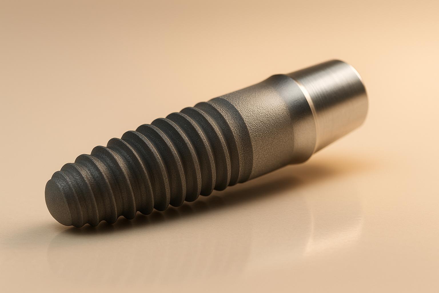
- Micro-scale modifications: These involve creating larger surface textures (1 to 100 micrometres) using methods like sandblasting and acid etching. They provide a rough surface that helps bone cells attach and grow.
- Nano-scale modifications: These focus on creating ultra-small features (1 to 100 nanometres) that interact with proteins and cells at a molecular level. Techniques like anodisation and chemical vapour deposition are commonly used.
Key differences:
- Micro-scale surfaces are cost-effective and have a long history of success but may trap bacteria and lack fine biological interaction.
- Nano-scale surfaces improve cellular behaviour and healing times but are more expensive and require advanced manufacturing.
Choosing the right type depends on the patient’s needs, with micro-scale modifications often preferred for routine cases and nano-scale modifications offering advantages in complex situations.
1. Micro-Scale Surface Modifications
Modification Methods
Micro-scale surface modifications involve creating tiny features on implant surfaces using well-established techniques commonly employed in implant production.
- Sandblasting: This method propels abrasive particles at high speeds onto the implant surface, producing an uneven texture. The resulting irregularities increase the surface area, improving tissue integration. The size of the abrasive particles used determines the depth and extent of the surface texture.
- Acid Etching: Acids are used to remove material from the surface, forming micro-pits and valleys. When combined with sandblasting, the technique is often referred to as SLA (Sandblasted, Large-grit, Acid-etched) treatment, which enhances surface roughness for better integration.
- Plasma Spraying: This technique applies a hydroxyapatite coating onto the implant, creating a bioactive surface that mimics the mineral composition of natural bone. The coating bonds to the titanium substrate, promoting integration with surrounding tissue.
- Machining and Threading: These methods create precise geometries and thread patterns on the implant surface. The resulting textures encourage mechanical interlocking with bone tissue, improving stability.
These modifications are specifically designed to improve cellular attachment and osseointegration, laying the groundwork for better mechanical stability.
Impact on Osseointegration
Micro-scale modifications play a key role in enhancing osseointegration. The irregular surface topography created by these methods provides a larger area for bone cells to attach. This expanded surface promotes better mechanical interlocking between the implant and the surrounding bone.
Osteoblasts (bone-forming cells) are more likely to adhere to micro-textured surfaces and produce higher levels of adhesion proteins. This improved protein adsorption forms a biological connection between the implant and the tissue, supporting early stability and integration.
Clinical observations have shown that implants with micro-scale modifications often exhibit a faster initial healing response compared to those with smooth surfaces. This can lead to earlier functional loading, which is particularly beneficial in clinical settings. These micro-scale enhancements will later be compared to nano-scale modifications to evaluate their relative effectiveness.
Clinical Evidence
Clinical studies have demonstrated that micro-scale modifications contribute to strong bone integration, which is essential for achieving stable, long-term results.
In fact, the improved osseointegration provided by these modifications has encouraged some clinicians to adopt faster loading protocols, enabling earlier restorative procedures compared to traditional timelines.
Additionally, there is evidence suggesting that micro-textured surfaces may be especially useful in cases of reduced bone density, as they can help enhance primary stability in softer bone.
Limitations
While these modifications offer clear benefits, they are not without drawbacks. The same surface irregularities that promote bone cell attachment can also trap bacteria, increasing the risk of peri-implant inflammation.
Deep surface textures can make it difficult to fully remove biofilms during routine maintenance, potentially complicating long-term care. Furthermore, achieving consistent results requires precise control over manufacturing processes, as variations in methods like sandblasting can impact the final surface characteristics and clinical outcomes.
Another consideration is cost. Advanced manufacturing techniques can be more expensive, and the fixed nature of these surface characteristics means careful planning is essential to ensure the chosen modification aligns with the specific clinical needs.
2. Nano-Scale Surface Modifications
Modification Methods
Nano-scale surface modifications involve creating features at the nanometre level using advanced techniques that go beyond traditional methods.
Anodisation is a common approach for generating titanium dioxide nanotubes on implant surfaces. By adjusting voltage and processing time, manufacturers can produce uniform nanotube arrays with diameters ranging from 15 to 120 nanometres. This surface structure enhances protein adsorption and improves cellular interactions at the molecular level.
Sol-gel coating applies thin films containing bioactive compounds such as calcium phosphate or silica. This method allows precise control over the surface’s chemistry and texture. The process forms a uniform layer that can embed growth factors or antimicrobial agents directly into the implant surface.
Chemical vapour deposition builds nano-structured coatings atom by atom, ensuring a consistent chemical composition and uniform biological response across the entire implant surface.
Plasma treatment changes the surface energy and chemistry without altering the underlying titanium structure. This technique creates hydrophilic surfaces, which improve protein adsorption and cellular attachment during the critical first hours after implantation.
These nano-scale methods are designed to create surfaces that support better molecular interactions, ultimately influencing osseointegration in a unique way.
Impact on Osseointegration
Nano-scale modifications have a direct impact on osseointegration by facilitating close interaction with proteins and cellular components, creating a more effective biological interface.
The nanoscale topography replicates the extracellular matrix, providing familiar attachment points for bone cells. This biomimetic environment encourages cells to behave as they would in natural bone tissue, promoting more consistent healing.
Nano-modified surfaces also enhance protein adsorption, with proteins like fibronectin and vitronectin showing a preference for binding to specific nanoscale features. These proteins create an ideal foundation for cellular attachment and growth.
Another advantage is the improved cellular signalling that these surfaces provide. The small-scale features influence cellular behaviour through mechanotransduction, where physical surface characteristics are translated into biological signals. This interaction guides cell function and differentiation, aligning with the goal of achieving better clinical outcomes.
Research shows that nano-modified surfaces can speed up the early stages of healing. Enhanced cellular attachment has been observed within the first 24 to 48 hours after implantation, which contributes to more predictable osseointegration.
Clinical Evidence
Clinical studies are beginning to back up the promising in vitro findings, showing that nano modifications can improve implant integration.
Research indicates faster healing with nano-modified surfaces, with some studies suggesting that these surfaces may support earlier loading protocols. For instance, successful outcomes have been reported at six to eight weeks, compared to the traditional 12-week healing period for conventional surfaces.
Histological analysis reveals closer bone-implant contact at the cellular level. Bone tissue integrates more tightly with the implant surface, resulting in stronger mechanical connections that could enhance long-term stability.
Nano-modified surfaces have also shown promise in more challenging cases, such as patients with poor bone quality or medical conditions that impair healing. The enhanced cellular interaction may partially offset reduced healing capacity in these situations, making these modifications particularly useful for complex cases.
Limitations
While nano-scale modifications offer clear advantages, there are challenges that limit their widespread use.
Manufacturing complexity and quality control are significant hurdles. These processes require advanced equipment and precise control, and standard inspection methods often fall short in detecting variations at the nanoscale.
The cost of production is another barrier. The sophisticated manufacturing and quality control processes make nano-scale modifications more expensive than conventional treatments.
Long-term stability of these features is a concern as well. Over time, normal physiological processes may degrade some nanoscale modifications, potentially reducing their effectiveness. Their fine structure makes them more prone to wear or dissolution compared to larger surface features.
Finally, there is limited clinical data available. While early results are encouraging, the lack of extensive long-term studies means that potential complications and outcomes over time are still being evaluated. The dental implant field typically requires robust, long-term evidence before adopting new technologies widely.
Maintenance is another consideration. Nano-modified surfaces may respond differently to cleaning procedures, requiring adjustments to care routines for both patients and practitioners. This adds another layer of complexity to their adoption in clinical settings.
What is the ideal implant surfaces Machine → AlO₂ Blasted Etched → HA → SBM → Anodized
sbb-itb-2be92ed
Advantages and Disadvantages
When considering the various methods of implant surface modification, it’s essential to weigh their benefits and limitations. Understanding these trade-offs helps clinicians select the most suitable implant for each patient.
Micro-Scale Modifications: Pros and Cons
Micro-scale modifications come with several well-documented benefits. They’re cost-effective to produce, thanks to established manufacturing techniques, and have a long history of use. This extensive track record provides a wealth of data on success rates and potential complications, enabling clinicians to predict outcomes with confidence and set realistic expectations for patients. Additionally, these surfaces are durable, maintaining their properties under normal physiological conditions over time.
However, the limitations of micro-scale modifications lie in their interaction with biological systems. Their larger surface features don’t mimic the natural extracellular matrix as effectively as nano-scale surfaces. This can lead to less efficient protein adsorption and a slower cellular response during the early healing stages, which may slightly delay the integration process.
Nano-Scale Modifications: Benefits and Limitations
Nano-scale modifications are designed to improve biological compatibility. These surfaces more closely replicate the natural bone environment, enhancing protein adsorption and cellular signalling. This biomimicry can accelerate healing and may even support faster loading protocols in certain cases. Such improvements are particularly valuable in complex clinical situations, like patients with poor bone quality or systemic health challenges, where enhanced cellular interaction can make a significant difference.
That said, nano-scale modifications come with their own set of challenges. Manufacturing these surfaces is more complex and costly, requiring specialised equipment. Quality control is also more demanding, as standard inspection methods may not effectively assess nano-scale features, potentially leading to variability between production batches. Additionally, there are concerns about the long-term stability of these delicate features, as well as the limited availability of long-term clinical data.
Comparative Analysis
The table below highlights the key differences between micro- and nano-scale modifications:
| Aspect | Micro-Scale Modifications | Nano-Scale Modifications |
|---|---|---|
| Manufacturing Cost | Lower, due to established processes | Higher, requiring specialised equipment |
| Quality Control | Standard methods are sufficient | Advanced techniques often needed |
| Clinical Evidence | Extensive long-term data available | Limited long-term studies so far |
| Biological Response | Supports effective osseointegration | Enhanced cellular interaction and response |
| Healing Time | Follows traditional healing timelines | Potential for faster healing in some cases |
| Surface Stability | Reliable over time | Possible concerns about long-term durability |
| Patient Suitability | Ideal for routine cases | Beneficial in complex or challenging situations |
Clinical Decision Considerations
The choice between micro- and nano-scale modifications depends on a balance of practical, biological, and patient-specific factors. Micro-scale modifications are often the go-to option for routine cases, especially when cost, durability, and a proven track record are priorities. They are particularly suitable for patients with good bone quality and normal healing capabilities.
On the other hand, nano-scale modifications may offer advantages in more complex cases, such as patients with poor bone quality or systemic health issues. The enhanced early biological response can be a game-changer in these scenarios. However, the higher costs and limited long-term data must be taken into account.
Interestingly, ongoing research is exploring hybrid surfaces that combine the strengths of both micro- and nano-scale modifications. These aim to optimise biological performance while keeping production practical and ensuring long-term reliability. Ultimately, the best decision comes down to evaluating the individual needs of the patient, the clinical situation, and economic factors, as both approaches can lead to successful outcomes when applied appropriately.
Conclusion
Drawing from established clinical practices and manufacturing insights, the comparison between micro-scale and nano-scale surface modifications highlights distinct advantages for each, with the best choice often depending on the clinical scenario and the individual needs of the patient.
Micro-scale modifications have a long track record of success, offering reliable osseointegration supported by years of clinical evidence. Their affordability and consistent outcomes make them a practical choice for routine implant procedures, particularly in patients with healthy bone quality. The predictable nature of these surfaces and the standardised quality control processes have cemented their role in dental practices across Australia.
On the other hand, nano-scale modifications bring a different set of benefits, focusing on enhanced biological compatibility through improved protein adsorption and cellular interactions. These surfaces are especially promising for complex cases, such as patients with compromised bone quality or systemic health issues that could impact healing. However, challenges remain, including higher production costs and the need for more long-term clinical data to confirm their effectiveness over time.
Looking ahead, the future of implant surface technology seems to be moving towards hybrid surfaces that merge the strengths of micro- and nano-scale modifications. These advanced designs aim to combine the biological benefits of nano-scale features with the proven reliability and cost-efficiency of micro-scale surfaces. Early studies suggest these hybrid options could offer a balanced solution, though further clinical research is essential to establish their long-term viability.
For dental practitioners in Australia, the current evidence supports a tailored approach to selecting implant surfaces. Micro-scale modifications are well-suited to most routine cases, while nano-scale surfaces may provide an edge in more challenging situations where faster healing is a priority. As hybrid technologies continue to develop and more data becomes available, their role in everyday dentistry is likely to grow.
Ultimately, advancements in surface modification are enhancing outcomes for patients, with both micro- and nano-scale approaches offering valuable tools for modern implant dentistry. Success, however, hinges on careful case selection, precise surgical techniques, and diligent post-operative care, ensuring that every approach contributes to the broader goal of optimising osseointegration and patient care.
FAQs
What is the difference between micro-scale and nano-scale surface modifications for dental implants, and how do they affect implant success?
Micro-scale surface treatments, such as sandblasted and acid-etched (SLA) surfaces, are widely recognised for enhancing osseointegration and improving implant stability. These treatments create a rough texture that strengthens the connection between the implant and the surrounding bone. Research backs their effectiveness, with survival rates reaching an impressive 99.2% in long-term studies.
Nano-scale modifications take a different approach, focusing on cellular interactions at a microscopic level. By working at the nanometre scale, these surfaces aim to improve bioactivity and encourage better integration with both soft and hard tissues. Although this area is still developing, early research points to the potential for nano-engineered surfaces to improve implant durability and clinical success.
Together, these advancements play a crucial role in the success of dental implants, with nano-scale techniques hinting at promising possibilities for the future of implant technology.
What should be considered when deciding between micro-scale and nano-scale surface modifications for dental implants?
When choosing between micro-scale and nano-scale surface modifications for dental implants, it’s essential to weigh their distinct impacts on bone integration and overall clinical results.
Nano-scale modifications aim to improve cellular interactions at the molecular level, which can support better osseointegration and potentially speed up the healing process. On the other hand, micro-scale modifications focus more on enhancing the implant’s mechanical stability and ensuring it anchors securely to the bone from the start. Each method affects how cells interact with the implant surface, and the choice between them depends on the specific clinical goals – whether it’s prioritising long-term stability or encouraging quicker recovery.
Your dentist will consider factors like your bone health, the implant location, and the desired outcomes to recommend the most suitable option for your needs. If you’re exploring dental implants, consult an experienced professional to learn about the latest advancements in implant technology that align with your situation.
How are new technologies combining micro-scale and nano-scale surface modifications to improve dental implants?
Emerging technologies are pushing the boundaries of dental implant design by incorporating micro-scale and nano-scale surface modifications. These advancements create dual-scale textures that closely resemble the natural structure of bone tissue, which helps improve cell attachment and supports osseointegration.
Techniques like femtosecond laser texturing and chemical treatments are being used to develop layered, intricate surfaces that enhance both stability and biological compatibility. The goal of these combined methods is to improve clinical outcomes, offering patients implants that are more dependable and effective.
Related Blog Posts
- How Titanium Grades Affect Implant Surface Modifications
- Surface Roughness and Osseointegration: Key Insights
- Osseointegration and Titanium Surface Design
- Preventing Implant Infections with Surface Engineering
Important Notice: Any surgical or invasive procedure carries risks. Before proceeding, you should seek a second opinion from an appropriately qualified health practitioner.
Individual results may vary. The information provided in this article is for educational purposes only and does not constitute medical advice.
Checkout Related Blogs


Get in touch with us
For more information, call us now to start feeling better. Or fill the form below to make appointment
The Latest News from Complete Smiles
How to Clean Clear Plastic Retainers
Checklist for Choosing Wearable Dental Devices
Checklist for Choosing Cloud AI Platforms in Dentistry
Complete Smiles Bella VistaAccepts All Major Health Funds, Including


