


Advances in Gum Disease Diagnosis Tools

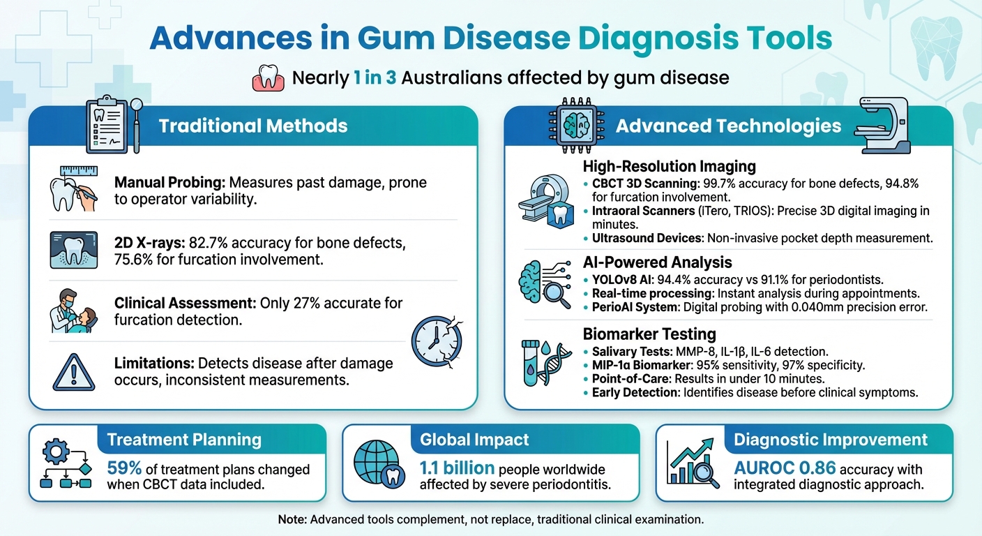
Gum disease affects nearly 1 in 3 Australians, yet diagnosing it early has been a challenge. Traditional methods like manual probing and X-rays often miss active disease, focusing instead on past damage. But new tools are changing that. From AI-driven imaging to salivary biomarker tests, dentists now have faster and more precise ways to detect and manage gum disease before it causes irreversible harm.
Here’s a quick look at what’s making a difference:
- 3D Imaging (CBCT): Provides near-perfect accuracy for detecting bone defects.
- AI in Diagnostics: Analyses scans with unmatched precision, reducing human error.
- Salivary Biomarkers: Non-invasive tests that identify early inflammation.
- Intraoral Scanners and Ultrasound: Offer detailed gum measurements without manual probing.
These technologies are already being used in Australian clinics to improve care. While costs and training remain hurdles, the shift towards early, proactive diagnosis is creating better outcomes for patients.
Traditional vs Advanced Gum Disease Diagnostic Tools Comparison
Your Perio Education Sidekick: Detecting and Treating Periodontal Disease with AI
High-Resolution Imaging Technologies
Modern imaging tools now provide 3D views of gum and bone health, revealing issues that traditional 2D X-rays often miss. This enhanced imaging capability supports the integration of digital and ultrasonic techniques, which are explored in the following sections.
Cone Beam Computed Tomography (CBCT)
CBCT technology stands out by offering clear 3D imaging, eliminating the problem of overlapping structures seen in 2D X-rays. It excels at identifying bone defects like craters, inter-radicular bone loss, and lingual/buccal marginal bone loss with exceptional accuracy [8][9].
For example, CBCT detects intrabony defects with a 99.7% accuracy rate, compared to just 82.7% for traditional intraoral radiography [9]. When it comes to furcation involvement (bone loss between tooth roots), CBCT achieves 94.8% accuracy, far surpassing the 75.6% detection rate of conventional methods. Clinical assessments, by contrast, are accurate only 27% of the time [9]. Research consistently highlights CBCT’s reliability in diagnosing infra-bony defects and furcation involvement [8].
This level of precision significantly influences treatment plans. Studies reveal that incorporating CBCT data alters treatment recommendations in 59% of cases compared to plans based solely on 2D imaging [9]. In Australia, the ALARA principle (As Low As Reasonably Achievable) should guide the use of CBCT scans, reserving them for complex cases like advanced furcation involvement, suspected endo-perio lesions, or planning regenerative surgeries [9][11]. The Australian Dental Association’s Policy Statement 6.22 outlines guidelines for the safe application of this technology [10].
Intraoral Scanners and Ultrasound Devices
Intraoral scanners, such as iTero and TRIOS, create high-resolution 3D digital images of teeth and gums in just minutes [3]. These scanners are more precise and consistent than traditional periodontal probes for measuring the width of keratinised gingiva [12]. When combined with CBCT, they provide a detailed view of the gingival dimension (the relationship between soft tissue and bone) without the need for invasive probing [12].
Studies comparing intraoral scanner data with CBCT measurements show a strong correlation (r = 0.790). However, traditional probing tends to overestimate pocket depth by approximately 0.82 mm, as noted by researchers Jin-Young Park and Hye-Min Chung:
"The similarity between the GD [Gingival Dimension] and PD [Probing Depth] measurements may suggest a possible tendency of overestimation when recording PD." – Jin-Young Park and Hye-Min Chung, Researchers [12]
Despite their benefits, intraoral scanners and cameras are not without limitations. They have lower specificity compared to traditional radiography, which can lead to more false positives [13]. Additionally, the accuracy of these tools heavily depends on the operator’s expertise, with sensitivity differences of up to 32% reported between users [13].
Ultrasound devices offer another non-invasive option for measuring pocket depth and gingival thickness [5]. Systems like the PerioScan go a step further by combining diagnosis with treatment, using ultrasonic tips to detect subgingival calculus while providing acoustic and visual feedback [1]. However, distinguishing between calculus and natural irregularities on root surfaces remains a challenge [1]. Beyond these tools, infrared thermography provides a novel way to detect inflammation, as outlined next.
Infrared Thermography
Infrared thermography works by detecting heat changes in inflamed tissues. Periodontal disease often causes temperature increases of 0.7°C to 3.0°C in affected areas compared to healthy gum tissue [1].
This method can identify "subclinical" periodontal disease – early inflammatory changes that occur before visible signs or radiographic bone loss appear. Unlike traditional probing, it is entirely non-invasive. Comparative studies show that specialised sublingual temperature probes like PerioTemp differ from infrared thermometers by only about 0.18°C [1].
However, the technology faces challenges in establishing a universal "normal" temperature, as this can vary significantly between individuals and specific tooth locations. External factors, like room temperature and a patient’s breathing, can also influence readings. While infrared thermography is a promising tool for detecting early inflammation, its variability means it is best used as a supplementary diagnostic method rather than a standalone solution.
Artificial Intelligence in Periodontal Diagnosis
AI-Assisted Imaging Analysis
Artificial intelligence is reshaping how gum disease is detected, with advanced systems now automating the analysis of radiographs and CBCT scans. By building on existing imaging tools, AI – particularly those using convolutional neural networks (CNNs) – pinpoints key anatomical landmarks to measure bone loss with precision [14][18].
One standout example is YOLOv8, a modern AI model capable of processing images in real time. It identifies bone loss around individual teeth and classifies periodontitis into stages I–IV based on the percentage of radiographic bone loss [14][16]. A study conducted in January 2025 at Fang Hospital tested YOLOv8 on 2,000 radiographs, achieving an impressive 94.4% accuracy and 100% sensitivity. In comparison, periodontists reached 91.1% accuracy and 90.6% sensitivity [14].
AI’s potential doesn’t stop there. Multimodal systems are taking diagnostics further by integrating data from various imaging sources. For instance, the PerioAI system combines intraoral scans with CBCT imaging to perform "digital probing." This method non-invasively measures the gingiva-to-bone distance with a precision error as low as 0.040 mm. In June 2025, this system was evaluated across 2,507 patients in multiple centres, showcasing its reliability [17].
"The integration of AI into periodontal care offers faster, more accurate, and comprehensive treatment, ultimately improving patient outcomes and alleviating healthcare burdens." – Jarupat Jundaeng et al., Mahasarakham University [14]
These advancements highlight the transformative role of AI in periodontal care, but they also prompt a closer look at the benefits and challenges these tools bring.
Benefits and Limitations of AI Tools
AI-driven tools have introduced a new level of precision and efficiency in periodontal diagnostics, but they’re not without their challenges. One of the key advantages is the standardised and objective assessments AI provides, which help reduce variability between clinicians [15][19]. Additionally, these systems can detect subtle changes in bone density that might be missed during manual evaluations [15][19].
That said, the effectiveness of AI tools is heavily reliant on the quality of imaging data. Issues like poor patient positioning or movement artefacts can compromise results [14]. Another challenge lies in specificity. While AI can be highly sensitive – detecting all cases of disease – it can also produce false positives. For example, in one clinical test, the AI achieved 100% sensitivity but 0% specificity, identifying every diseased case but also flagging healthy cases incorrectly [14].
| Feature | AI-Assisted Tools | Traditional Methods |
|---|---|---|
| Objectivity | Standardised algorithms ensure consistent results | Results vary depending on the clinician |
| Speed | Processes images in real time during appointments | Relies on time-intensive manual assessment |
| Early Detection | Captures subtle changes in bone density | Often identifies disease at later stages |
While AI tools are powerful, they are best used as aids rather than replacements for clinicians. For Australian dental practices adopting these technologies, maintaining professional oversight remains essential – particularly when verifying negative results. Additionally, investing in high-quality equipment and staff training is critical to ensure accurate interpretation of AI outputs [15].
"AI-powered tools eliminate operator variability by standardising measurements and analyses, enhancing diagnostic consistency across clinicians." – Raafat Musief Sarakbi et al., Ajman University [15]
sbb-itb-2be92ed
Biomarkers for Gum Disease Detection
While imaging technology and AI improve structural assessments, molecular biomarkers provide a deeper understanding of the biochemical processes driving periodontal disease.
Salivary Diagnostic Tests
Saliva, containing over 1,166 proteins, offers a non-invasive way to detect gum disease before visible symptoms appear [20][22]. Modern diagnostic tools can identify biomarkers linked to tissue damage and inflammation.
Some of the key biomarkers for early detection of periodontitis include MMP‑8, MIP‑1α, IL‑1β, IL‑6, and Haemoglobin [20][21]. For instance, MMP‑8, an enzyme released by neutrophils that breaks down collagen, is a reliable marker for active tissue destruction, with sensitivity rates between 65% and 87% [20]. Meanwhile, MIP‑1α has demonstrated impressive diagnostic accuracy, boasting 95% sensitivity and 97% specificity in identifying bone resorption [20].
A study conducted by the University of Kentucky (2009–2013) involving 209 participants found that combining biomarkers, such as IL‑1β and IL‑6, significantly enhanced diagnostic precision. The research revealed that an IL‑1β concentration of ≥28 pg/mL in saliva correlated with a 4.27-fold increased risk of being clinically diagnosed with periodontitis [23].
Recent advancements in point-of-care (POC) testing have made these biomarkers more accessible during dental visits. Tools like lab-on-a-chip devices and lateral-flow immunoassays enable practitioners to measure biomarkers like active MMP‑8 chairside, allowing for quicker clinical decisions [4][20][21]. However, as of late 2023, there are no FDA-approved salivary diagnostic tests specifically designed to assess periodontal disease risk [24].
"The purpose of a ‘futuristic’ periodontal diagnosis in the near future will be to diagnose periodontal disease before it becomes clinically detectable in order to stop its progression early by the use of biomarkers." – Carlo Cafiero et al. [20]
Building on these innovations, metabolomic profiling is emerging as a tool for even more precise early detection through detailed metabolic analysis.
Metabolomic Profiling and Current Research
Metabolomic profiling involves analysing small-molecule metabolites in saliva, using advanced techniques like NMR, LC‑MS, and GC‑MS to uncover complex metabolic patterns [25]. Research has linked certain metabolites from glycerophospholipid and protein breakdown to severe periodontitis [25].
Some of the key metabolites associated with periodontal inflammation include cadaverine, 5‑oxoproline, histidine, carnitine, and fatty acids such as arachidonate. Scientists have catalogued 853 distinct metabolites in human saliva, creating a valuable resource for developing diagnostic tools [24].
Another exciting development is the ability to culture periodontal bacteria in synthetic environments, which allows researchers to identify early-warning bacterial metabolites. This is particularly important given the widespread impact of chronic periodontitis, which affects nearly 50% of the global population, with severe cases in about 10% [25].
"Metabolomics serves as the keystone for designing biomolecule-specific biosensors, which can be used for the detection and identification of bacterial-specific metabolites (biomarker) in saliva." – Metabolomics Journal [25]
Although metabolomic profiling holds great potential for monitoring disease activity, it currently cannot pinpoint inflammation at specific tooth sites. This limitation means traditional clinical measurements remain essential [4]. In Australia, dental practices are encouraged to view these advanced tools as valuable complements to conventional periodontal examinations.
Integrating New Diagnostic Tools into Clinical Practice
Combining Traditional and Modern Techniques
Diagnosing gum disease effectively requires a mix of tried-and-true methods alongside cutting-edge technology. For instance, manual probing with standardised stainless-steel probes remains a cornerstone of diagnosis. Applying a steady 0.25 N pressure ensures accuracy without causing tissue damage or false positives [26]. To maintain consistency, clinicians can calibrate their probing force using a digital electronic balance [26].
In Australia, many dental practices are embracing a hybrid approach, combining traditional examinations with AI-powered platforms like DTX Studio. These systems bring together X-rays, 3D scans, and clinical photos into one streamlined digital interface. This integration not only speeds up the detection of bone loss and infections but also supports personalised treatment plans. For example, advanced 3D imaging tools paired with digital diagnostic platforms enable clinicians to classify gum disease using the 2017 World Workshop system, which assigns a "Stage" (severity) and "Grade" (risk of progression) based on a combination of clinical and radiographic data [26].
"The structure of precision periodontics… consists of a multidimensional diagnosis, the resulting stratification of patients into subgroups, and treatment approaches according to the characteristics of each subgroup." – Takeshi Kikuchi, Department of Periodontology, Aichi Gakuin University [7]
While traditional methods reveal past tissue damage, combining them with point-of-care biomarker tests can provide insight into current disease activity [7][4]. Tools like the Florida Probe and pa-on Parometer take this a step further by automating data entry through voice-activated software. This automation reduces recording errors while maintaining the consistent probing force necessary for reliable measurements [1].
That said, adopting these technologies isn’t without its challenges. As we’ll explore, implementing these tools requires careful planning to address both logistical and financial hurdles.
Practical Adoption Considerations
Cost and complexity are two major obstacles when it comes to integrating advanced diagnostic tools into Australian dental practices. High upfront costs for equipment like CBCT scanners and electronic probes, combined with the need for ongoing staff training, can strain resources [1][27]. A phased approach can help – starting with simpler tools that offer immediate benefits, such as intraoral scanners like iTero or TRIOS, and gradually moving to more advanced options like biomarker kits [1].
Compliance is another critical factor. Advanced imaging devices, particularly CBCT scanners, must meet national radiation safety guidelines, and all diagnostic tools must receive approval from the Therapeutic Goods Administration (TGA) [1][3]. Proper training is equally important, not just for operating the equipment but also for interpreting digital data and 3D scans accurately. For example, clinicians need to be skilled at identifying the cemento-enamel junction (CEJ) as a fixed reference point for CAL measurements to avoid miscalculating attachment loss [26].
When selecting salivary tests, it’s essential to focus on biomarkers validated by multiple studies, such as MMP-8, IL-1β, and Porphyromonas gingivalis [4]. However, the lack of standardisation in saliva sampling protocols and diagnostic methods can make it challenging to integrate these tests consistently across clinics [27].
These hurdles underscore the need for more streamlined diagnostic solutions, which we’ll delve into next.
Future Directions in Gum Disease Diagnostics
As practices work to integrate existing technologies, emerging innovations are set to transform gum disease diagnostics further. The concept of "P4 Periodontics" – Predictive, Preventive, Personalised, and Participatory – is gaining traction in Australia [7]. One exciting development is the use of microfluidic lab-on-a-chip technology in chairside biomarker kits. These devices deliver results in under 10 minutes, allowing real-time monitoring of salivary proteins and treatment responses [4].
"Saliva is an optimal biological fluid to serve as the diagnostic tool for periodontitis. The collection of saliva is safe, noninvasive, and simple." – Suk Ji and Youngnim Choi, Frontiers in Cellular and Infection Microbiology [4]
Remote monitoring is another game-changer. Patients can upload photos and participate in virtual check-ins, reducing the need for frequent in-office visits [3]. Digital workflows are also advancing, with in-house 3D printing, such as the Asiga Max 2, enabling the creation of precise surgical guides for implants based on CBCT data [3]. Additionally, intraoral scanners combined with smile design software allow clinicians to present patients with digital simulations of potential treatment outcomes, enhancing the participatory aspect of care [3][7].
With nearly 46% of adults aged 30 and older affected by periodontal disease [26], these technological advancements offer new opportunities for early detection and tailored treatment plans across Australian dental practices.
Conclusion
The approach to diagnosing gum disease is evolving, shifting focus from documenting past damage to identifying active inflammation and predicting disease progression. This is a significant step forward, especially considering that severe periodontitis impacts approximately 1.1 billion people globally [28].
Advancements like high-resolution imaging, AI-driven platforms, and salivary biomarker analysis are enabling earlier detection of inflammation and microbial shifts – often before any clinical symptoms appear [2]. These tools go beyond traditional methods like probing and X-rays, which primarily reflect historical damage, offering the potential for timely intervention.
The growing complexity of periodontal diagnostics has highlighted the need for integrated approaches. As noted in Infection Journal:
"The complexity of these interactions necessitates moving beyond conventional diagnostics towards integrated, advanced technologies." – Infection Journal [28]
By combining traditional probing techniques with modern digital tools, diagnostic accuracy is significantly improved. For instance, Australian research has shown that integrating methods such as screening questions and radiographic data can lead to a higher diagnostic accuracy, with an AUROC score of 0.86 [6].
Looking ahead, the concept of Precision Periodontics – customising care based on an individual’s genetic, microbial, and lifestyle factors – offers promising opportunities for better disease management [7]. As Australian dental practices embrace these advanced, evidence-based tools, the emphasis should remain on practical integration. This combination of traditional and modern diagnostics not only facilitates early detection and improved treatment outcomes but also enhances overall patient quality of life.
FAQs
How are AI-driven tools enhancing the diagnosis of gum disease?
AI-powered tools are transforming the way gum disease is diagnosed. By leveraging advanced deep-learning models, these tools analyse high-resolution images like intra-oral scans and cone beam computed tomography (CBCT) to detect and classify bone loss and pocket depth with impressive accuracy. This approach minimises the inconsistencies often found in manual assessments.
Additionally, AI technology incorporates biomarker analysis, which plays a key role in identifying periodontitis earlier and with greater accuracy. This early detection empowers dental professionals to create tailored treatment plans more quickly, leading to better patient outcomes and promoting long-term oral health.
What are the advantages of using salivary biomarkers to detect gum disease early?
Salivary biomarkers provide a simple and pain-free approach to spotting gum disease in its early stages. By examining saliva, dental experts can uncover indicators of periodontal inflammation long before any serious symptoms arise. It’s a fast and hassle-free method, making it perfect for keeping track of oral health over time.
Catching gum disease early with salivary biomarkers means dental care can be tailored to the individual. This approach not only improves how gum disease is managed but also helps avoid complications, ensuring better overall oral health.
What challenges do dentists face when adopting new technologies for diagnosing gum disease?
Dentists encounter various hurdles when trying to incorporate advanced diagnostic tools for gum disease into their practices. One of the biggest obstacles is the steep cost of equipment like cone-beam CT scanners or optical imaging systems. This can be particularly challenging for smaller clinics with limited budgets. On top of that, these technologies often demand specialised training to use and interpret correctly, which adds both time and expense.
Another issue is the difficulty of integrating new technologies into existing workflows. Many systems aren’t designed to work seamlessly together, which can lead to fragmented data management and inefficiencies. Additionally, while biomarkers found in saliva or gingival crevicular fluid hold potential for diagnosing gum disease, their reliability and standardisation are still under scientific scrutiny. This makes it tricky for dentists to adopt them widely.
Despite these challenges, some clinics, such as Complete Smiles Bella Vista, are starting to embrace these tools as part of their evidence-based approach to care. By doing so, they aim to maintain high standards of patient safety and effectiveness while navigating the complexities of these advancements.
Related Blog Posts
- Salivary Tests for Early Detection of Gum Disease
- AI in Dentistry: Predicting Periodontal Disease
- Advances in Salivary Biosensors for Periodontal Care
- How AI Wearables Detect Early Gum Disease
Important Notice: Any surgical or invasive procedure carries risks. Before proceeding, you should seek a second opinion from an appropriately qualified health practitioner.
Individual results may vary. The information provided in this article is for educational purposes only and does not constitute medical advice.
Checkout Related Blogs


Get in touch with us
For more information, call us now to start feeling better. Or fill the form below to make appointment
The Latest News from Complete Smiles
How to Clean Clear Plastic Retainers
Checklist for Choosing Wearable Dental Devices
Checklist for Choosing Cloud AI Platforms in Dentistry

Complete Smiles Bella VistaAccepts All Major Health Funds, Including


