


How Surface Modifications Improve Implant Biocompatibility

Surface modifications make titanium implants more compatible with bone tissue, improving their success rates in dental and orthopaedic applications. By altering the implant’s texture, chemistry, and molecular structure, these changes enhance the bond between the implant and bone, while also reducing complications like inflammation and implant failure.
Key Takeaways:
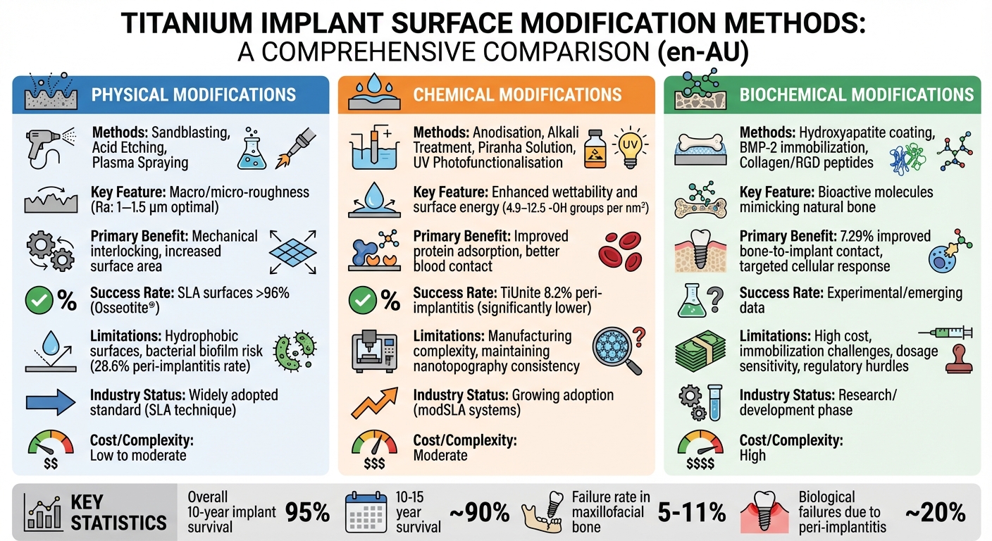
- Physical modifications: Techniques like sandblasting, acid etching, and plasma spraying create rough surfaces to increase bone adhesion and mechanical stability.
- Chemical modifications: Methods such as anodisation and hydrophilic treatments improve protein adsorption, blood interaction, and reduce bacterial growth.
- Biochemical modifications: Coatings with bioactive molecules like collagen, BMP-2, and hydroxyapatite mimic natural bone properties, encouraging better integration.
Important Stats:
- 95% of dental implants last over 10 years.
- Acid-etched surfaces like Osseotite® achieve over 96% success rates.
- SLA-treated surfaces (Sandblasted, Large-grit, Acid-etched) are the industry standard due to their optimal roughness (Ra: 1–1.5 μm) for bone growth.
While these modifications improve implant performance, challenges like peri-implantitis and manufacturing consistency remain. Research is ongoing to develop advanced surfaces that balance bioactivity, durability, and cost-effectiveness.
What Techniques Modify Implant Material Surfaces?
Physical Surface Modifications
Transforming smooth titanium implants into textured surfaces has revolutionised their integration with bone tissue. By introducing texture, these surfaces promote mechanical interlocking, which is key to successful bone-implant bonding. Techniques like sandblasting, acid etching, and plasma spraying are commonly used to create varying degrees of roughness, laying the groundwork for enhanced osseointegration.
Sandblasting, Acid Etching, and Plasma Spraying
Sandblasting, also known as grit blasting, involves propelling ceramic particles such as aluminium oxide (Al₂O₃) or titanium dioxide (TiO₂) at high speeds onto the implant’s surface. This process produces macro-roughness while introducing a slight negative charge, which can attract calcium ions and bone-forming cells [5][8].
Acid etching exposes the implant surface to strong acids like hydrochloric, sulphuric, or hydrofluoric acid. This creates micro-pits ranging from 0.5 to 2 μm, significantly increasing the surface area available for cell adhesion [5]. For example, commercially available implants like Osseotite® utilise this method and have shown long-term clinical success rates exceeding 96% [5].
Plasma spraying employs a plasma torch to deposit molten materials – typically titanium or hydroxyapatite – onto the implant. This process forms a thick, porous coating approximately 30 μm deep, which enhances the bone-to-implant interface [5][7]. The porous layer is rich in Ti–OH residues, which assist in protein absorption and encourage bone growth.
Many manufacturers combine these methods to maximise their benefits. For instance, the SLA (Sandblasted, Large-grit, Acid-etched) treatment has become a widely adopted industry standard. It creates roughness on both macro and micro levels, which research suggests is most effective when the average surface roughness (Ra) measures between 1 and 1.5 μm [6]. This engineered texture plays a critical role in influencing osteoblast activity, leading to better bone integration.
Effects on Osseointegration
These physical modifications do more than just increase surface area – they actively enhance cellular responses, which are essential for implant success. By creating a scaffold-like texture, the surface encourages osteoblasts (bone-forming cells) to migrate, adhere, and multiply. As A. Jemat highlights:
"Significant surface roughness played an important role in providing effective surface for bone implant contact, cell proliferation, and removal torque" [7].
Rough surfaces guide cell movement and provide a stable platform for osteoblast adhesion, which triggers their proliferation and differentiation [5]. This increased surface area, combined with mechanical interlocking, leads to higher removal torque, making implants more resistant to dislodging forces.
However, there are some limitations to these physical modifications. Microrough surfaces can become hydrophobic, which may reduce their ability to attract blood during the initial healing phase compared to chemically treated hydrophilic surfaces [3]. Additionally, the negative charge generated by sandblasting diminishes over time, and there’s a risk of residual blasting particles remaining embedded if the surface isn’t properly cleaned. Despite these challenges, physical surface modifications remain a cornerstone of modern implant technology, especially when paired with chemical treatments to enhance both surface texture and wettability.
Chemical Surface Modifications
Building on the mechanical texturing discussed earlier, chemical modifications take implant surfaces a step further by altering their molecular makeup. These changes don’t just provide a rough structure for tissues to grow on – they actively interact with surrounding tissues to encourage better integration.
Altering Surface Chemistry
Chemical treatments are designed to influence how proteins and cells interact with the implant’s surface. One way this is achieved is by increasing the number of hydroxyl groups (-OH) on the surface. These groups act as docking points for proteins like fibronectin and integrins, which are crucial for cell adhesion and signalling [1][9].
Anodisation is a key technique that uses an electrochemical process to thicken the titanium dioxide (TiO₂) layer and create micropores. This method has proven effective, as seen with TiUnite implants, which showed only an 8.2% occurrence of peri-implantitis due to improved osseointegration [5]. The thicker oxide layer helps retain blood clots and provides a stable platform for bone to grow.
Another approach is alkali treatment with sodium hydroxide (NaOH), which forms a negatively charged sodium titanate layer. This layer attracts positively charged calcium ions (Ca²⁺) from body fluids, which then bind with phosphate ions to form a bone-like apatite layer [8]. This process mimics how bone naturally mineralises, speeding up integration. For even greater hydroxylation, Piranha solution, a mix of sulphuric acid and hydrogen peroxide, is used. This treatment produces an impressive density of reactive -OH groups, ranging from 4.9 to 12.5 groups per nm² [1][9].
Hydrophilic Treatments
Over time, titanium surfaces exposed to air accumulate carbon contamination, a phenomenon known as "ageing." This makes the surfaces hydrophobic (water-repellent) and reduces their bioactivity. Hydrophilic treatments counteract this by boosting surface energy and wettability, which enhances tissue response. As researchers F. Rupp and colleagues explain:
"Hydrophilic implants have been proven to improve the initial blood contact, to support the wound healing and thereby accelerating the osseointegration" [3].
High wettability allows for rapid adsorption of blood proteins while preserving their natural structure. This is critical because denatured proteins can provoke inflammatory reactions [1]. By improving protein conditioning, hydrophilic surfaces encourage quicker recruitment and differentiation of osteoblasts, shortening the time needed for bone integration.
These surfaces also help guard against bacterial colonisation. Their nanoscale features, combined with high surface energy, create an environment that is less inviting to bacteria compared to traditional hydrophobic surfaces [11]. Techniques like UV photofunctionalisation, oxygen plasma treatment, and storage in aqueous solutions prevent carbon build-up and maintain bioactivity until the implant is placed. Commercial systems such as modSLA (modified Sandblasted, Large-grit, Acid-etched) are produced under nitrogen and stored in liquid to preserve their hydrophilic properties [10]. This chemical preparation sets the stage for the bioactive coatings discussed in the next section.
Biochemical Surface Modifications
Biochemical modifications take chemical enhancements a step further by adding molecules that replicate natural tissue components. By incorporating substances naturally present in bone and connective tissue, these bioactive coatings help reduce the implant’s "foreign body" nature and create an environment that promotes better integration with surrounding tissue.
Bioactive Coatings: Collagen, BMP-2, and Hydroxyapatite
Bioactive coatings are designed to imitate the natural tissue environment, encouraging bone cell growth and proliferation. One key material, hydroxyapatite (HA) – the primary mineral in bone – forms a chemical bond with bone tissue, stimulating bone formation. However, its limited bonding strength to titanium can cause cracking or peeling during implant placement, potentially leading to inflammation [14].
Bone Morphogenetic Protein-2 (BMP-2), an FDA-approved growth factor, is another important bioactive agent. It promotes mesenchymal stem cells to differentiate into osteoblasts, the cells responsible for bone formation. Research indicates that bioactive surfaces incorporating BMP-2 can improve bone-to-implant contact by an average of 7.29% [13]. However, finding the right dosage is tricky; using too little or too much can lead to immature bone formation, which reduces the effectiveness of bioactivity [15]. As Nansi López-Valverde and colleagues explain:
"The process of peptide [or BMP] immobilization on Ti implant surfaces can be a complex process… the biological activity of certain peptides would be reduced by the immobilization process" [15].
Type I collagen, which mimics the extracellular matrix, enhances cell adhesion. When combined with BMP-2, it synergistically boosts osseointegration, laying the groundwork for further advancements in growth factor and extracellular matrix protein modifications.
Growth Factors and Extracellular Matrix Proteins
Building on bioactive coatings, biochemical modifications also utilise growth factors and extracellular matrix (ECM) proteins to fine-tune cellular responses. ECM proteins are essential for tissue regeneration. For instance, fibronectin and type I collagen contain the Arg-Gly-Asp (RGD) motif, a specific amino acid sequence that connects a cell’s actin cytoskeleton to the implant surface. These connections activate signalling pathways that control cell proliferation, movement, and differentiation.
Titanium surfaces modified with RGD peptides and self-assembled monolayers (SAMs) show notable improvements in bone healing. These biochemically enhanced surfaces speed up calcification by osteogenic cells, while SAMs provide stable anchoring layers, ensuring bioactive agents remain securely attached to even the most complex implant geometries [9][12].
sbb-itb-2be92ed
Research Evidence on Improved Osseointegration
Clinical Results from Dental Implant Studies
Clinical studies consistently show that surface modifications in dental implants lead to impressive survival rates. Currently, implants boast a 10-year survival rate of approximately 95% [4]. For SLA-treated surfaces, this success rate climbs even higher, exceeding 96% [5]. These results have cemented modified surfaces as a standard in clinical practice.
One key finding is that an average surface roughness (Ra) of 1–1.5 μm appears to be optimal for bone formation [6]. Additionally, bioactive coatings, such as those incorporating BMP-2, have been shown to enhance early bone-to-implant contact [15].
However, these advancements are not without challenges. Modified surfaces have been linked to a peri-implantitis rate of 28.6%, significantly higher than the 7.4% observed with turned surfaces [4]. Interestingly, specific commercial modifications like the porous anodised TiUnite surface demonstrate a much lower peri-implantitis incidence of 8.2% [5]. This suggests that not all surface modifications carry the same level of risk. While these clinical outcomes highlight significant progress, they also expose the need for ongoing research to address existing limitations.
Research Gaps and Future Directions
Even with high success rates, a portion of dental implants – between 5% and 11% – still fail to achieve proper osseointegration in maxillofacial bone [5]. This points to unresolved challenges in the field. As F. Rupp from Dental Materials explains:
"Manufacturing of multi-scaled complex surfaces including distinct nanotopographies, wetting properties, and stable cleanliness is still a technical challenge and far away from being reproducibly transferred to implant surfaces" [3].
One major issue is the variability in research methods. Studies often differ in their models, follow-up durations, and measurement criteria, making it difficult to draw consistent conclusions. This highlights the pressing need for standardised, long-term human studies – ideally with follow-up periods exceeding 10 years – to validate findings from animal research [15][5].
Looking ahead, there is growing interest in developing "smart" multifunctional surfaces. These advanced surfaces aim to not only encourage bone growth but also combat bacterial infections [14]. Such innovations could pave the way for the next generation of dental implants.
Comparing Surface Modification Methods
Comparison of Physical, Chemical, and Biochemical Surface Modification Methods for Titanium Implants
Comparison Table of Methods
After examining physical, chemical, and biochemical surface modifications in detail, this section offers a concise comparison of these techniques. Each method has its own strengths and limitations, helping to explain why techniques like SLA have become industry benchmarks while others are still in experimental stages.
For implants, physical modifications such as sandblasting and acid etching are widely used to alter surface texture. These methods increase the surface area, improving mechanical interlocking with bone. The SLA (Sandblasted, Large-grit, Acid-etched) technique is particularly notable, as it combines macro-roughness from blasting with micro-pits from etching, making it the go-to standard [5]. Its versatility allows application on complex implant shapes [2][9]. However, the downside is that rougher surfaces can encourage bacterial biofilm accumulation, which may heighten the risk of peri-implantitis [4].
On the other hand, chemical modifications aim to improve surface energy and wettability rather than focusing solely on texture. Methods such as anodisation and hydrophilic treatments enhance protein adsorption and improve initial blood interaction [1][3]. While these techniques offer clear benefits, creating multi-scaled surfaces with precise nanotopographies and maintaining their cleanliness remains a significant hurdle. As Professor F. Rupp highlights:
"Manufacturing of multi-scaled complex surfaces including distinct nanotopographies, wetting properties, and stable cleanliness is still a technical challenge and far away from being reproducibly transferred to implant surfaces" [3].
Meanwhile, biochemical modifications take a more targeted approach by immobilising bioactive molecules – such as collagen, BMP-2, or hydroxyapatite – onto the implant surface. These modifications aim to stimulate specific biological responses, including antibacterial effects [1][16]. However, they come with challenges like higher costs, technical difficulties in achieving stable immobilisation, and stricter regulatory requirements [3][16]. For example, hydroxyapatite coatings show promising initial bioactivity but may lose their bonding strength to titanium over time when exposed to body fluids [5].
Ultimately, the choice of method depends on balancing clinical outcomes with manufacturing feasibility and cost. Mechanical and wet chemical techniques dominate industrial applications due to their simplicity and effectiveness [2][9], while biochemical methods continue to advance as researchers address their technical challenges.
Conclusion: How Surface Modifications Improve Implant Success
The evolution of implant surface technology has transformed dental implant success rates, with roughened textures, hydrophilic properties, and bioactive coatings playing a key role. These advancements accelerate healing, improve mechanical stability, and lower the risk of infection, making implants more reliable and effective.
Clinical evidence backs these improvements. For instance, acid-etched implants like Osseotite® boast over 96% long-term success rates, while overall implant survival hovers around 90% after 10–15 years [5]. These impressive statistics highlight the importance of precise surface engineering, particularly for patients with lower bone density or those needing faster loading protocols.
Barbara D. Boyan, PhD, sheds light on this progress:
"Implant surfaces that mimic the inherent chemistry, topography, and wettability of native bone have shown to provide cells in the osteoblast lineage with the structural cues to promote tissue regeneration" [10].
Hydrophilic surface modifications are especially noteworthy. They enhance early protein adsorption, promote healing, and reduce inflammation by encouraging macrophages to adopt an anti-inflammatory M2 state instead of a pro-inflammatory M1 state [10].
However, even with these technological breakthroughs, proper care and maintenance are critical. While advanced surface designs significantly enhance osseointegration, poor oral hygiene can still lead to complications like peri-implantitis, which is responsible for around 20% of biological implant failures [5]. For patients considering dental implants at clinics such as Complete Smiles Bella Vista, understanding that these innovations work best alongside diligent hygiene practices is essential.
The transition from basic machined surfaces to sophisticated multi-functional modifications marks a major leap in modern dentistry. As research delves further into smart surfaces and nanobiotechnology, the future holds even greater potential for improving implant outcomes and patient satisfaction.
FAQs
How do surface modifications improve the success and longevity of dental implants?
Surface modifications, including controlled roughening, hydrophilic treatments, and specialised coatings, are key to improving the integration and longevity of dental implants. By designing a textured surface at the micron scale (around 0.4–8.7 µm), these implants encourage stronger bone cell attachment and growth. This speeds up osseointegration, providing the stability needed to reduce micromovement – an issue that can otherwise result in bone loss over time.
Hydrophilic, or ‘wet’, surfaces enhance the adsorption of blood proteins, which helps create stronger cell adhesion and reinforces the connection between the implant and surrounding bone. On top of that, chemical or biological coatings, such as hydroxyapatite or antimicrobial layers, add further benefits. They improve the implant’s ability to support bone growth while reducing the risk of bacterial colonisation and peri-implantitis, a common cause of implant failure.
These technological advancements not only ensure a stronger initial bond between the implant and the surrounding tissue but also protect against long-term problems like infections or mechanical wear. Clinics like Complete Smiles Bella Vista leverage these innovations to provide patients with durable, high-quality dental implant solutions tailored to their specific needs.
What are the key differences between physical, chemical, and biochemical surface modifications in implants?
Improving the interaction between implants and surrounding tissue often involves a mix of physical, chemical, and biochemical surface modifications, each contributing in its own way.
Physical modifications focus on altering the surface texture or roughness of the implant without changing its chemical makeup. Techniques like sand-blasting, laser texturing, and plasma-spraying are commonly used to create a rough surface. This roughness helps the implant form a stronger mechanical bond with bone tissue.
Chemical modifications work by changing the surface chemistry, often through the addition of functional groups. Methods such as acid-etching, oxidation, or applying bioactive coatings are typical here. These changes encourage better protein adhesion and cell attachment, helping the implant integrate more effectively with the body.
Biochemical modifications go a step further by incorporating biologically active molecules, like peptides or growth factors, onto the implant’s surface. These additions not only guide cellular responses to improve osseointegration but can also offer antimicrobial protection.
In real-world applications, these techniques are often combined. For example, an implant might feature a roughened surface (physical modification) paired with chemical coatings and biochemical agents. This combination enhances biocompatibility, ensures better integration, and supports long-term stability.
Why is a hydrophilic surface important for dental implants?
A hydrophilic implant surface significantly boosts how well an implant interacts with surrounding tissues, making it more compatible with the body. It promotes protein adsorption, a key step for cell attachment, and enhances the activity of osteoblasts – the cells that drive bone formation.
This improved cellular activity supports osseointegration, the process where the implant firmly bonds with the bone. This results in greater stability and long-term success. By creating ideal conditions for healing, hydrophilic treatments play a vital role in the effectiveness of modern dental implants across Australia.
Related Blog Posts
- Surface Roughness and Osseointegration: Key Insights
- Osseointegration in Titanium Implants: How It Works
- Surface Modifications for Better Osseointegration
- Titanium Implant Surface Modifications for Osseointegration
Important Notice: Any surgical or invasive procedure carries risks. Before proceeding, you should seek a second opinion from an appropriately qualified health practitioner.
Individual results may vary. The information provided in this article is for educational purposes only and does not constitute medical advice.
Checkout Related Blogs


Get in touch with us
For more information, call us now to start feeling better. Or fill the form below to make appointment
The Latest News from Complete Smiles
How to Clean Clear Plastic Retainers
Checklist for Choosing Wearable Dental Devices
Checklist for Choosing Cloud AI Platforms in Dentistry
Complete Smiles Bella VistaAccepts All Major Health Funds, Including


