





Advances in Bone-Implant Interface Research
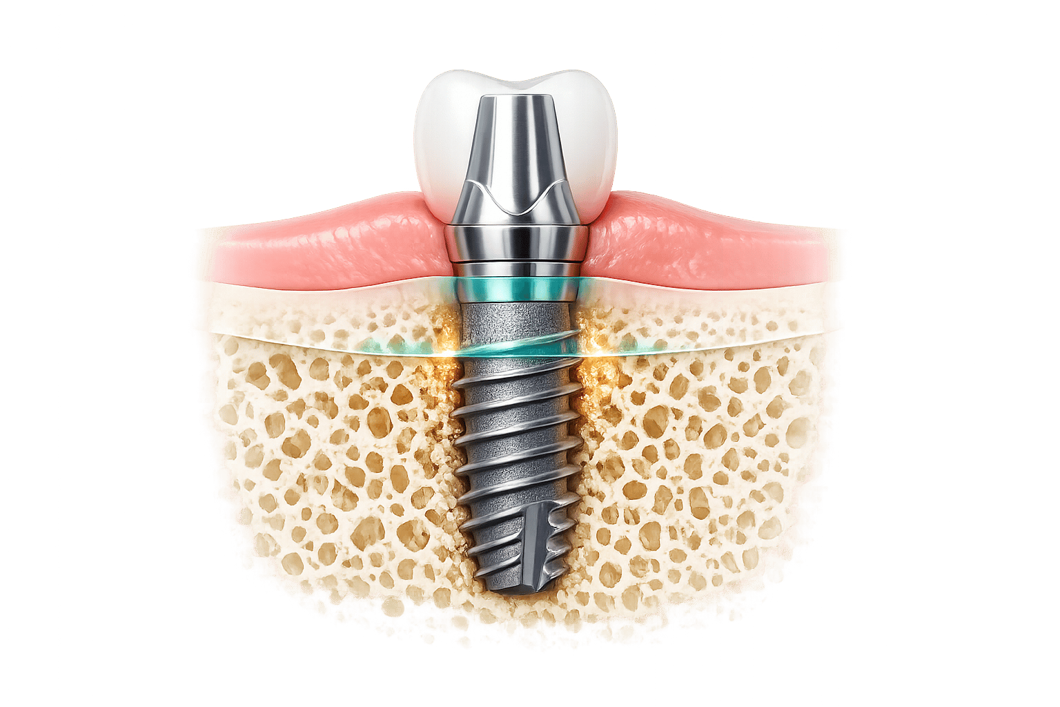
Osseointegration, or the connection between bone and implants, is vital for the success of dental and orthopaedic devices. While titanium-based implants have high success rates, challenges like implant loosening and peri-implantitis persist. Newer methods, including bioactive coatings and tissue engineering, aim to improve integration by promoting bone regeneration and reducing complications.
Key Points:
- Titanium Implants: Mechanically stable but rely on physical bonding with bone.
- Surface Modifications: Techniques like sandblasting and acid etching improve bone adhesion.
- Tissue Engineering: Uses scaffolds, growth factors, and cells to mimic natural bone.
- Emerging Techniques: Include drug-loaded nanotubes, ion-doped coatings, and bio-inspired layers.
While traditional methods remain reliable, tissue engineering shows promise for creating "live" implants that interact biologically with the body. However, high costs and regulatory challenges limit its current application.
1. Traditional Osseointegration Methods
Bone Integration Mechanisms
The biological process behind traditional osseointegration unfolds in a predictable sequence. Right after an implant is placed, proteins from blood and surrounding fluids coat the titanium surface within nanoseconds. This triggers blood clot formation, activates the immune system, and attracts mesenchymal stem cells, which eventually differentiate into osteoblasts. As S Parithimarkalaignan from the Department of Prosthodontics explains:
"Osseointegration is a direct bone-to-implant contact without intervening soft tissue." [8]
This direct bone contact happens through two pathways. Contact osteogenesis involves new bone forming directly on the implant’s surface, while distance osteogenesis sees bone growing from the edges of the surgical site toward the implant. The process typically takes 3 to 6 months and involves the formation of initial woven bone, followed by lamellar bone, which adapts to mechanical loads over time [5][8].
Initially, stability is achieved mechanically, but biological fixation strengthens it over time. However, bone tissue is highly sensitive to heat; even a one-minute exposure to 47°C can cause tissue death, making surgical precision absolutely critical [8][9].
These biological responses highlight the importance of surface modifications to improve implant integration further.
Surface Modification Techniques
Traditional approaches to improving bone integration focus on altering the implant surface using mechanical, chemical, and physical methods. Techniques like sandblasting, acid etching, and plasma spraying create textures at both micro and macro scales, improving cell adhesion and bone growth.
- Sandblasting: High-speed alumina particles create macro-roughness (10–30 µm), offering anchor points for bone.
- Acid Etching: Strong acids like HCl or H₂SO₄ produce micropores (1–3 µm) that promote osteoblast differentiation.
- Plasma Spraying: This method applies molten droplets to the surface, increasing surface area and forming three-dimensional structures for bone ingrowth [9].
The SLA (Sand-blasted, Large-grit, Acid-etched) technique, introduced by Straumann in 1997, remains the most widely used commercial method. It creates both macro-irregularities and micro-cavities, enhancing protein adsorption and cell adhesion [2][9]. Other notable commercial systems include Astra Tech‘s TiOblast®, Nobel Biocare‘s TiUnite®, and BioHorizons‘ Laser-Lok®, each offering unique surface modifications [9].
| Technique | Primary Effect | Common Materials |
|---|---|---|
| Sandblasting | Increases surface roughness/area | Alumina particles |
| Acid Etching | Creates micropores (1–3 µm) | HCl, H₂SO₄ |
| Plasma Spraying | Adds bioactive coating | Hydroxyapatite (HA) |
| Anodisation | Forms TiO₂ nanotubes/nanopores | Titanium Dioxide |
| Laser Texturing | Precise topographical patterning | Titanium Alloy |
Surfaces with Sa values below 0.5 µm risk fibrous encapsulation rather than bone integration. Moderately rough surfaces (Sa 1.0–2.0 µm) are ideal for proper bone growth and mechanical interlocking [8]. The connection between bone and titanium is incredibly close – collagen filaments are separated from the titanium oxide surface by a proteoglycan layer only 20–40 nm thick [8].
These surface modifications are key to the high clinical success rates of traditional implants.
Clinical Adoption and Impact
Traditional osseointegration methods have proven their reliability in clinical settings. Back in 1986, researchers Branemark and Albrektsson reviewed all dental implants placed in their facility over one year and found a 96.5% success rate in the mandible after five years. They credited this success to precise surgical techniques and the use of commercially pure titanium [8]. More recently, a 10-year study involving 511 dental implants with micro- and submicron-scale roughness reported a survival rate of 98.9% [10].
From 2010 to 2019, titanium-based implants saw a sharp rise in usage, growing from 5% to 46%, while PEEK implants declined due to their tendency to form fibrous tissue rather than bond with bone [10]. Despite these advancements, challenges remain. Around 20% of patients develop peri-implantitis within five years of implant placement, according to the third European Association for Osseointegration consensus conference in 2012 [3]. After 11 years, about 25% of patients and 16% of implants are affected by this inflammatory condition [3][9].
These issues stem from titanium’s bioinert nature – it forms only physical bonds with bone, which are less stable than the chemical bonds aimed for in newer bioactive approaches [3]. This has driven the development of tissue engineering techniques to address these limitations.
This is how a DENTAL IMPLANT integrates in the BONE – Osseointegration ©
2. Tissue Engineering Methods
Tissue engineering takes bone regeneration a step further by actively promoting healing, rather than just passively integrating with existing bone.
Bone Integration Mechanisms
A key strategy in tissue engineering is the "Quad" approach, which combines scaffolds, advanced fabrication techniques, regenerative cells, and growth factors to enhance bone growth and vascularisation [1]. This approach activates cellular pathways like Wnt/β-catenin and YAP/TAZ, which drive the expression of bone-related genes [4].
Michael B. Berger from Virginia Commonwealth University highlights the importance of this process:
"Osseointegration is the robust biological cascade that occurs after a biomaterial is placed in or adjacent to native bone tissue." – Michael B. Berger, Department of Biomedical Engineering, Virginia Commonwealth University [10]
Advanced biomaterials also play a role in modulating the immune response, steering it towards healing and reducing the risk of fibrous tissue formation [10]. For instance, in a 2022 study, Moncal and colleagues used bioink loaded with rat bone marrow stem cells to treat craniomaxillofacial defects. The results were striking: after six weeks, around 40% of bone tissue had formed with 90% coverage, compared to just 10% new bone and 25% coverage in untreated defects [1].
To further improve integration, surface modifications are employed to optimise the interaction between implants and bone.
Surface Modification Techniques
Surface engineering focuses on modifying implant surfaces at various scales [10][4]. One effective method is anodic oxidation, which creates highly organised TiO₂ nanotubes. Smaller nanotubes (approximately 30 nm) have proven more effective in promoting bone integration and reducing inflammation compared to larger ones (80 nm) [2]. These nanotubes can also serve as carriers for drugs or antibacterial agents, such as silver nanoparticles [3].
Another approach, ion doping, involves adding trace elements to implant coatings. For example:
- Zinc-doped coatings: Boost osteoblast proliferation by 25% and improve cell adhesion by 40%.
- Magnesium-doped surfaces: Increase alkaline phosphatase activity by 38% and lead to a 4.5-fold rise in cell proliferation.
- Copper-doped coatings: Show antibacterial effectiveness of 99.45% against Staphylococcus aureus and 98.65% against Escherichia coli [5].
Polydopamine-assisted coatings offer a bio-inspired alternative, forming stable hydroxyapatite layers at room temperature. These coatings outperform traditional high-temperature plasma-sprayed versions without the risk of thermal degradation [2]. In one study, researchers applied a dual-drug coating of curcumin and vitamin K2 on hydroxyapatite-coated implants, which significantly improved osseointegration within just five days in a rat model [3].
| Technique | Primary Benefit | Key Application |
|---|---|---|
| Anodic Oxidation | Creates TiO₂ nanotubes for drug delivery | Antibacterial and bone growth support |
| Ion Doping (Zn, Mg, Sr) | Enhances cell activity and adhesion | Improved proliferation and healing |
| Polydopamine Coating | Stable bioactive layer at room temperature | Enhanced protein adsorption |
| Laser Cladding | Forms strong ceramic bonds | Bioactive surface integration |
Clinical findings also back these advancements. Implants with hydrophilic (water-attracting) surfaces have shown significantly better bone formation – 14.8% at two weeks and 48.3% at four weeks – compared to traditional microroughened ones [10].
Soft Tissue Stability
While bone regeneration is vital, soft tissue integration is equally important. The difference in rigidity between bone and soft tissue can complicate healing [13]. Soft tissue interfaces often lack sufficient cells and blood vessels, making natural regeneration harder than in well-vascularised bone [7][13].
Fibrous encapsulation is a major challenge, as it blocks bone ingrowth [4]. Recreating the natural gradient from bone to soft tissue is particularly difficult because this transitional zone rarely regenerates after injury [13].
Current Osteoporosis Reports emphasises the importance of this interface:
"The interface plays a critical role in stabilising and distributing the mechanical stress between soft tissues (e.g., cartilage and tendon) and bone." – Current Osteoporosis Reports [13]
Nanotopographical modifications can help by improving the attachment of human gingival fibroblasts and epithelial cells, which are crucial for creating a secure biological seal in dental implants [3]. However, the cells at these interfaces are often terminally differentiated, meaning they lack the regenerative capacity of younger, progenitor cells, making full integration a persistent challenge [13].
Clinical Adoption and Impact
Tissue engineering methods are gradually being adopted in clinical settings, though they remain relatively new compared to traditional techniques. The focus has shifted from merely fixing and restoring bone structure to creating "live" tissue that mimics natural bone environments [6].
Research is advancing towards "smart" implants that combine multiple functions, such as vascularisation, antibacterial properties, and immune response modulation, into a single design [6]. Additionally, 3D printing is enabling the creation of patient-specific scaffolds that merge bioactive glass with polymers, improving both strength and biological compatibility [14].
Despite these advancements, challenges like aseptic loosening still occur. For example, a study on the Less Invasive Stabilisation System (LISS) for distal femoral fractures found that 23 out of 107 repairs required revisions, with four cases linked to implant loosening [7].
Tissue engineering presents exciting possibilities, such as unlimited material availability and the ability to chemically bond with bone rather than relying solely on mechanical fixation [3][14]. These developments align with the growing demand for advanced, biologically integrated solutions in clinical practice.
sbb-itb-2be92ed
Strengths and Weaknesses
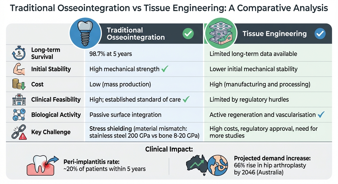
Traditional Osseointegration vs Tissue Engineering Methods Comparison
Traditional osseointegration and tissue engineering each bring their own benefits and challenges to the table, offering distinct approaches to improving implant integration and functionality.
Traditional methods rely on the mechanical interlocking of the implant with bone. This approach provides immediate stability and boasts a strong clinical track record. For example, rough surface titanium implants demonstrate a 98.7% survival rate over 5.2 years [16]. Additionally, these implants are cost-efficient due to their suitability for mass production [15]. However, their reliance on physical rather than chemical bonds with bone can lead to long-term issues. One notable drawback is stress shielding, caused by the mismatch in modulus between materials – stainless steel at around 200 GPa versus bone at 8–20 GPa – leading to bone resorption over time [16]. Tissue engineering, on the other hand, aims to address these mechanical limitations by focusing on biological integration.
Tissue engineering takes a biological approach, promoting healing through vascularisation and bone regeneration. Preclinical studies show encouraging results for bone regeneration [1]. However, this method comes with its own set of obstacles. As highlighted by Materials Science and Engineering C:
"The clinical approach of Bone Tissue Engineering has not taken place on a large scale yet, due to the need of more in depth studies, its high manufacturing costs and the difficulty to obtain regulatory approval" [11].
Similarly, Frontiers in Bioengineering and Biotechnology remarks:
"Self-sufficient solutions that enable complete tissue integration and homeostasis remain elusive" [1].
These challenges – complex regulatory pathways, high costs, and the need for further research – limit the current clinical application of tissue engineering.
When compared, traditional implants benefit from established, cost-efficient production methods, whereas tissue engineering involves expensive components like growth factors, cell processing, and specialised fabrication. As a result, traditional osseointegration remains the standard in clinical care, while tissue engineering is still primarily experimental.
Here’s a side-by-side comparison of the two approaches:
| Feature | Traditional Osseointegration | Tissue Engineering |
|---|---|---|
| Long-term Survival | 98.7% at 5 years [16] | Limited long-term data available |
| Initial Stability | High mechanical strength | Lower initial mechanical stability |
| Cost | Low (mass production) [15] | High (manufacturing and processing) [1] |
| Clinical Feasibility | High; established standard of care | Limited by regulatory hurdles [1] |
| Biological Activity | Passive surface integration | Active regeneration and vascularisation [1] |
Conclusion
Traditional osseointegration and tissue engineering take distinctly different paths when it comes to bone-implant integration. Traditional methods still dominate clinical practice, largely due to their reliability and consistently high survival rates. However, their dependence on mechanical fixation rather than fostering biological integration can sometimes lead to long-term complications.
Tissue engineering, on the other hand, focuses on encouraging biological regeneration using the "Quad" approach. This method combines scaffolds, regenerative cells, morphogens, and biofabrication to replicate the natural structure and function of bone. As Guang Zhu from the University of Sydney explains:
"The ability of an implant to achieve osseointegration to a large extent determines its long-term success in the body" [3].
These two approaches represent the evolving landscape of implant dentistry, each with its own strengths and challenges.
The demand for advanced implant solutions is only expected to grow. By 2046, primary hip arthroplasty procedures in Australia are predicted to increase by 66% [3]. Clinics like Complete Smiles Bella Vista, which currently provide dental implant services, may adopt tissue engineering advancements such as 3D-printed scaffolds and bioactive coatings. These innovations could be especially beneficial for high-risk patients, including those with conditions like diabetes or osteoporosis [12].
Although tissue engineering is still in the experimental phase due to regulatory hurdles [17], developments in patient-specific scaffolds and smart biomaterials suggest a shift is on the horizon. As research progresses, these technologies are likely to move from the lab into clinical practice, potentially transforming implant dentistry in Australia over the coming decades.
FAQs
What are the key challenges associated with traditional titanium implants?
Titanium implants, while widely used, come with a set of challenges that can affect their performance and longevity. A major concern is the risk of implant loosening, which happens when the implant doesn’t form a strong, stable bond with the surrounding bone – a process known as osseointegration. The success of this bonding largely depends on the implant’s surface properties, which sometimes fail to support the necessary cellular activity.
Another issue is the potential for infection at the implant site. Even though titanium is biocompatible, infections can still undermine the implant’s long-term success. These challenges underscore the importance of exploring advanced solutions, like tissue engineering, to improve the reliability and durability of these implants.
What role does tissue engineering play in improving bone-implant integration?
Tissue engineering plays a vital role in improving bone-implant integration by developing bioengineered scaffolds that mimic the structure and characteristics of natural bone. These scaffolds provide a supportive framework for cellular growth, helping to regenerate bone tissue around the implant site.
This method encourages a more organic healing process, which in turn enhances the stability and durability of implants. It presents an exciting alternative to conventional techniques, offering improved outcomes for patients.
What challenges exist in regulating tissue-engineered implants?
Ensuring tissue-engineered implants are safe, effective, and reliable comes with its share of challenges. One major hurdle is the need for rigorous testing to confirm properties like biocompatibility, mechanical stability, and long-term functionality within the human body. Since these implants often interact with complex biological processes – like vascularisation and immune responses – thorough preclinical and clinical evaluations are crucial.
Another significant issue is the variability between tissue-engineered products, which complicates standardisation and quality control. Regulatory bodies demand consistent manufacturing protocols and detailed documentation to ensure both safety and reproducibility. As the field advances with technologies like smart scaffolds and bio-responsive materials, regulators face the task of updating frameworks to accommodate these developments while keeping patient safety at the forefront.
Ultimately, collaboration is key. Researchers, clinicians, and regulatory agencies must work closely to streamline approval processes and ensure these innovations can transition smoothly into clinical use.
Related Blog Posts
- Osseointegration and Titanium Surface Design
- Osseointegration in Titanium Implants: How It Works
- Surface Modifications for Better Osseointegration
- Titanium Implant Surface Modifications for Osseointegration
Important Notice: Any surgical or invasive procedure carries risks. Before proceeding, you should seek a second opinion from an appropriately qualified health practitioner.
Individual results may vary. The information provided in this article is for educational purposes only and does not constitute medical advice.
Checkout Related Blogs




Get in touch with us
For more information, call us now to start feeling better. Or fill the form below to make appointment
The Latest News from Complete Smiles
How to Clean Clear Plastic Retainers
Checklist for Choosing Wearable Dental Devices
Checklist for Choosing Cloud AI Platforms in Dentistry

Complete Smiles Bella VistaAccepts All Major Health Funds, Including





