


Modified Widman Flap: Step-by-Step Guide
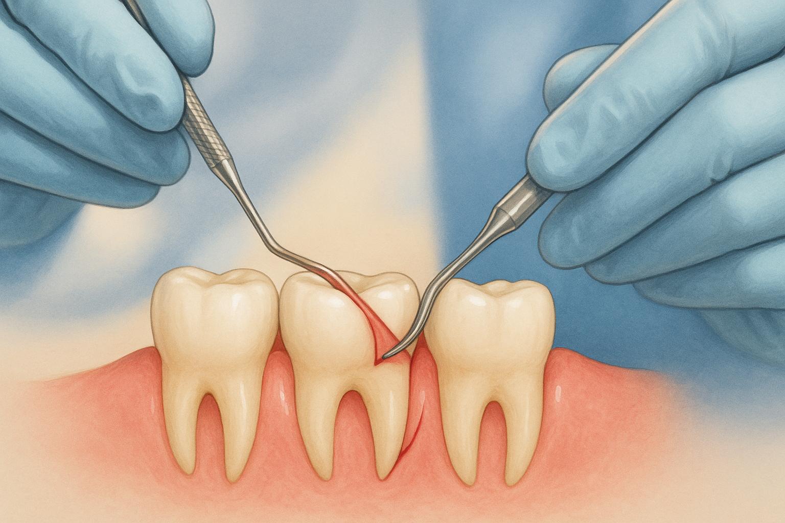
The Modified Widman Flap is a precise periodontal surgery aimed at cleaning tooth roots while preserving gum and bone structure. It’s ideal for treating periodontal pockets deeper than 5 mm that don’t respond to non-surgical treatments like scaling and root planing. This method, developed in 1974 by Ramfjord and Nissle, is widely used in Australia for managing periodontitis, especially in areas where preserving gum aesthetics is important.
Key Points:
- Purpose: Treat periodontal disease by removing subgingival plaque and calculus.
- Best for: Moderate to severe periodontitis with pockets over 5 mm.
- Steps: Incisions, flap reflection, tissue removal, root cleaning, suturing, and optional dressing.
- Benefits: Minimal tissue removal, reduced sensitivity, and improved healing.
- Limitations: Not suitable for cases requiring significant bone reshaping.
The procedure involves detailed preoperative planning, effective use of anaesthesia, and careful postoperative care to ensure optimal healing and long-term oral health. It’s a reliable option for patients prioritising gum health and aesthetics.
Preoperative Preparation
Patient Assessment
A detailed patient assessment is essential to confirm the suitability of the procedure and identify any factors that may influence healing. This step lays the groundwork for planning and guides the initial therapy phase.
The periodontal examination evaluates probing depths, attachment loss, and bleeding on probing to determine the severity of periodontal disease. Areas with pocket depths greater than 5 millimetres that have not responded to non-surgical treatment are typically ideal for surgical intervention.
Radiographic evaluation is another critical component of surgical planning. Periapical and bitewing radiographs provide insight into bone levels, root morphology, and furcation involvement, helping clinicians tailor the surgical approach.
A thorough medical history review is vital to identify systemic conditions that may affect healing or influence anaesthetic options. Conditions like diabetes, cardiovascular disease, or bleeding disorders may require special precautions and collaboration with the patient’s GP. In Australia, this team-based approach ensures both safety and optimal outcomes.
The patient’s smoking status and overall health also play a significant role in periodontal healing. Documenting these factors is important for discussions about informed consent and helps set realistic expectations for treatment results.
Initial Therapy
After the assessment, initial therapy focuses on preparing the patient for surgery. This phase aims to optimise tissue health and minimise bacterial load.
Scaling and root planing is performed to remove subgingival plaque and calculus, reducing inflammation and bacterial contamination in the surgical area. This non-surgical treatment also allows clinicians to assess tissue response and confirm the need for surgery.
Patients are provided with clear oral hygiene instructions, including proper brushing techniques, interdental cleaning, and the use of antimicrobial rinses. Consistent plaque control before and after surgery is crucial for successful outcomes.
A healing period of 4–6 weeks is recommended to allow for collagen maturation and improved flap adaptation[5]. During this time, clinicians monitor the patient’s compliance and tissue response through follow-up visits and plaque scoring.
Pre-surgical instructions are also given, covering dietary recommendations and medication guidelines. Clear communication about what to expect helps reduce patient anxiety and ensures they are fully prepared for the procedure.
Instruments and Anaesthesia
With the patient prepared and tissues optimised, attention shifts to surgical instruments and anaesthesia. Proper preparation ensures a smooth procedure and patient comfort.
Essential instruments include precise scalpel blades (#15 or #15C), periodontal curettes, tissue scissors, periosteal elevators, and suture materials. These tools are key to efficient flap reflection, debridement, and adaptation.
All instruments must be sterilised following Australian infection control standards, and sterile technique should be maintained throughout the procedure. Having backup instruments on hand helps avoid disruptions and keeps the surgery running smoothly.
Local anaesthesia is administered according to Australian guidelines, often using Lidocaine with adrenaline (1:100,000 or 1:200,000), with adjustments made for patients with contraindications.
The anaesthetic technique involves infiltration around the surgical site to ensure complete numbness before making incisions. Adequate anaesthesia not only keeps the patient comfortable but also supports precise surgical techniques by minimising movement and reducing tissue bleeding.
A pre-surgical checklist should be used to confirm that instruments are ready, anaesthesia is appropriate, and emergency protocols are in place. This ensures a well-organised and efficient surgical process.
Modified Widman Flap
Step-by-Step Surgical Technique
The Modified Widman Flap is performed through a structured five-step process designed to preserve tissue and encourage effective healing. Each step is carefully executed, building on the previous one, with precise incisions and attention to anatomical details.
Step 1: Initial Incisions
The procedure begins with an internal bevel incision, placed 0.5–1 millimetres from the gingival margin, following the natural scalloped shape of the teeth [1][3]. To achieve the correct angle, the scalpel blade is held parallel to the tooth’s long axis.
This incision is crafted to provide access to the periodontal pocket while retaining as much keratinised tissue as possible. Maintaining the thickness of the papilla is essential for both aesthetics and healing [1].
Using a #15 or #15C blade, the surgeon creates a bevelled incision to remove the diseased pocket lining while preserving the external gingival architecture. The blade is typically angled at about 45 degrees to the tooth surface, ensuring the flap can be properly adapted later in the procedure. Once this is completed, the flap is ready for reflection.
Step 2: Flap Reflection and Secondary Incisions
Following the initial incision, a limited full-thickness flap is reflected using a periosteal elevator. The reflection is kept minimal, just enough to allow access to the root surfaces and underlying bone.
Next, sulcular incisions are made along the tooth surfaces to detach any remaining tissue attachments. These incisions connect seamlessly with the initial bevel incision, creating a continuous tissue separation.
Finally, interdental incisions are performed to sever the tissue collars between the teeth, providing clean access while maintaining the integrity of the reflected flap.
Step 3: Granulation Tissue Removal and Root Debridement
Once the flap is reflected and tissue collars are removed, attention shifts to removing granulation tissue. All inflamed and infected tissue is carefully excised using curettes and tissue scissors. This step is critical for eliminating bacterial contamination and ensuring a clean surgical field.
Following tissue removal, hand instruments and ultrasonic scalers are used to clean the root surfaces. This involves removing calculus, biofilm, and contaminated cementum, leaving a smooth surface. The area is then irrigated with sterile saline to clear debris and improve visibility, ensuring thorough debridement for effective healing and long-term stability [1][2].
Step 4: Flap Adaptation and Suturing
The flap is repositioned to ensure it adapts closely to the tooth surfaces and interproximal areas. Proper adaptation is key to avoiding exposed bone, minimising postoperative complications, and encouraging healing [1][2].
If necessary, the flap can be thinned using sharp dissection to improve its fit.
Interrupted direct sutures are placed in each interdental space using fine suture material, such as 4-0 or 5-0 [1]. These sutures should hold the flap securely without excessive tension, allowing for comfortable healing and maintaining adequate blood flow. Once suturing is complete, the surgical site is prepared for the final step.
Step 5: Application of Periodontal Dressing
The final step involves optionally applying a periodontal dressing to protect the surgical site. Whether or not to use a dressing depends on factors like the extent of the surgery, patient comfort, and the clinician’s judgement [1].
If applied, the dressing should cover the surgical area without interfering with the patient’s bite or causing discomfort. In Australia, dressings are typically supplied in metric-measured kits and should be prepared according to the manufacturer’s guidelines.
The dressing helps protect the site, reduces discomfort, and stabilises the flap during the initial healing phase. Patients should be given clear instructions on caring for the dressing and informed about when it should be removed, which is usually within 7–10 days.
sbb-itb-2be92ed
Postoperative Care and Healing
Taking proper care after a Modified Widman Flap surgery is crucial for a smooth recovery. During this healing phase, following your dentist’s instructions closely and staying alert to any unusual symptoms can make all the difference.
Immediate Postoperative Instructions
The first 24 hours after surgery are critical. To protect the blood clot and promote healing, avoid forceful rinsing or touching the surgical site. Pain can usually be managed with non-steroidal anti-inflammatory drugs (NSAIDs) like ibuprofen, unless unsuitable for you, in which case paracetamol may be recommended. To reduce swelling, apply a cold pack to your cheek in 10–15 minute intervals throughout the first day.
It’s also essential to avoid strenuous activities for 24–48 hours and steer clear of smoking and alcohol, as both can interfere with tissue healing. Stick to soft, cool foods and stay hydrated, but avoid hot foods and drinks during the initial recovery period.
Oral hygiene needs to be modified as well. Continue brushing gently in areas unaffected by surgery, and clean the surgical site with a soft brush or cotton swab as directed by your dentist. A 0.12–0.2% chlorhexidine rinse may be prescribed for 1–2 weeks to control plaque and lower the risk of infection. Avoid alcohol-based mouthwashes to prevent irritation, and hold off on flossing near the surgical area until your dentist confirms it’s safe. Following these steps carefully will help set the stage for a successful recovery.
Follow-Up Appointments
Your first follow-up visit typically happens 7–10 days after surgery. During this appointment, sutures will be removed, and your dentist will check how the healing process is progressing. Additional check-ups may be scheduled over the next 2–4 weeks to monitor tissue recovery and ensure effective plaque control. A more detailed evaluation is usually done around three months after surgery to assess periodontal health and long-term healing.
At these visits, your dentist will examine the tissue’s colour and texture, measure pocket depths, and provide ongoing advice for maintaining oral hygiene. The timing and frequency of follow-ups may vary depending on your individual healing process and risk factors. These appointments are also key for spotting and addressing any early signs of complications.
Expected Healing and Complications
Soft tissue healing begins within the first 1–2 weeks, as the wound surface forms a new layer of epithelium. Connective tissue attachment and further maturation continue over 4–6 weeks, with full periodontal healing – such as tissue reattachment and reduced pocket depths – taking up to three months. Mild swelling, bleeding, and discomfort are normal and should gradually subside.
However, it’s important to watch for complications. Persistent or heavy bleeding can often be managed by applying gentle pressure with clean gauze, but if it doesn’t stop, contact your dentist immediately. Signs of infection – like increased pain, swelling, pus, or fever – also require urgent attention and may need antibiotic treatment. Though uncommon, more serious issues like flap dehiscence or tissue death can occur and may require specialist care. If you notice sudden swelling, severe pain, trouble swallowing or breathing, or if the surgical dressing or sutures come loose prematurely, contact your dentist right away.
Patients are given clear emergency contact details and information about Medicare or private health insurance coverage for follow-up visits and suture removal.
Dental practices like Complete Smiles Bella Vista play a key role in recovery by providing detailed aftercare instructions, scheduling timely follow-ups, and offering direct access for urgent concerns. Their approach ensures effective pain management, thorough oral hygiene guidance, and close monitoring for complications, all aimed at promoting the best possible recovery.
Benefits and Limitations
Understanding the strengths and constraints of the Modified Widman Flap is essential for its effective clinical application. While it offers several advantages over other periodontal surgical methods, it does have specific limitations that influence its use in certain cases.
Comparison with Other Techniques
The Modified Widman Flap stands out for its ability to deliver precise debridement while minimising tissue trauma[2][3]. Compared to more invasive techniques, it preserves alveolar bone, reduces postoperative sensitivity, and maintains aesthetic outcomes[2][3]. Additionally, it promotes close postoperative adaptation of healthy connective tissue to the tooth surface, which supports better healing and long-term periodontal stability[1][4].
However, the technique isn’t without its challenges. Its design inherently limits access for osseous recontouring, making it less suitable for cases requiring significant bone reshaping or extensive pocket elimination[1][2]. It can also fall short in completely removing deep granulation tissue in complex defects. Furthermore, precise surgical skills are crucial to avoid flap trauma or improper adaptation, which could compromise the outcomes[1][2].
| Technique | Pocket Depth Reduction | Attachment Gain | Soft Tissue Damage | Bone Exposure | Aesthetics | Best Used For |
|---|---|---|---|---|---|---|
| Modified Widman Flap | Good | Good | Minimal | Minimal | Good | Moderate pockets, aesthetic areas |
| Undisplaced Flap | Good | Moderate | Moderate | Moderate | Moderate | Deep pockets, recontouring needed |
| Access Flap (Open Flap) | Moderate | Moderate | Minimal | Minimal | Good | Access for debridement |
Long-term studies suggest that the Modified Widman Flap achieves comparable or slightly better results in pocket depth reduction and attachment gain compared to undisplaced and access flaps. It also offers the added benefit of less tissue trauma and better preservation of keratinised gingiva[1][3]. Postoperative outcomes, including healing and patient comfort, are generally positive, with minimal root exposure and favourable aesthetic results[2][4]. Research and expert consensus consistently show that this technique provides stable long-term results for pocket depth reduction and attachment level maintenance, often matching or exceeding other conservative flap techniques[1].
Potential complications, such as delayed healing and temporary sensitivity, are relatively rare and manageable. Proper surgical technique, careful flap adaptation, and postoperative care – such as the use of chlorhexidine rinses and pain relief medications – are key to addressing these issues[1][2].
Patient Selection Factors
The success of the Modified Widman Flap also depends on selecting the right patients. It is most effective for individuals with moderate periodontal pockets who need thorough root debridement but do not require extensive bone reshaping[1][2]. Its tissue-preserving nature makes it particularly suitable for anterior teeth and patients with high aesthetic priorities[2][4].
Patients with a favourable tissue biotype tend to experience better outcomes. However, the severity of periodontal disease is a critical factor – while the technique works well for moderate periodontitis, severe cases with deep angular defects may be better addressed with regenerative procedures or more aggressive flap techniques. Additionally, the patient’s oral hygiene habits and commitment to maintenance care are crucial for achieving optimal healing and long-term success. Those with difficulty managing plaque control might need additional education, support, or alternative treatment options.
Australian dental practices incorporate the Modified Widman Flap into broader periodontal care strategies, ensuring treatments are tailored to individual needs. The approach aligns with Australia’s strict infection control standards and includes patient education that meets local health literacy levels, making it a practical option for many Australians seeking periodontal care.
Conclusion
The Modified Widman Flap stands out as a valuable surgical technique in periodontal care, offering Australian patients an effective way to manage chronic periodontitis while protecting healthy tissue and preserving aesthetics. Since its introduction, this method has consistently delivered reliable results in clinical settings.
By thoroughly cleaning root surfaces and reducing pocket depths with minimal tissue damage, the procedure promotes better healing and long-term outcomes. Studies show that patients experience shallower pocket depths and improved attachment levels over time[1]. Its tissue-conserving nature also minimises postoperative sensitivity and enhances healing, making it especially beneficial for front teeth in areas where appearance matters most[2].
The procedure’s success hinges on both surgical expertise and careful postoperative care. Ongoing professional periodontal support is essential for maintaining the gains achieved through surgery and ensuring lasting oral health[1][2][4].
In Australia, clinics like Complete Smiles Bella Vista incorporate this technique into their treatment plans, adhering to the country’s rigorous infection control standards while delivering predictable and effective results for patients seeking periodontal therapy.
Achieving and maintaining healthy gums requires teamwork. While the Modified Widman Flap provides a strong starting point for healthier gums, long-term success depends on the patient’s commitment to good oral hygiene, regular professional cleanings, and consistent follow-up care.
FAQs
Why is the Modified Widman Flap procedure recommended for managing moderate to severe periodontitis?
The Modified Widman Flap procedure is a targeted surgical approach used to manage moderate to severe periodontitis. It focuses on tackling deep periodontal pockets and enhancing gum health by allowing access to areas that regular cleaning methods can’t reach.
During the procedure, inflamed tissue and plaque deposits hidden beneath the gumline are carefully removed. This meticulous cleaning of root surfaces and affected areas sets the stage for better healing and infection control. By reducing the depth of periodontal pockets, it helps protect teeth and the surrounding structures, contributing to improved oral health and overall functionality.
What makes the Modified Widman Flap procedure unique compared to other periodontal surgeries in terms of recovery and results?
The Modified Widman Flap procedure is a targeted periodontal surgery aimed at treating gum disease by giving dentists better access to the root surfaces and deep periodontal pockets. What sets this procedure apart is its focus on preserving as much healthy gum tissue as possible while carefully removing infected areas. Because it’s less invasive than some other surgical options, patients often find it comes with less discomfort and quicker healing times.
Recovery can vary from person to person, but most patients report only minimal swelling and discomfort. Following proper post-operative care is crucial – this includes keeping up with good oral hygiene and closely following your dentist’s advice. If you’re considering this procedure, speak with your dental professional to determine if it’s the right fit for your situation.
What should be considered when deciding if the Modified Widman Flap procedure is suitable for a patient?
The Modified Widman Flap procedure is often considered for individuals dealing with moderate to advanced gum disease, especially when non-surgical treatments haven’t provided sufficient results. Its suitability relies on various factors, including the patient’s oral health status, the severity of the periodontal condition, and the desired treatment outcomes.
Before recommending this procedure, a dental professional will carefully assess several aspects, such as the condition of the gum tissue, the extent of bone loss, and the patient’s ability to maintain proper oral hygiene after surgery. A strong commitment to post-operative care is crucial for the success of this treatment. Be sure to discuss your specific situation with your dentist or periodontist to determine if this approach is the right fit for you.
Related Blog Posts
- Flapless Implant Surgery: Benefits and Process
- Suturing Protocols for Coronally Advanced Flaps
- Research on Crown Lengthening Benefits
- 5 Steps of Flap Surgery Explained
Important Notice: Any surgical or invasive procedure carries risks. Before proceeding, you should seek a second opinion from an appropriately qualified health practitioner.
Individual results may vary. The information provided in this article is for educational purposes only and does not constitute medical advice.
Checkout Related Blogs


Get in touch with us
For more information, call us now to start feeling better. Or fill the form below to make appointment
The Latest News from Complete Smiles
How to Clean Clear Plastic Retainers
Checklist for Choosing Wearable Dental Devices
Checklist for Choosing Cloud AI Platforms in Dentistry
Complete Smiles Bella VistaAccepts All Major Health Funds, Including


