


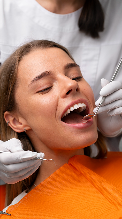
Optical Imaging Tools for Gum Disease Diagnosis

Gum disease affects millions in Australia, with 30.1% of adults diagnosed with periodontitis in 2017–18. Early detection is crucial to prevent severe health issues like heart disease and stroke. Traditional methods like X-rays often miss early signs, but advanced optical imaging tools are changing the game.
Key Tools for Gum Health:
- Optical Coherence Tomography (OCT): Non-invasive, high-resolution imaging to detect early gum changes.
- Gum Cameras: Live visuals of sub-gum areas for precise cleaning and diagnosis.
- Laser Light Scanning: Gentle detection of hidden plaque and tooth decay.
- Simple Light Tests: Quick yes/no detection of hard build-up on teeth.
- Fluorescence Spectroscopy: Highlights plaque maturity using light-induced autofluorescence.
Why Optical Imaging Matters:
- Better Accuracy: Detects issues earlier than traditional methods.
- Non-invasive: Safer and more comfortable for patients.
- Improved Outcomes: Helps dentists create precise treatment plans.
Quick Comparison Table:
| Technology | Best For | Advantages | Limitations |
|---|---|---|---|
| OCT | Early gum disease detection | Non-invasive, detailed imaging | High cost, limited depth (1–3 mm) |
| Gum Cameras | Sub-gum cleaning & diagnosis | Clear visuals, precise cleaning | Time-consuming, operator-dependent |
| Laser Light Scanning | Hidden decay & plaque detection | Gentle, no cutting required | May need additional confirmation |
| Simple Light Tests | Hard build-up detection | Fast, easy to use | Less accurate in wet conditions |
| Fluorescence Spectroscopy | Plaque maturity assessment | No staining, safe for children | Affected by blood or restorations |
The Future of Gum Care:
Optical imaging is transforming dental care in Australia, offering safer, more accurate ways to diagnose and treat gum disease. While costs and training remain barriers, these tools are set to become standard in dental practices nationwide.
Julia Walther: Optical coherence tomography in the oral cavity
1. Optical Coherence Tomography (OCT)
Optical Coherence Tomography (OCT) is a top tool for spotting gum disease. It uses near-infrared light to make very clear cross-section pictures of gum tissues, letting doctors see what’s under the gum. This way, not only is the image quality great, but it also makes the check-up easy on the patient.
Non-Invasive Check-Up
A key trait of OCT is that it does not invade the body. Unlike older ways that might poke around or take bits of tissue, OCT uses near-infrared light on the tissue. It reads how strong and fast the light comes back to make sharp images [2].
This method is smooth and painless for patients. The light goes into the tissue up to 2 millimetres, enough to check most gum structures without touching [2].
OCT is very good for watching how gums heal after surgery. Doctors can look at the healing gums after beauty fixes without bothering the new tissue [3].
Clear, High-Quality Images
The top-notch clarity from OCT sets it apart. It can show details as tiny as 1–15 micrometres, similar to what experts see in labs. This lets doctors spot tiny changes in gums that other tools might miss [3].
OCT does many types of scans like A-scans, B-scans, and C-scans, giving a full picture of the tissue [2].
With this high-tech tool, OCT can spot early changes in gums, often before they show up in usual checks. By studying the ways light shifts in the tissue, it sees changes [2].
Using it for Gum Health
OCT is handy for diagnosing gums. It checks sulcus depth accurately in both healthy and sick spots, giving good info for planning treatment [3]. This is helpful since old tools might not be as accurate or might hurt a bit.
It’s also good at showing how much gum disease has moved along [3]. Plus, OCT can outline gum tissue shapes and find hard bits above and below the gum [3]. All this helps doctors make better, sharper treatment plans.
Another plus is that OCT can show clear details of gingival tissues and tooth films [3]. This gives doctors a deep look at bacteria and affected areas, making treatments more aimed and helpful.
Limits in Use
OCT has lots of pluses but also some downsides. The short reach of the near-infrared light means it can’t go deeper than 1–3 millimetres, mainly just skin-deep structures like the top skin and films, not the deeper gum parts.
The gear itself is another big block. OCT tools cost a lot and are often too big, which limits how often they are used in teeth care places. Right now, most kits are made to scan the front teeth, which cuts down how much they can be used [4].
Looking at bigger spots can take a lot of time too, as many pics need to be taken to see the whole area. This can make the check-ups longer, which may tire both the patient and the person doing the work [4].
Also, the type of tissue can change how well OCT works. Things like how thick the enamel and dentin are, the state of the tooth, and any added materials can mess with how light is taken in, making it hard to read the pics. Materials that bend light the same way can also make pics look the same, which makes it tough to tell parts apart.
Even so, new tech steps are starting to tackle these issues. Newer OCT setups with longer waves (above 1,300 nanometres) are getting better at going deeper, and more made-for-the-job, smaller systems are being made to make the tech easier to use every day [4].
2. Gum Camera Look
Using a small camera, gum camera look lets doctors check under the gum line without having to cut open the gums. This tool helps dentists see parts that are hidden, making it easier to find and manage gum disease early on.
Soft Way In
A big plus of this method is that there’s no need to cut into the gums. It uses a thin light stick that goes into the gum. Together with cleaning and root smoothing, it helps slow down gum disease. The camera lets dentists see the root while cleaning, making sure they do a full job. Studies show fewer infections linger when using the gum camera compared to typical non-cut methods.
This gentle way also stands out with its high-tech picture feature.
Clear Pictures
The pictures from gum camera look are very clear. For example, the DeVA-1 2G camera can show live sharp images at 100 times the size of tiny spaces and roots under the gums. This lets dentists see and take out plaque, stone, and bugs well. Usual cleaning without seeing can miss up to 40% of bad stuff. The big zoom from the camera makes sure cleaning is deep and full.
Spotting Problems
Thanks to its clear pictures, gum camera look is great for spotting hidden issues. It can find things like tooth rot, root cracks, open fillings, and more under the gums. This is especially good for people with deep gum spaces or those getting care for ongoing gum trouble.
It also helps a lot with tooth implants. For example, too much cement on implants leads to problems 81% of the time. Using the camera to take off extra cement has helped many, with 74% of treated implants showing no signs of trouble after.
Hard Parts in Practice
Even with its perks, gum camera look has some limits. Studies said that when compared to usual methods, there’s no big change in gum healing or how much the gum sticks to the teeth. Also, these ways take more time and need doctors to be very good at reading the images. But as the tool gets better, these issues might become less of a worry, making it a big part of new gum care methods.
3. Laser Light Show for Teeth
The laser light shows on teeth use soft laser beams, often at a light size of 655 bits, to spot hidden things like tooth build-up and early tooth rot. By turning on the tooth’s own glow, this light show lets us see parts that are hard to check with old ways.
Soft Way In
A key plus of the laser light show is that it is all soft on the body. There is no need to cut or take out good bits, which keeps the gums whole while looking at them. It’s so gentle that there’s often no need for sleep meds [5].
How Well It Shows and Sees
This way gives good detail for finding hidden build-up under the gums. A study in Lasers in Med Science showed it was right 76.2% of the time in spotting this, better than another way, which was right only 68.2% of the time. Also, it was more likely to work the same way each time, with scores of 0.71 versus 0.54 in the other way [7].
These points show it works well, and is a big help for gum check-ups.
Use in Checking Gums
The laser light show is great for checking gums without cutting. It finds hidden build-up and early rot, which are often missed in normal look-overs. This clears a path for better care plans and acts as a no-rays partner to old X-rays.
Hard Parts and Thoughts
Though it helps a lot, the laser light show is not perfect. It may be wrong sometimes, which could lead to extra treatments if used alone [6]. To cut down risks, doctors should use this way with other check-up ways. While the light level is safe, right machine setup and learning for users are key for right results. We need stronger studies to fully get what it can do in many care places [6].
4. Simple Light Tests
Simple light tests offer a clear, yes-or-no way to spot hard buildup under the gums. They use two types of light – red and near-infrared – to tell apart the hard buildup from the healthy part of the tooth root. For example, tools like DetecTar check the light that bounces back from the tooth. This is then matched with saved data using a process to see if there is buildup. This method is a fast and sure add-on to other sight tools.
No Harm Way
The device has a tip like a common gum tool, making sure it does not hurt the gums. It tells you right away with a green light and a sound when buildup is found. This quick hint lets people know as it goes on, making it more hands-on and easy to get.
How Well It Spots Buildup
Unlike sight tools that show clear images, simple light tests only focus on yes-or-no spotting. Studies prove it to be more right than old touch tests, hitting a spot-on rate of 79% over 60% [8].
Use in Gum Checks
This tech is really good for finding hard buildup under the gum that might be missed in normal checks. It does a steady job in spotting buildup on single-root teeth, often doing better than on teeth with many roots [8]. Tests also show it is true to rely on, with match scores of 0.54 versus 0.39 for touch tests [8].
Problems in Clinics
Even with its good points, simple light tests have some downsides. They need special gear, and the work can take longer than old ways [9]. Also, fluids like spit, blood, or wash stuff can mess up the light tests, needing a clean and dry work area [7]. When put side by side with other sight ways, like laser tests, simple light tests have shown a bit less right (68.2% versus 76.2%) and steady (0.54 versus 0.71) results [7]. Knowing what it does well and not is key for using it right in clinics.
sbb-itb-2be92ed
5. Fluorescence Spectroscopy
Fluorescence spectroscopy stands out as a valuable optical technique for identifying and measuring dental plaque. This method works by detecting the natural autofluorescence of plaque on teeth. When exposed to a specific wavelength of light, microbial porphyrins in the plaque emit a red glow, which can be observed and measured by dental professionals.
Non-invasiveness
One of the key benefits of fluorescence spectroscopy is its gentle approach. It doesn’t harm tooth enamel or other hard tissues [11], nor does it require the use of probes or dyes. Instead, it relies on light-induced autofluorescence, making it completely non-invasive and safe for repeated use during treatment [10]. This is particularly important for children, as the technique can safely accommodate age- and infection-related changes in tooth fluorescence [14].
Another advantage is that it avoids the aesthetic drawbacks of traditional plaque detection methods, such as staining caused by disclosing solutions [10]. This makes it an appealing option for both patients and clinicians when assessing plaque for periodontal evaluations.
Applications in Periodontal Diagnosis
Fluorescence spectroscopy operates on a straightforward principle: porphyrins in dental plaque emit red fluorescence when exposed to a specific wavelength of light [10]. The intensity of this red glow corresponds to the thickness and maturity of the plaque, with brighter signals indicating more developed and potentially harmful deposits [12]. This feature allows dental professionals to pinpoint areas of concern with greater precision.
Research has shown that mature plaque, which often harbours anaerobic bacteria like Fusobacterium, Treponema, Tannerella, and Prevotella, can develop rapidly within just four days and is closely linked to conditions like gingivitis and periodontitis [12]. Quantitative light-induced fluorescence (QLF) builds on this principle by providing a non-invasive way to detect plaque while offering an objective measure of oral hygiene [12]. Studies confirm that red fluorescence is a reliable indicator of mature plaque, eliminating the need for additional staining [12]. This is particularly noteworthy given that periodontitis ranks as the sixth most common disease globally [12], with prevalence rates in some areas reaching as high as 99% for both periodontitis and gingivitis [13].
Limitations in Clinical Use
While fluorescence spectroscopy offers many advantages, it’s not without its challenges. Factors like blood, bacteria, and stained dental restorations can interfere with the accuracy of fluorescence readings [15]. For example, materials used in fillings, crowns, and other restorations may lead to false positives. Similarly, conditions like prolonged drying, the presence of pits and fissures, bacterial plaque, and calculus can affect the reliability of results [15].
Other practical issues include the time required for the process, which can be longer than traditional methods for detecting subgingival calculus. Operator technique also plays a significant role, as variability can impact results. Additionally, while the method requires inserting a device into the gingival pocket, it doesn’t generate images, which limits its visual utility. Blood within the pocket can further complicate the process, prompting recommendations to rinse the area with saline during measurements – a step that may add to the workload in busy dental settings.
Comparison of Optical Imaging Tools
Optical imaging tools vary in resolution, applications, strengths, and limitations, making them suitable for different clinical scenarios. Here’s a breakdown of their key features:
| Imaging Tool | Resolution | Key Applications | Primary Advantages | Notable Limitations |
|---|---|---|---|---|
| Optical Coherence Tomography (OCT) | ~20 µm | Cross‐sectional imaging of soft and hard tissues; 3D reconstruction | Non‐invasive, real‐time monitoring, no ionising radiation | High equipment costs; limited penetration depth (1–3 mm) |
| Periodontal Endoscopy | Visual inspection | Direct visualisation of subgingival calculus deposits | Faster treatment; enhanced root surface illumination | Requires insertion into gingival pockets; operator‐dependent |
| Laser Fluorescence Imaging | Molecular detection | Identifies subgingival calculus via emission wavelengths | Greater sensitivity and specificity than tactile probing | Accuracy affected by blood and dental restorations |
| Differential Reflectometry | Surface analysis | Detects calculus via light reflection patterns | Non-contact method | Limited clinical validation data |
| Fluorescence Spectroscopy | Molecular fluorescence | Plaque detection and evaluation of plaque maturity | Non-invasive; no staining needed | Influenced by blood; longer processing time |
Performance and Cost Considerations
Clinical performance varies across these tools. For instance, periodontal endoscopy significantly reduces treatment time, cutting it from 26.34±5.63 minutes per tooth to 15.23±3.35 minutes, compared to 12.56±3.50 minutes without endoscopic support [9]. Similarly, laser fluorescence imaging shows clear differentiation, with sound root surfaces measuring 6.2±6.5 arbitrary units versus 57.7±34.1 for subgingival calculus [9].
Cost is another major factor for Australian practices. Advanced systems demand substantial upfront investment, with annual maintenance costs ranging from A$1,500 to A$3,000. Swept-source OCT, while offering faster imaging and better resolution, comes at a higher price point [18][9].
Safety Benefits Over Radiography
Unlike traditional radiographic methods, optical imaging tools eliminate exposure to ionising radiation. For example, OCT provides non-invasive, non-destructive imaging, making it safer for repeated use during treatment. This is a significant advantage over radiography and dental CT, which involve radiation exposure [16].
Challenges to Consider
Despite their benefits, these technologies face challenges. High equipment costs, extended operation times, and factors like blood, bacterial interference, or metallic restorations can affect accuracy. Additionally, many studies on these tools remain in vitro, highlighting the need for more clinical research [9].
When choosing an optical imaging tool, consider the specific clinical requirements, budget limitations, and the level of diagnostic detail needed for effective periodontal treatment planning. Each tool brings unique capabilities to the table, helping clinicians achieve better outcomes.
Clinical Practice in Australia
In Australia, dental clinics are increasingly incorporating optical imaging into periodontal diagnosis. This shift reflects a growing emphasis on advanced technology in dental care, though the extent of its use varies across different practices. Building on earlier breakthroughs in optical imaging, these tools are enhancing both the precision of diagnoses and the effectiveness of treatment planning.
Current Technology Adoption Trends
The adoption of optical imaging technology in Australian dental clinics has gained significant momentum, particularly with the rise of AI-powered diagnostic platforms. For instance, in November 2024, Avant Dental highlighted that tools like Pearl’s Second Opinion® have boosted early decay detection rates and increased patient case acceptance by as much as 30% [21].
"It’s about delivering the best patient care. Early detection allows dentists to offer less invasive treatments and prevent more serious issues later. This benefits dentists by providing valuable insights and helps patients better understand their needs through AI visuals, leading to earlier and more frequent treatment acceptance." – Sheela Roth, Director of Clinical Operations at Pearl [21]
Diagnostic Accuracy Improvements
One of the key challenges in periodontal care has been the variability in diagnosis among practitioners. Studies indicate that diagnostic concurrence across clinicians examining the same radiographs is less than 50%. However, AI-powered optical imaging tools are helping to close this gap. Clinics utilising these platforms have reported a 30% rise in case acceptance within the first 90 days, alongside improvements in diagnostic accuracy by up to 37%, ensuring more consistent results across different practitioners [21].
Advanced Practice Integration
Australian dental practices are now adopting a more comprehensive approach by combining multiple optical imaging technologies. For example, Complete Smiles Bella Vista, led by Dr. James Hanna, integrates advanced diagnostic tools into their periodontal care and general dentistry services. These technologies complement traditional diagnostic methods, enabling the detection of subtle changes in periodontal tissues that might otherwise go unnoticed [19].
Beyond basic imaging, clinics are embracing three-dimensional tissue analysis. Technologies like Optical Coherence Tomography (OCT) deliver high-resolution cross-sectional images comparable to histology, while laser-induced fluorescence provides real-time numerical assessments of calculus deposits. These advancements allow practitioners to monitor outcomes of grafting procedures and track responses to treatments with greater precision [19].
Patient Communication and Safety Benefits
One of the standout advantages of optical imaging adoption in Australia is its impact on patient communication. Studies reveal that around 64% of patients struggle to understand traditional radiographs. Tools equipped with AI-enhanced visualisations help bridge this gap, making it easier for patients to grasp their diagnosis and treatment options. This not only improves treatment acceptance but also aligns with Australia’s stringent safety standards [21][19].
"Overall, optical methods can support traditional periodontal diagnosis and improve treatment planning and clinical periodontal care." – Fardad Shakibaie and Laurence Walsh [19]
Future Outlook and Training Requirements
The future of optical imaging in Australian dental practices looks promising, with experts predicting widespread adoption across the profession within the next decade.
"It’s hard to imagine that in five to 10 years from now you’ll walk into a practice that doesn’t utilise AI. I think it will become a natural part of the dental workflow." – Makenzie Harris, Director at Gamma Tech [21]
However, the successful integration of these technologies depends heavily on proper training. As Prof. William C. Scarfe aptly noted:
"I think Australia is at the forefront of access to technology, too. But like the United States, Europe and anywhere else in the world, it doesn’t mean you’re competent in the use of the technology. Just because you have a Ferrari doesn’t mean you can drive a Ferrari. So my role these days is to educate drivers." [20]
Conclusion
Optical imaging technologies are transforming how periodontal diseases are diagnosed, tackling long-standing challenges faced by dental professionals. With over a billion people worldwide affected by periodontal disease [17], the urgency for precise and early detection has never been greater.
Traditional diagnostic tools often fall short, detecting bone loss only after 30%-50% of resorption has occurred, which means early stages of the disease are frequently overlooked [1]. Optical imaging, however, offers a radiation-free alternative that delivers accurate diagnoses [1].
The improvements in diagnostic precision are striking. For instance, AI-driven tools have shown impressive results – DeepLabv3+ achieved sensitivity and specificity rates of 0.92 and 0.94, respectively [22]. These technologies also allow for the detection of inflammatory changes before bone loss becomes visible, giving practitioners a critical window to intervene early. Techniques like OCT provide detailed, high-resolution images of periodontal structures without the need for ionising radiation [1].
Modern periodontal care now requires a multidisciplinary approach, combining soft and hard tissue treatments. This demands a thorough, three-dimensional understanding of the periodontal apparatus [1]. Optical imaging delivers the detailed, multi-dimensional insights necessary for effective treatment planning [1].
AI-enhanced diagnostics are also paving the way for more advanced care. From analysing 2D radiographs to aiding in 3D crown reconstructions, AI is becoming an integral part of dentistry. For practitioners, adopting these tools means more precise treatment planning and better outcomes for patients [22].
For Australian dental practices, incorporating optical imaging technology is not just an opportunity to stay at the forefront of innovation – it’s a commitment to providing the best possible care. By overcoming the limitations of traditional diagnostic methods, these advancements promise to improve oral health outcomes and elevate the standard of periodontal care across communities.
FAQs
How does optical coherence tomography (OCT) help in detecting gum disease earlier than traditional X-rays?
Optical coherence tomography (OCT) provides detailed, high-resolution cross-sectional images of the gums and nearby tissues, allowing dentists to spot early signs of gum disease that might be missed with other methods. Unlike traditional X-rays, OCT excels at capturing soft tissue details, offering a clearer view of potential issues.
Another advantage of OCT is that it doesn’t use ionising radiation, making it a safer choice for regular monitoring and early detection. By enabling timely diagnosis, this imaging technology can lead to more effective treatments and better long-term oral health.
What challenges do dental clinics face when adopting optical imaging technologies for gum disease diagnosis?
Dental clinics often encounter a range of hurdles when bringing optical imaging technologies into their practice. One major challenge is the upfront cost – both in terms of purchasing the equipment and investing in comprehensive staff training. On top of that, team members may resist adopting new methods, which can slow down the transition.
Technical roadblocks also play a part. For instance, ensuring these new tools work seamlessly with current systems while maintaining high diagnostic standards can be tricky. Many clinics also don’t have dedicated in-house IT teams, instead relying on external providers. This reliance can add another layer of complexity to the integration process.
To make the adoption smoother, clinics should focus on careful planning, prioritise thorough training for their staff, and choose technologies that fit well with their current workflows.
How does fluorescence spectroscopy help in identifying mature dental plaque, and what are its challenges in clinical use?
Fluorescence spectroscopy offers a useful method for identifying mature dental plaque by detecting its red autofluorescence. This red glow is often linked to plaque that has advanced to a stage where it could contribute to periodontal disease. The technique provides a non-invasive way to evaluate plaque maturity, making it a helpful tool for early diagnosis and planning treatment.
That said, there are some challenges. The method can struggle in areas with limited visibility, such as spots affected by malocclusion or shading. Additionally, certain devices might underestimate plaque levels, and the approach isn’t fully validated for every clinical scenario. More research is needed to improve its accuracy and expand its use in everyday dental practice.
Related Blog Posts
- Periodontal Pocket Depth: Measurement Techniques
- AI Research in Oral Disease Detection: Key Findings
- Advances in Gum Grafting Materials and Techniques
- Radiographic Staging of Periodontal Diseases
Important Notice: Any surgical or invasive procedure carries risks. Before proceeding, you should seek a second opinion from an appropriately qualified health practitioner.
Individual results may vary. The information provided in this article is for educational purposes only and does not constitute medical advice.
Checkout Related Blogs


Get in touch with us
For more information, call us now to start feeling better. Or fill the form below to make appointment
The Latest News from Complete Smiles
How to Clean Clear Plastic Retainers
Checklist for Choosing Wearable Dental Devices
Checklist for Choosing Cloud AI Platforms in Dentistry
Complete Smiles Bella VistaAccepts All Major Health Funds, Including


